
Aquaculture, being a crucial sector around the world, is a major aspect of food security and is an essential source of affordable and high-quality proteins for the growing global population (Saleh et al., 2025). Diseases and environmental hazards are major hurdles for sustainable fish production and generate substantial financial losses with rising productivity in aqua-culture (Okasha et al., 2025). Rising intensity of fish farming also results in greater susceptibility of fish to a large number of pathogens, which results in environmental stresses and may significantly restrict growth and induce infectious diseases of aquatic animals (Farooq et al., 2025). In aquaculture industry, antibiotics were used for improving growth and controlling bacterial disease (Hoseinifar et al., 2024), but they are no longer advised as the first line of treatment (Oliveira et al., 2025). This is due to the numerous health hazards associated with the usage of these compounds, including the development of resistant bacteria, the direct or indirect spread of drug resistance to people (Choi et al., 2025). As a result, it is now more important than ever to establish an environmentally friendly compound (Mashhadizadeh et al., 2024). The interest in nutritional management is also rising in the aquaculture feed sector, particularly regarding the use of natural feed additives as a replacement for antibiotics (Tian et al., 2024; Fu et al., 2025).
The employment of natural origin-based feed additives, including probiotics, prebiotics, phytochemicals, and acidifiers, might improve the growth rate and survival ratio of fish by improving the utilization of the diet, the diversity of gut microbiota, the immunoresistance, and the resistance to disease (Raslan et al., 2025).
Currently, the use of probiotics represents a significant role as an alternative to chemical drugs and antibiotics and disinfectants in controlling microbial populations and preventing diseases in fish farming (Islam et al., 2025). Probiotics have been used in fish farming as a potential alternative to bacterial infections, which enhances the activity of the immune system in different species of fish (Fadel et al., 2025). The Lactobacillus genus, which has many of the most effective probiotic bacteria and was first explored in 1780, has emerged as one of the widely used types of probiotics to inhibit pathogens (Majeed et al., 2025; Thuy, 2025). Lactobacillus casei probiotic, as feed additive, boosts growth and the immunity of fish to pathogens by enhancing intestinal morphology and microbial populations (Choi et al., 2025).
Resveratrol, a non-flavonoid polyphenol extracted from grape skin and leaves, peanuts, pine nuts, Chinese cinnamon, and many other plants (Tian et al., 2024), is another type of feed additive that has been widely used in fish feed due to its antioxidant, anti-inflammatory, and metabolic-regulating properties (Ye et al., 2023; Fu et al., 2025) and has shown positive effects on growth performance, gut health, and immunity (Tian et al., 2024; Yang et al., 2024).
Grass carp (Ctenopharyngodon idella) is an economical herbivorous fish. They are a widely cultivated freshwater fish species worldwide due to their high nutritional value, rapid growth rate, relatively low rearing costs, and affordable price to consumers (Xie et al., 2024; Shen et al., 2025).
According to several studies, feeding different types of probiotics alone or in combination showed positive effects on growth performance (Xie et al., 2024; Preena et al., 2025; Choi et al., 2025). Therefore, it appears that combining the herbal compound resveratrol and the probiotic Lactobacillus casei in fish diets can lead to a wide range of biological activities, synergistic effects, and faster stimulation of physiological reactions in fish. So, this study was conducted to evaluate the potential effects of resveratrol and probiotics (L. casei) alone or in combination on growth performance, antioxidant capacity, growth-related genes, and resistance to Aeromonas hydrophila in grass carp.
Material and methods
Ethical approval
The experimental animal care and use committee of the University of Kharazmi (IR.KHU.REC.1403.097) approved all procedures of the current study.
Experimental diets
Lactobacillus casei were obtained from the Iranian Biological Resource Center (Tehran, Iran) and after being transferred to the laboratory, they were first cultured in de Man, Rogosa, and Sharpe (MRS) broth media at 37°C for 24 hours. Then, a re-culture was cultured at 37°C for 20 hours and subsequently, L. casei cells centrifuged, and the cell pellet was washed twice with sterile normal saline to approximately 1 × 10⁸ CFU/mL (Aini et al., 2024). Resveratrol (RES) was purchased from commercial company (Sigma Aldrich, CAS number: 501-36-0). To experimental diets, a basal diet was formulated (Adineh et al., 2021) and then supplemented by 1 × 10⁸ CFU/mL L. casei and 0.3 g RES per kg diet (Zheng et al., 2017). Four experimental diets were prepared including un-supplemented diet (control, T0), 1 × 10⁸ CFU/mL L. casei (T1), 0.3 g resveratrol per kg diet (T2), and combination of probiotic and RES (T3).
Fish and rearing conditions
To carry out this project, 300 fish with an average weight of 4.89±0.08 g were initially obtained from the warm-water fish breeding center (Karaj, Iran) and transferred to the research hall of the Faculty of Biological Sciences, Kharazmi University, where they were stored in three 200-L tanks. The fish were acclimated to rearing conditions for two weeks and fed with control diet. After two weeks, the fish were weighed individually and a total of 240 were distributed in twelve 100-L tanks with a density of 20 fish per tank and assigned to four treatments and three replications. The fish were fed with four experimental diets including T0: control-without additives; T1: containing L. casei; T2: containing resveratrol; T3: containing a combination of both, all for a duration of 56 days. During feeding trial, fish were fed three times every day (at 8:00, 12:00, 16:00) in a satiated state. Uneaten food was collected one hour after each feeding, after drying at 60°C, which was used in the food consumption calculations. The tanks were supported by continuous aeration and subjected to daily siphoning to remove waste, whereby 50% of the water was drained and replaced with fresh water each day before the first feeding time. Physicochemical parameters were regularly monitored: pH, 7.5±0.2; temperature, 28.4±1.3°C; dissolved oxygen level, 7.4±0.3 mg/L; and photoperiod, 12L:12D. After 56 days, the feeding was stopped, the fish were anesthetized by clove powder (200 mg/L) and weighed individually to evaluate growth performance.
Sampling
To analyze mucosal immune factors, three fish from each tank (nine fish per treatment) were placed separately in polyethylene bags containing 2 ml of chloride sodium (50 mM) and the mucus was collected by gently rubbing, and the mucus from the three fish was centrifuged (1500 g for 10 min) and then the supernatants were transferred to sterile tubes to measure immune factors (Darvishi et al., 2022). For antioxidant factors, three fish per tank (nine fish per treatment) were randomly selected and anesthetized with clove powder (200 mg L−1 water, Adineh et al., 2021) and livers were removed and stored in nitrogen tank and transferred to a −80°C. Also, the liver of three other fish (nine fish per treatment) was sampled to examine the expression of genes related to growth. It was immediately stored in a nitrogen tank and transferred to a −80°C.
Immune and antioxidant factors
Total protein (TP) content in skin mucus samples was determined following the method of Lowry (1951). The concentration of total immunoglobulin (Ig) in skin mucus was assessed using the procedure described by Siwicki and Anderson (1994), where Ig levels were calculated by subtracting the protein concentration of the sample before and after treatment with polyethylene glycol. Mucosal lysozyme (LYZ) activity was analyzed using the turbidimetric technique outlined by Ellis (1990). In this assay, 50 μl of mucus sample was combined with 2 ml of a bacterial suspension prepared in citrate-phosphate buffer, and the reduction in absorbance was monitored at 450 nm over a 15-minute period using an ELISA reader. LYZ activity for each sample was determined with reference to a standard curve generated using egg white lysozyme (Sigma-Aldrich). The pro-tease activity of fish mucus was measured by the azocasein method. In this method, a certain amount of mucus was added to a 0.7% azocasein solution and the mixture was incubated for 20 hours at 30°C with continuous stirring. Then, the reaction was stopped by adding trichloroacetic acid and the mixture was centrifuged at 15,000 g for 5 minutes to separate the supernatant. After adding sodium hydroxide (1N), the absorbance of the solution was measured at a wavelength of 450 nm. The protease activity of the samples was calculated relative to the positive control, a 5 mg/ml trypsin solution with 100% activity.
The liver tissues of each fish were individually thawed on ice and the tissues were homogenized with 1:9 saline, centrifuged, then the supernatant was taken as the tissue homogenate. Antioxidant markers were evaluated in liver tissue, including malondialdehyde (MDA) levels and the enzymatic activities of superoxide dismutase (SOD), glutathione peroxidase (GPX), and catalase (CAT), all measured using commercial diagnostic kits (Pars Azmoun, Iran).
Growth gene expression
Total RNA was extracted from liver tissue of fish (nine fish per treatment) using the TRI-zol reagent (Yekta Tajhiz Azma, Tehran, Iran) following the manufacturer’s protocol. Briefly, each tissue sample was homogenized in 0.7 mL of RNX Plus solution and incubated at room temperature for 7 min, followed by the addition of 200 µL chloroform. After vigorous mixing and incubation for 3 min, samples were centrifuged at 14,000 g for 15 min at 4°C. The clear supernatant was transferred to a new tube, mixed with 1 mL of cold isopropanol, and incubated at −20°C for 30 min. RNA was pelleted by centrifugation at 14,000 g for 10 min, washed with 1 mL of 70% ethanol, and centrifuged at 8000 g for 5 min at 4°C. The pellet was air-dried under a clean hood and dissolved in 50 µL RNase-free water at 55°C for 5 min. Extracted RNA was stored at −70°C until further use.
RNA concentration and purity were measured with a NanoDrop 2000 spectrophotometer, and RNA integrity was confirmed by 1% agarose gel electrophoresis. Only RNA samples with A260/A280 ratios between 1.8 and 2.0 were used for cDNA synthesis. First-strand cDNA was synthesized using the YT cDNA synthesis kit (YT5400; Yektatajhiz, Tehran, Iran) according to the manufacturer’s instructions.
Gene expression analysis of Ghrelin and IGF-1 was performed using SYBR Green-based quantitative real-time PCR with gene-specific primers (Table 1). GAPDH was used as the reference gene, and its stability across treatments was confirmed prior to analysis. Primer efficiencies were evaluated using standard curves and were within the acceptable range. All qPCR reactions were performed in technical triplicate, and amplification specificity was confirmed via melting-curve analysis.
Table 1.
Primer sequence of genes
| Gene | Primer | Primer efficiency (%) | Accession number |
|---|---|---|---|
| Ghrelin | F: 5′ CGCTCTTTCCTTATGTCTCG 3′ R: 5′ TCATCCTCTTTAATCACTGGG 3′ | 96.3 | XM_051896386.1 |
| IGF | F: 5′ GTGGAGACAGGGGCTTTTA 3′ R: 5′ AAAGCAGCATTCGTCCACA 3′ | 95.4 | XM_051891268.1 |
| GAPDH | F: 5′ CCATCACAGCCACACAGAAG 3′ R: 5′ TGGAGGCTGGGATAATGTTC 3′ | 102.7 | XM_051865633.1 |
The qPCR cycling program consisted of an initial denaturation at 95°C for 10 min, followed by 40 cycles of 95°C for 15 s (denaturation), 57°C for 40 s for IGF-1 and GAPDH and 55°C for Ghrelin (annealing), and 72°C for 20 s (extension). A final melting curve analysis was performed from 57 to 95°C (IGF-1 and GAPDH), 55 to 95°C (Ghrelin) for 10 s to verify amplification specificity. Ct values were normalized to the reference gene, and relative gene expression was calculated using the 2–ΔΔCt method, with results expressed relative to the control group.
Disease resistance
The pathogenic strain Aeromonas hydrophila (IBRC-M 10814) used in this experiment was obtained from the Iranian Biological Resource Center (IBRC). According to the IBRC records, the strain had been taxonomically confirmed through 16S rRNA gene sequencing. For challenge preparation, the bacterium was cultured in tryptic soy broth (TSB) and incubated at 28°C for 24 h under continuous shaking (150 rpm) to obtain bacteria in the logarithmic growth phase. The culture was then harvested by centrifugation at 850 g for 15 min, washed twice with sterile phosphate-buffered saline (PBS), and resuspended in PBS to achieve the desired concentration.
To confirm the viable bacterial concentration, serial ten-fold dilutions were prepared and plated on tryptic soy agar (TSA) using the spread-plate method. Plates were incubated at 25°C for 24 h, and colonies were counted to verify the actual 1 × 10⁸ CFU mL–1 suspension used for the challenge.
After the 8-week feeding trial, eight fish from each tank were challenged via intraperitoneal (IP) injection, which is a standard method for A. hydrophila challenge in carp. Each fish received 0.1 mL of the verified bacterial suspension using a 1-mL insulin syringe fitted with a 26-gauge needle, injected into the ventral abdominal cavity, slightly anterior to the pelvic fins. The challenge dose was selected based on previously published studies using A. hydrophila in grass carp (Shohreh et al., 2025)
During the 14-day challenge period, fish were maintained under the same environmental conditions used during the feeding trial: water temperature 28.4±1°C, dissolved oxygen above 7.4±0.3 mg L−1, pH 7.2–7.6, and total ammonia nitrogen below 0.02 mg L−1. Water quality was monitored daily, and fish were not fed during the first 48 h post-infection to reduce stress and avoid confounding physiological responses; thereafter, they were fed once daily at a maintenance ration.
Fish were observed twice daily for mortality and clinical signs. Typical signs included hemorrhagic lesions on the skin and fin bases, lethargy, loss of equilibrium, exophthalmia, abdominal distension, and darkening of body coloration, which are characteristic of motile aeromonad septicemia. Mortality was confirmed by the absence of opercular movement and lack of response to tactile stimulation.
Data analysis
For data analysis, the normality of the distribution of variables was first checked using the Shapiro–Wilk test. Then, the Levene’s test was used to check the homogeneity of variances to ensure that the variance between groups was the same. After confirming the assumptions of normality and homogeneity of variance, the data were compared using the one-way ANOVA test. In cases where a significant difference was observed between the groups (P<0.05), the Duncan’s multiple range test was used to determine the difference between the means of the groups.
Results
Growth performance
Significant differences were observed in growth factors between the control and treatments fed with supplemented diets (Table 2). The supplemented diets (T1, T2, and T3) increased the final weight (FW), weight gain (WG), and specific growth rate (SGR) of the fish, and the highest value was obtained in the treatment that was fed L. casei and RES simultaneously (T3) (Table 2, P<0.05). Supplemented diets had a significant effect on fed conversion ratio (FCR) and the lowest rate was observed in the T3 (Table 2, P<0.05).
Table 2.
Growth performance of grass carp fed with probiotic Lactobacillus casei and resveratrol (RES) for 56 days
| Initial weight (g) | Final weight (g) | Weight gain (g) | FCR | SGR | Survival rate (%) | |
|---|---|---|---|---|---|---|
| T0 | 4.89±0.08 a | 10.39±0.10 a | 5.51±0.07 a | 3.27±0.04 c | 1.35±0.02 a | 100 |
| T1 | 4.93±0.09 a | 11.06±0.22 ab | 6.12±0.16 ab | 2.95±0.08 b | 1.44±0.02 ab | 100 |
| T2 | 4.84±0.14 a | 11.34±0.12 bc | 6.50±0.10 bc | 2.77±0.04 ab | 1.52±0.04 ab | 100 |
| T3 | 4.87±0.11 a | 11.84±0.19 c | 6.95±0.26 c | 2.60±0.10 a | 1.58±0.06 b | 100 |
Immune and antioxidant factors
Lysozyme activity (Figure 1 A) increased significantly (P<0.05) in T2 and T3 treatments compared to the control group (T0), while T1 treatment showed a slight but non-significant (P>0.05) increase compared to the control. Protease activity (Figure 1 B) also showed a similar pattern, so that T2 and T3 treatments had significantly higher levels of protease compared to the control (P<0.05). The total immunoglobulin (Figure 1 C) level in T2 treatment was the highest and was significantly higher than T0 and T1 (P<0.05), while T3 was at an intermediate level between T1 and T2. The concentration of total protein (Figure 1 D) in the treatment groups showed a significant increase compared to the control and reached its highest level in T2 and T3 treatments (P<0.05).
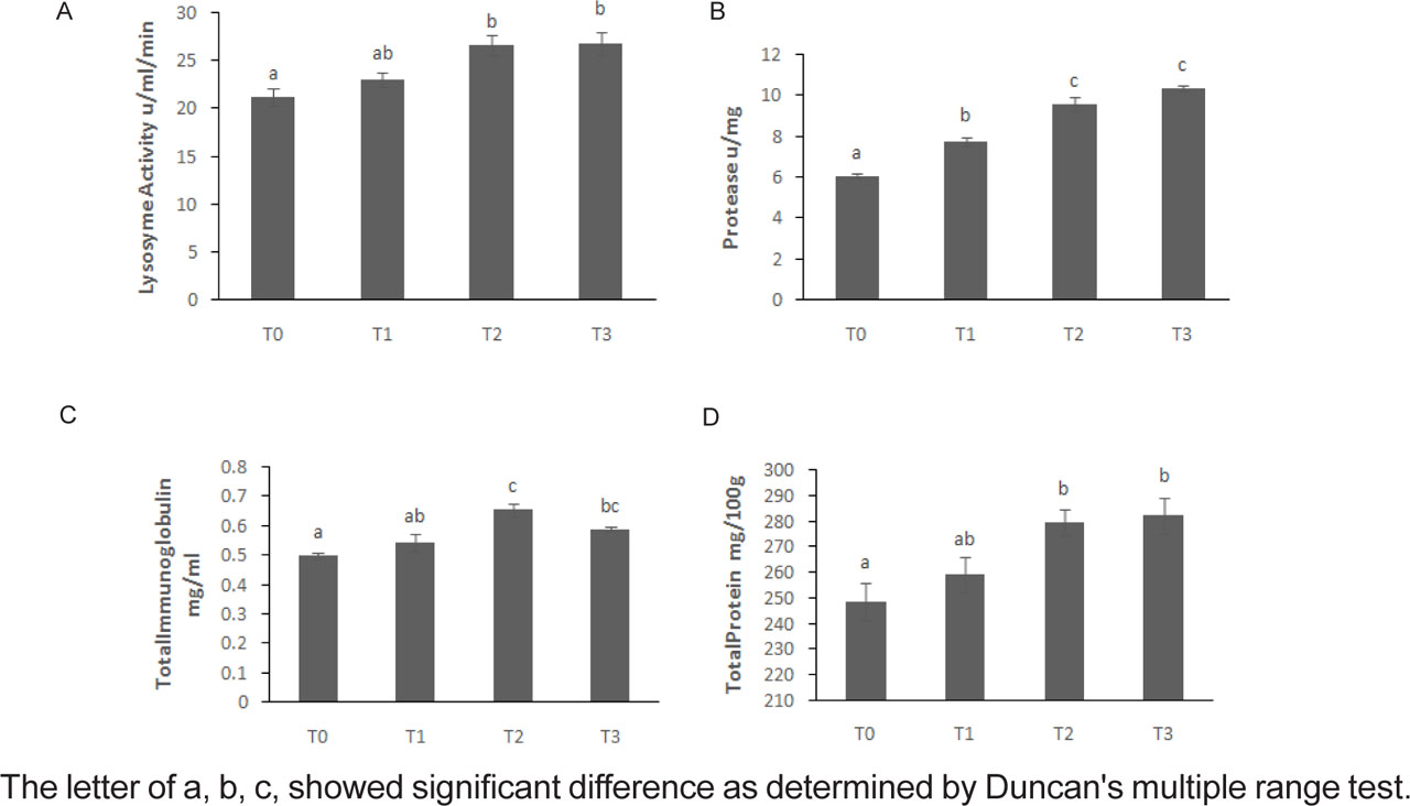
Figure 1.
Mucosal immune components of the grass carp after 56 days of feeding with experimental diets, Lactobacillus casei and resveratrol. T0: basic food as control; T1: 1 × 10⁸ CFU/mL Lactobacillus casei; T2: 0.3 g resveratrol/kg; T3: Lactobacillus casei + resveratrol.
CAT (Figure 2 A) activity in T2 and T3 treatments was significantly higher than the control group (T0) (P<0.05), while T1 did not show a significant change. The level of MDA (Figure 2 B) was significantly decreased in T2 and T3 treatments compared to the control group (P<0.05). GPx (Figure 2 C) activity increased, with the highest level observed in T3 treatment (P<0.05). SOD (Figure 2 D) activity increased in the treatments compared to the control group, and the lowest value was observed in T0 (P<0.05).
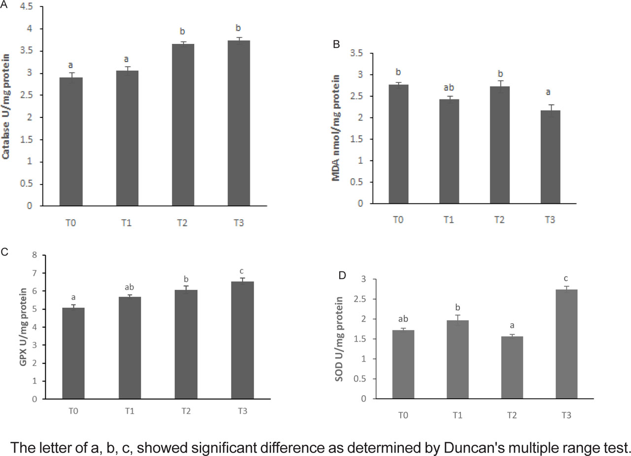
Figure 2.
Liver antioxidant enzyme activity and oxidative stress indices (MDA) in the grass carp after 56 days of feeding with experimental diets, Lactobacillus casei and resveratrol. T0: basic food as control; T1: 1 × 10⁸ CFU/mL Lactobacillus casei; T2: 0.3 g resveratrol/kg; T3: Lactobacillus casei + resveratrol.
Gene expression
The expression of Ghrelin (Figure 3 A) in the control group (T0) showed the lowest value, while the T1, T2 and T3 treatments significantly increased the expression of this gene compared to the control (P<0.05). For the IGF1 (Figure 3 B), the trend of increasing expression is also clear; the control group (T0) had the lowest expression, the T1 and T2 treatments both showed a significant increase compared to the control, and the T3 treatment had the highest expression and was significantly different from all groups (P<0.05).
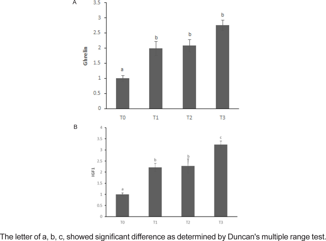
Figure 3.
Growth gene expression in the grass carp after 56 days of feeding with experimental diets, T0: basic food as control; T1: 1 × 10⁸ CFU/mL Lactobacillus casei; T2: 0.3 g resveratrol/kg; T3: Lactobacillus casei + resveratrol.
Disease resistance
Based on the survival curve (Figure 4), the mortality rate in the control group (T0) increased faster than in the other treatments. In contrast, the T3 treatment showed the lowest mortality rate and the longest lifespan during the experimental period (Table 3). The T1 and T2 treatments also caused a relative reduction in mortality compared to the control, but their effect was less than that of T3.
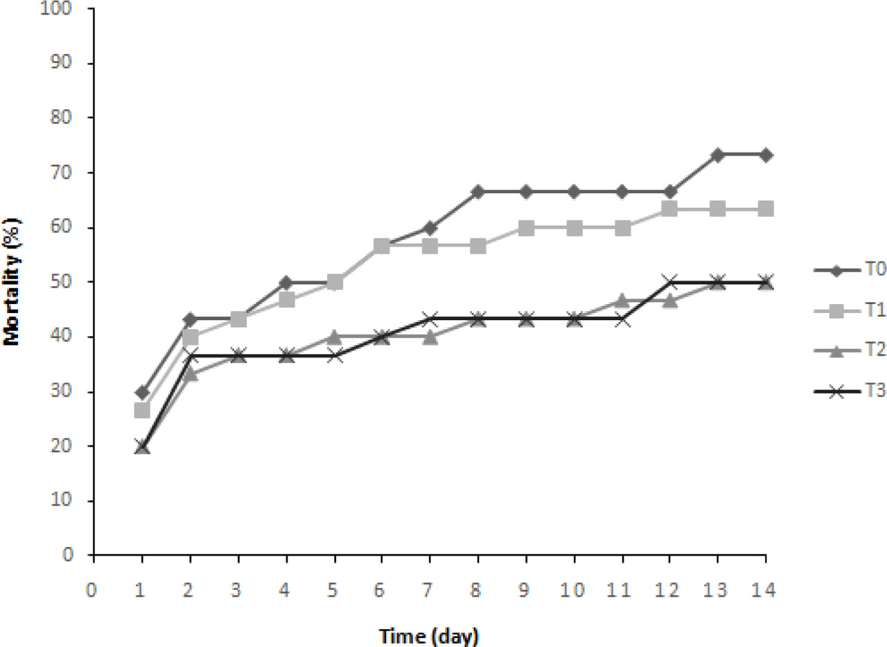
Figure 4.
Cumulative mortality rate of grass carp after 56 days of feeding with Lactobacillus casei and resveratrol. T0: basic food as control; T1: 1 × 10⁸ CFU/mL Lactobacillus casei; T2: 0.3 g resveratrol/kg; T3: Lactobacillus casei + resveratrol.
Table 3.
Effect of probiotic Lactobacillus casei and resveratrol on mortality rate and the relative percentage of survival (RPS) of grass carp after 14 days post-challenge by Aeromonas hydrophila
| Treatment | Mortality rate (%) | Survival rate (%) | RPS |
|---|---|---|---|
| T0 | 73.33 b | 26.67 a | – |
| T1 | 63.33 ab | 36.77 ab | 10.00 |
| T2 | 50.00 a | 50.00 b | 28.57 |
| T3 | 50.00 a | 50.00 b | 28.67 |
Discussion
Recently, the use of dietary supplements, including probiotics and plant derivatives, has become widespread in aquaculture (Gabr et al., 2023; Preena et al., 2025). In this study, the use of L. casei and resveratrol alone or in combination efficiently improved the growth performance (WG, FW, FCR, and SGR) compared with the control group. The results showed that the combination of L. casei and resveratrol had a higher performance compared to the other groups. Consistently, the use of resveratrol and the probiotic Lactobacillus acidophilus (Gabr et al., 2023, and resveratrol and two other probiotics, namely Lactobacillus acidophilus and Bifidobacterium bifidum (Naderi Farsani et al., 2021) enhanced growth performance and feed conversion in common carps and rainbow trout, respectively. Similarly, the use of resveratrol was found to significantly affect the improvement of growth performance and feed conversion in snakehead fish, Channa argus (Tian et al., 2024). The results showed that the lower the value of the feed conversion ratio, the less the feed consumed to grow the fish (Ahmed et al., 2025 a, b). The lower feed conversion ratio in the current study by fish fed the probiotic L. casei and RES indicates improved feed efficiency and better nutrient utilization, similar to El-Haroun et al. (2006), Islam et al. (2021), and Gule and Geremio (2022), who reported that the use of probiotics reduces feed waste and enhances growth performance in Nile tilapia (O. niloticus). In support, Choi et al. (2025) demonstrated that the supplementation of probiotics in the diet of juvenile olive flounder resulted in lower FCR. In addition, the dietary inclusion of RES as a natural supplement has also been reported to enhance growth performance and increase feed utilization rates (Tian et al., 2024; Yang et al., 2024). Consequently, based on the above, the application of both probiotics and resveratrol in the grass carps aided in the promotion of growth performance by the increase of the beneficial bacteria count in the digestive tract of the host (Gabr et al., 2023; Naderi Farsani et al., 2021), hence resulting in enhanced digestion and absorption processes (Preena et al., 2025). Furthermore, the lactic acid bacteria in the host’s gut suppress the growth of the pathogens in the digestive tract (Choi et al., 2025).
The antioxidant system is the primary defense mechanism of fish cells affected by oxidative stress (Gabr et al., 2023). The findings of the present study revealed a significant rise in the concentration of the anti-oxidant enzymes (SOD, CAT, and GPX) in the probiotic and resveratrol-treated group, alone or in combination, compared to the control group (Naderi Farsani et al., 2021). Moreover, the combination of the probiotic L. casei and resveratrol-supplemented diet exhibited higher antioxidant potential compared to the other group, as reflected by the significantly higher concentration of SOD, CAT, and GPX and the lower concentration of MDA. In addition, the immune factors of mucus were measured in grass carp fed a probiotic and resveratrol-supplemented diet, alone or in combination, and the results revealed a higher concentration of lysozyme, TP, and immunoglobulin in fish fed a probiotic and resveratrol-supplemented diet compared to the control group. These findings agree with some research papers about the immune-stimulating effect of the phytogenic compounds and probiotics (Chizhayeva et al., 2022; Gabr et al., 2023). For instance, Babakarami et al. (2025) found that probiotics have a significant efficacy to enhance the levels of the immune factors lysozyme and immunoglobulin in fish. Many studies have warranted the use of probiotics and herbal supplements as immunostimulatory agents in fish (Naderi Farsani et al., 2021; Gabr et al., 2023), through boosting the antioxidant defense mechanism and the immune system (Aydın and Barbas, 2020; Elumalai et al., 2020; Naderi Farsani et al., 2021). This may have been due to the ability of feed supplements to enhance antioxidant activity through the improvement of the immune response and gut microflora.
In this study, the fish receiving probiotics and resveratrol separately and together had an increase in the level of growth hormone and IGF-1 compared to the control group. These findings are in agreement with Kuebutornye et al. (2020), Back et al. (2020) and Choi et al. (2025), wherein they found that the use of probiotic supplements caused an increase in the expression of genes encoding for the growth hormone in fish. Similarly, Jang et al. (2023) and Choi et al. (2025) found that adding the probiotic Bacillus sp. into the diet of red sea bream caused an improvement in the level of the growth hormone; this improved the level of growth performance as well. Hence, this might similarly contribute as a mechanism through which L. casei enhances the level of grass carp’s growth parameters in this study.
In fish farms, bacterial infections can cause significant mortality (Raslan et al., 2025). Therefore, immunostimulants are primarily used to strengthen the immune system and enhance resistance to infectious diseases. In this study, consuming probiotics and resveratrol, either alone or in combination, increased the resistance of grass carp to A. hydrophila more than those fed a basal control diet 14 days after infection. The mortality rate was higher in the control group compared to the other dietary treatment groups. In contrast, the survival rate and percentage survival (RPS%) were higher in the groups treated with the supplemented feed compared to the control group. These results agree with Reyes-Becerril et al. (2021) and Babakarami et al. (2025) who found that the use of probiotics in fish feed increased resistance to pathogenic infections with A. hydrophila. Similarly, Raslan et al. (2025) reported that dietary probiotics improved Nile tilapia fingerlings resistance to pathogenic infections with Aeromonas veronii. Abou-El-Atta et al. (2019) found that the use of the dietary probiotic Lactobacillus plantarum improved the resistance of Nile tilapia to Aeromonas sobria infection. Therefore, it is recommended to use diets supplemented with a combination of probiotics and resveratrol in aquaculture, as they increase fish immunity, reduce oxidative stress, and maintain healthy gut microbiota, leading to resistance to pathogenic infections.
Conclusions
Based on this study, it was found that probiotics Lactobacillus casei and resveratrol individually or in combination have had positive impacts on grass carp in terms of growth performance, antioxidant ability, genes involved in growth, and alleviating challenges posed by bacterial pathogens A. hydrophila. Based on this data, it could be concluded that inclusion of the resveratrol and probiotics complex in aquatic feed could be an efficient substitute for antibiotics. More studies should be conducted to determine the role of probiotics and resveratrol compound values on fish and resistance to sickness.
Notes
Acknowledgments
This research was supported by the University of Kharazmi and partially supported by Chiang Mai University.