
Rainbow trout, Oncorhynchus mykiss, is a key species in the aquaculture industry, prized for its economic value, rapid growth rate, and adaptability. According to FAO (2023), global trout production has surpassed 950,000 tons annually. However, disease outbreaks pose a significant challenge in this sector, as they do in other areas of aquaculture (Woo and Bruno, 2011). Most fish pathogens are opportunistic and can be managed through effective management practices. Notably, Aeromonas hydrophila, Streptococcus iniae, and Yersinia ruckeri are three well-documented opportunistic pathogens that impact trout farms (Woo and Bruno, 2011).
When disease outbreaks occur on a fish farm, the common recommendation is to use antibiotic-containing feeds. However, sick fish often lose their appetite and do not consume enough feed to receive the necessary daily dosage of antibiotics (Hoseini and Yousefi, 2019). Furthermore, the overuse of antibiotics has led to the emergence of resistant pathogens, which further diminishes the effectiveness of antibiotic treatments (Nhinh et al., 2021). Consequently, one of the primary strategies for mitigating disease-related losses in aquaculture is to enhance fish health to prevent outbreaks.
Feed supplements in aquaculture offer numerous benefits that improve fish health, growth, and overall production efficiency (Lee et al., 2015). These supplements frequently contain bioactive compounds with immunostimulant and antioxidant properties, which can bolster the immune system of fish and reduce the likelihood of disease, resulting in healthier and more resilient fish populations. Probiotics, in particular, play a crucial role in promoting fish well-being in aquaculture, ultimately enhancing production efficiency, disease management, and environmental sustainability (Hoseinifar et al., 2018, 2021).
Yeasts are recognized as promising probiotics in aquaculture, valued for their nutritional benefits and bioactive compounds that enhance animal feed (Czerucka et al., 2007). The significantly larger size of yeast cells compared to bacterial cells may explain why even a modest introduction of yeast [104 colony-forming units (cfu)/g] in feed can yield positive effects in the host (del Valle et al., 2023). By occupying the fish gut, yeasts reduce available space for pathogenic microbes. Additionally, their cell wall components, such as beta-glucan and mannan, have been shown to promote gut development and bolster host immunity (Meena et al., 2013; Torrecillas et al., 2014).
Among eukaryotic microorganisms, Saccharomyces cerevisiae is the most extensively studied, owing to its widespread application in the food, alcoholic beverage, and pharmaceutical industries (Tovar-Ramírez et al., 2004, 2010). This species has gained popularity in aquaculture due to its health-promoting effects on various cultured species. Extensive research has focused on the use of yeast in aquaculture, highlighting its nutritional advantages and its capacity to enhance growth and health responses in fish (Abass et al., 2018; Cid García et al., 2020; El-Bab et al., 2022). The inclusion of S. cerevisiae or its derivatives in the diet of rainbow trout has been shown to improve gut mucosal transcription and immunity (Rawling et al., 2021; Richard et al., 2021).
However, S. cerevisiae comprises numerous strains, each exhibiting specific characteristics across different species. For instance, studies by Waché et al. (2006) and Sheikhzadeh et al. (2016) demonstrated that S. cerevisiae var. boulardii CNCM I-1079 was more effective than the S. cerevisiae strain NCYC Sc 47/g in stimulating immune capacity, digestive tract development, and disease resistance in rainbow trout. This underscores the need to investigate new strains in aquaculture for their potential benefits. Therefore, the present study aimed to evaluate the effects of S. cerevisiae strain PTCC5052 as an in-feed probiotic on immunological and antioxidant parameters, gut microbial communities, and stress responses in rainbow trout fingerlings.
Material and methods
Yeast culture
Yeast was obtained in lyophilized form from the Iranian Research Organization for Science and Technology (Tehran, Iran). It was cultured on potato dextrose agar for 24 h before being inoculated onto nutrient agar for mass production. After 48 h of culturing on nutrient agar, the cells were harvested and suspended in sterile physiological saline at a concentration of 1 × 10⁸ cfu per mL. This stock was utilized for feed supplementation within 24 h.
Feed preparation, fish rearing, and stress induction
Juvenile rainbow trout (10 g) were sourced from a private supplier in Mazandaran province. Ninety fish were distributed across nine aquaria, each containing 45 L of dechlorinated tap water. The fish were fed either a control diet (Faradaneh Co., Shahrekord, Iran) or the control diet supplemented with yeast at concentrations of 1 × 10⁷ or 1 × 10⁸ cfu/g (Zargham et al., 2011). Feed supplementation was performed by top-dressing and 3% gelatin was used to avoid leaching (Hassanalizadeh Chari et al., 2020). Each feed was administered to three aquaria at a daily rate of 3% of the total biomass for a duration of 30 days. The aquaria were siphoned daily to remove fish waste, and 30% of the water was replaced with clean water. Water parameters, including temperature (15.3±0.29°C), pH (7.69±0.19), dissolved oxygen (6.89±0.36 mg/L), and total ammonia (0.98±0.13 mg/L), were measured daily using digital equipment as previously described (Yousefi et al., 2025).
At the conclusion of the feeding trial, four fish from each aquarium were sampled (as detailed below), while the remaining fish were subjected to a 24-hour crowding stress. To induce stress, the fish from each aquarium were captured and transferred to a new tank containing 6 liters of water, equipped with aeration. After 24 hours of being held under stress, the fish were sampled again (four fish per aquarium).
Sampling procedure
At the time of sampling, two fish were captured from each aquarium using a dip net and placed in a eugenol bath for 1 min to induce anesthesia. Following this, heparinized syringes were employed to withdraw blood samples from the caudal vein. One portion of the blood samples was immediately used for leukocyte counting, while the other portion was centrifuged for 10 min at 2800 × g to obtain plasma, which was then stored at –20°C for further analysis. After blood sampling, the fish were euthanized by severing the spinal cord, and their abdominal cavities were opened to dissect a piece of the liver, which was frozen in liquid nitrogen. These samples were subsequently stored at –70°C for later analysis.
Another set of two fish from each aquarium was captured and anesthetized as described above. The skin mucus was collected by gently scraping a spatula from the head to the tail of the fish. Following this, the fish were euthanized as previously described, and their abdominal cavities were opened under aseptic conditions. A segment of the hindgut was dissected and used immediately for microbial analysis.
Analysis
White blood cell (WBC) counts were performed according to the method outlined by Dacie and Lewis (1996), using a diluting solution composed of trisodium citrate and formalin. WBC differential counts were conducted after preparing blood smears and staining them with Giemsa (Blaxhall, 1972).
Plasma cortisol levels were determined using a commercial kit (Monobind Co., USA), with inter- and intra-assay variations of 6.28% and 3.98%, respectively. Plasma glucose, alanine aminotransferase (ALT), and aspartate aminotransferase (AST) levels were determined using commercial kits from Zistchem Co. (Tehran, Iran) on a microplate reader (DANA 3200, Tehran, Iran).
Plasma and mucus lysozyme activities were determined according to Ellis (1990) using Micrococcus luteus in a phosphate buffer pH 6.2. The decrease in optical density was measured for 5 min at 500 nm and each 0.001 decrease was considered as one unit of lysozyme. Mucus soluble protein levels were quantified according to Lowry et al. (1951) and were used to normalize lysozyme activity.
Plasma and mucus alternative complement (ACH50) activity were measured using sheep erythrocyte as target (Yano, 1992). The samples were first diluted in veronal buffer containing EGTA and magnesium to obtain a 5% sample solution. This dilute was mixed with sheep erythrocyte for 90 min and hemolysis rate was determined against a 100% hemolyzed solution.
The liver samples were homogenized in two volumes of phosphate buffer at pH 7.0. The homogenates were then centrifuged for 15 min at 18,928 × g, and the supernatants were used to measure antioxidant parameters. Superoxide dismutase (SOD), glutathione peroxidase (GPx), reduced glutathione (GSH), and malondialdehyde (MDA) levels were determined using commercial kits from Zellbio GmbH (Germany). Catalase (CAT) activity was assessed according to the method described by Goth (1991). In brief, samples were mixed with hydrogen peroxide and incubated for 60 seconds, allowing CAT to decompose the hydrogen peroxide. The remaining hydrogen peroxide then reacted with ammonium molybdate, producing a yellow color that was measured at 450 nm. Soluble protein levels in the liver homogenate were quantified according to Lowry et al. (1951) and were used to normalize the activity of the antioxidant enzymes.
To evaluate the total viable microbes, lactic acid bacteria (LAB), and Vibrio sp. in the gut, samples were freshly homogenized in sterile physiological saline at dilution factors ranging from 10−1 to 10−7. The diluted samples were cultured on nutrient agar (for total viable cell), De Man–Rogosa–Sharpe agar (for LAB) or thiosulfate–citrate–bile salts–sucrose agar (for Vibrio sp.) for 48 h and cfu per plate was determined.
The bactericidal activity of the plasma and mucus samples was assessed against A. hydrophila, S. iniae, and Y. ruckeri, all provided by the Iranian Research Organization for Science and Technology (Tehran, Iran). Briefly, a suspension of each bacterium was prepared in physiological saline to achieve an optical density of 0.1–0.15 at 600 nm. The bacterial suspensions were then mixed with an equal volume of the fish plasma and mucus samples and incubated at room temperature for 1 h. Following incubation, the mixtures were serially diluted (10−1 to 10−6) and cultured on tryptic soy agar (for A. hydrophila and Y. ruckeri) and the same medium supplemented with 5% blood (for S. iniae) for 24 h. Bactericidal activity was determined based on the bacterial growth observed in the sample plates compared to the control plates (which did not contain plasma or mucus).
Statistical analysis
The data were assessed for normal distribution using the Shapiro-Wilk test and for homogeneity of variance with the Levene test. Following confirmation of these assumptions, a one-way ANOVA was conducted, accompanied by Duncan’s test, to identify significant differences among the treatments at a significance level of P<0.050. When there was a significant difference between two treatments, the effect size was determined by Cohen’s d. All analyses were performed using SPSS version 22.
Results
There were no significant differences in fish growth performance and feed efficiency (Table 1) or in blood leukocyte counts (Table 2) among the various treatments.
Table 1.
Growth performance and feed efficiency of rainbow trout fed the control diet or diets supplemented with 107 (Y7) and 108 (Y8) cfu/g yeast. Data are mean ± SD of three replicates
| Initial weight (g) | Final weight (g) | SGR (%/d) | WG (%) | FCR | |
|---|---|---|---|---|---|
| Control | 10.1±0.06 | 19.9±0.40 | 2.24±0.05 | 96.0±2.98 | 1.02±0.03 |
| Y7 | 10.1±0.15 | 19.8±0.59 | 2.25±0.06 | 96.3±3.54 | 1.01±0.04 |
| Y8 | 10.0±0.15 | 19.5±0.21 | 2.22±0.08 | 94.7±4.80 | 1.03±0.05 |
| P-value | 0.966 | 0.641 | 0.863 | 0.866 | 0.855 |
| F-value | 0.467 | 0.479 | 0.151 | 0.148 | 0.161 |
Table 2.
Blood leukocytes of rainbow trout fed the control diet or diets supplemented with 107 (Y7) and 108 (Y8) cfu/g yeast. Data are mean ± SD of three replicates
| WBC (1000 cell/µL) | Lymphocyte (%) | Neutrophil (%) | Monocyte (%) | |
|---|---|---|---|---|
| Control | 18.0±7.94 | 90.7±0.58 | 7.33±0.88 | 2.00±1.00 |
| Y7 | 21.7±7.51 | 90.3±3.79 | 7.67±2.52 | 2.33±1.53 |
| Y8 | 19.3±4.93 | 89.7±2.08 | 8.00±1.00 | 2.33±1.53 |
| P-value | 0.812 | 0.887 | 0.903 | 0.943 |
| F-value | 0.216 | 0.123 | 0.103 | 0.059 |
Dietary yeast supplementation had significant effects on the gut microbial communities of the fish. The Y7 treatment resulted in a notable decrease in the total viable cell count in the gut compared to the control group. Both yeast treatments exhibited significantly lower counts of Vibrio sp. and higher counts of LAB compared to the control, with the lowest Vibrio sp. count associated with the Y8 treatment (Table 3).
Table 3.
Intestinal total bacteria, LAB and Vibrio sp. counts of rainbow trout fed the control diet or diets supplemented with 107 (Y7) and 108 (Y8) cfu/g yeast. Data are mean ± SD of three replicates
| Total viable cell (logcfu/g ww) | LAB (logcfu/g ww) | Vibrio sp. (logcfu/g ww) | |
|---|---|---|---|
| Control | 7.03±0.31 b | 2.84±0.10 a | 2.73±0.21 c |
| Y7 | 6.07±0.31 a | 3.33±0.27 b | 2.07±0.15 b |
| Y8 | 6.60±0.26 ab | 3.43±0.26 b | 0.80±0.13 a |
| P-value | 0.019 | 0.038 | <0.001 |
| F-value | 8.16 | 5.95 | 106 |
| Cohen’s d | Control vs. Y7 = 3.14 | Control vs. Y7 = 2.65 | Control vs. Y7 = 3.71 |
| Control vs. Y8 = 3.22 | Control vs. Y8 = 11.6 | ||
| Y7 vs. Y8 = 9.01 |
Dietary yeast supplementation significantly influenced all antioxidant parameters except CAT activity. Specifically, hepatic SOD activity increased significantly in the Y7 treatment compared to the control. Additionally, hepatic GPx and GSH levels significantly increased, while MDA levels significantly decreased in both the Y7 and Y8 treatments compared to the control (Figure 1).
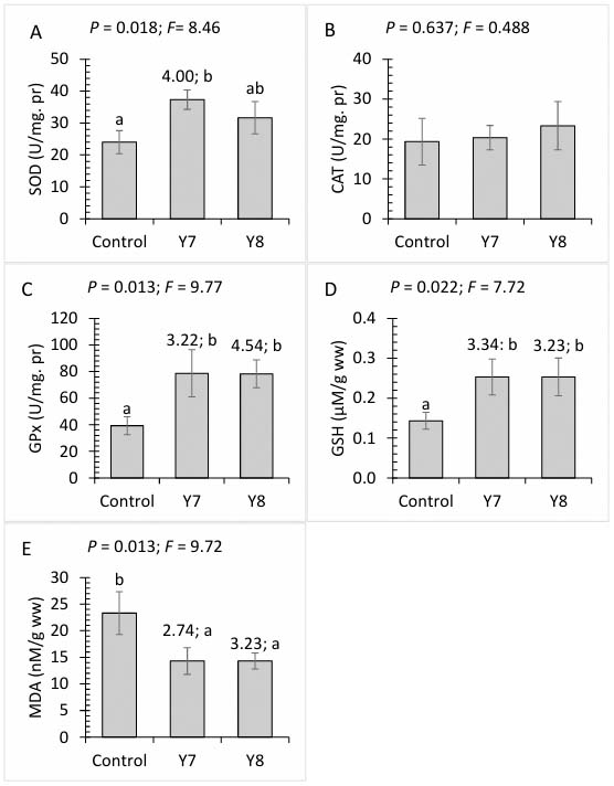
Figure 1.
Hepatic superoxide dismutase (A), catalase (B), glutathione peroxidase (C), reduced glutathione (D) and malondialdehyde (E) of rainbow trout fed the control diet or diets supplemented with 107 (Y7) and 108 (Y8) cfu/g yeast. Data are mean ± SD of three replicates. Different letters above the bars show significant differences among the treatments (Duncan). Values above the bars show effect size (Cohen’s d) of the treatment compared to control
No significant changes were observed in plasma and mucus ACH50 activities among the treatments. However, plasma lysozyme levels significantly increased in both the Y7 and Y8 treatments, compared to the control group, while mucus lysozyme levels increased significantly only in the Y7 treatment (Figure 2).
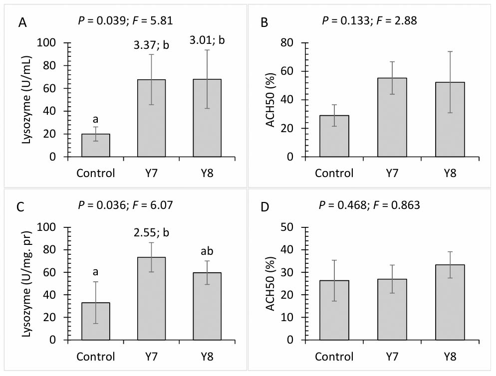
Figure 2.
Lysozyme and ACH50 activities in plasma (A and B) and skin mucus (C and D) of rainbow trout fed the control diet or diets supplemented with 107 (Y7) and 108 (Y8) cfu/g yeast. Data are mean ± SD of three replicates. Different letters above the bars show significant differences among the treatments (Duncan). Values above the bars show effect size (Cohen’s d) of the treatment compared to control
Dietary treatments did not significantly affect plasma bactericidal activities against A. hydrophila and Y. ruckeri, but they did significantly enhance bactericidal activity against S. iniae. Furthermore, both yeast treatments demonstrated significantly higher mucosal bactericidal activity against A. hydrophila. Although no significant changes were observed in mucosal bactericidal activity against S. iniae, the Y7 treatment exhibited significantly higher bactericidal activity against Y. ruckeri compared to the control group (Figure 3).
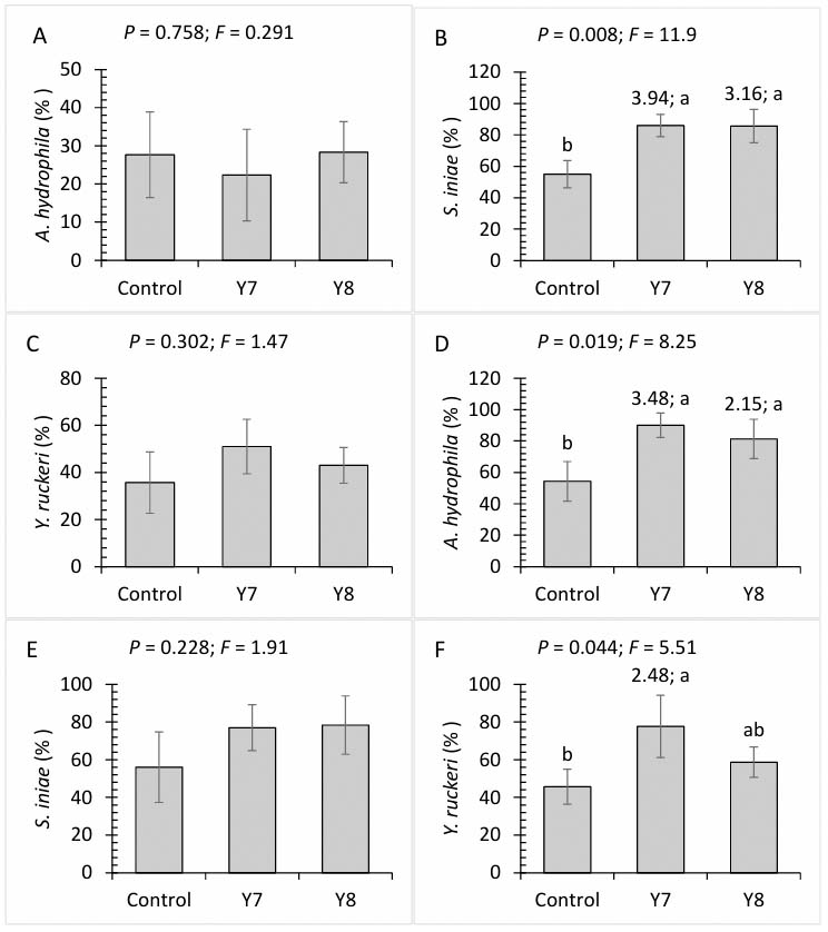
Figure 3.
Bactericidal activities in plasma (A-C) and skin mucus (C-F) of rainbow trout fed the control diet or diets supplemented with 107 (Y7) and 108 (Y8) cfu/g yeast. Data are mean ± SD of three replicates. Different letters above the bars show significant differences among the treatments (Duncan). Values above the bars show effect size (Cohen’s d) of the treatment compared to control
Interaction effects between dietary yeast and stress were noted for plasma cortisol and glucose levels, as well as hepatic ALT and AST activities. Prior to stress, these parameters showed no significant differences among the treatments. However, after 24 h of crowding stress, plasma cortisol and glucose levels, along with hepatic AST activity, significantly increased in all treatments, although the yeast treatments exhibited significantly lower values compared to the control. Hepatic ALT levels significantly increased in the control group following stress, while no such elevation was observed in the yeast treatments (Table 4).
Table 4.
Biochemical responses of rainbow trout fed the control diet or diets supplemented with 107 (Y7) and 108 (Y8) cfu/g yeast and exposed to 24-h crowding stress. Data are mean ± SD of three replicates
| Cortisol (ng/mL) | Glucose (mg/dL) | ALT (U/mg pr) | AST (U/mg pr) | ||
|---|---|---|---|---|---|
| Before stress | Control | 32.0±7.94 a | 44.0±7.55 a | 14.7±2.52 a | 29.3±5.68 a |
| Y7 | 30.3±8.08 a | 44.7±4.93 a | 15.3±3.21 ab | 29.0±6.56 a | |
| Y8 | 33.0±7.21 a | 43.7±4.61 a | 15.0±2.65 ab | 26.7±4.51 a | |
| After stress | Control | 165±11.5 c | 141±25.4 c | 43.0±7.54 c | 77.7±13.0 c |
| Y7 | 104±13.5 b | 97.0±5.20 b | 21.0±4.36 ab | 54.3±6.51 b | |
| Y8 | 99.7±10.5 b | 98.0±11.5 b | 23.0±3.00 b | 55.3±6.51 b | |
| P-value | Yeast | <0.001 | 0.014 | 0.002 | 0.0.25 |
| stress | <0.001 | <0.001 | <0.001 | <0.001 | |
| Interaction | <0.001 | 0.013 | 0.001 | 0.047 | |
| F-value | Yeast | 20.0 | 6.29 | 11.7 | 5.09 |
| stress | 369 | 137 | 48.7 | 89.8 | |
| interaction | 19.7 | 6.40 | 12.9 | 3.98 | |
| Cohen’s d | |||||
| Control before–after | 4.70 | 5.90 | 5.63 | 5.17 | |
| Y7 before–Y7 after | 5.08 | 10.3 | - | 3.88 | |
| Y8 before–Y8 after | 6.10 | 6.72 | - | 5.20 | |
| Control after–Y7 after | 4.88 | 2.90 | 3.69 | 2.39 | |
| Control after–Y8 after | 5.90 | 2.45 | 3.79 | 2.29 | |
Discussion
The present study showed that dietary yeast supplementation had no significant effects on growth performance of the fish. These results are in line with a previous study on stripped seabass, Morone chrysops × M. saxatilis, fed on diets supplemented with 10–40 g/kg brewer’s yeast for eight weeks (Li and Gatlin, 2003). However, the roles of yeast strain and dosage should not be ruled out. For example, adding baker’s yeast at a dose of 10 g/kg for six weeks to diet of Galilee tilapia, Sarotherodon galilaeus, at doses of 30–70 g/kg (Abdel-Tawwab et al., 2010) or for 84 days to diet of Nile tilapia, Oreochromis niloticus (Abass et al., 2018) significantly improved the fish growth performance. Furthermore, yeast strain P13 at a dose of 104 cfu/g administered for 4 weeks significantly improved growth performance of orange-spotted grouper, Epinephelus coioides (Chiu et al., 2010). Based on the present results, S. cerevisiae strain PTCC 5052 is not a growth-promoting probiotic in rainbow trout under the present conditions of the experiment. However, further studies on the same yeast and fish species, but different period of administration, dosages and fish life stage are recommended.
The non-specific immune system of fish plays a crucial role in defending against pathogens, making it vital for disease management in aquaculture. Enhancing this immune response can help reduce disease outbreaks (Rauta et al., 2012). Key immune cells like granulocytes (neutrophils and monocytes) are important for immediate defense through phagocytosis and antimicrobial production, while eosinophils contribute to inflammation (Singh et al., 2014; Chen et al., 2022). Although lymphocytes are essential for long-term immunity, their slower response highlights the need for strong non-specific defenses (Ainsworth, 1992). Improving these immune components can lead to healthier fish and more sustainable aquaculture practices. We found no significant changes in leukocyte number and composition as a result of dietary yeast supplementation. These results are in line with Abdel-Tawwab et al. (2020) who reported no change in leukocytes in Nile tilapia after an eight-week feeding on diet supplemented with 2 g/kg yeast, although higher supplementation levels showed significant effects. Dietary 10 g/kg yeast had no significant effects on blood total leukocyte in common carp, Cyprinus carpio, after 15, 45 and 60 days (Gopalakannan and Arul, 2010).
Lysozyme and complement proteins are important soluble immune components in fish blood, which have been routinely studied in aquaculture researches. Lysozyme, an antibacterial agent, rises in response to bacterial infections in fish, helping to eliminate pathogens. It also enhances the phagocytosis process in granulocytes, which is crucial for the early removal of these pathogens (Song et al., 2021). The complement system is crucial to the immune system, performing essential functions like cell lysis, opsonization, and inflammation within the innate immune response. Additionally, it connects the innate and adaptive immune systems by stimulating B cells to generate antibodies (Bavia et al., 2022). We found that dietary yeast can increase lysozyme in plasma and skin mucus, which are in line with previous studies on other fish such as hybrid striped bass (Li and Gatlin, 2003), orange-spotted grouper (Chiu et al., 2010) and Nile tilapia (Abass et al., 2018). Hence, it can be speculated that yeast supplementation may increase disease resistance in rainbow trout. Supporting this, we found significant improvement in bactericidal activities against common pathogens of rainbow trout in the plasma and skin mucus. This improvement can be related to the elevation in lysozyme activity and/or increase in the levels of other antimicrobial agents.
The gut microbiota, composed of various microorganisms, is essential for fish health and aquaculture (Talwar et al., 2018). It enhances immune function, nutrient absorption, and resistance to pathogens, thereby promoting growth and overall well-being (Reverter et al., 2018). Maintaining a balanced gut microbiota is crucial for sustainable aquaculture, as disruptions can negatively affect fish health. We found an increase in the gut beneficial bacteria (LAB) and a decrease in the gut harmful bacteria (Vibrio sp.), suggesting this strain of yeast can modulate gut microbial balance toward a health structure. Similarly, Feng et al. (2022) found a decrease in gram-negative and pathogenic bacteria and an increase in gram-positive and beneficial bacteria (e.g. Lactobacillus sp., Bacillus sp. and Bifidobacter sp.) in the gut of largemouth bass, Micropterus salmoides, after dietary supplementation with yeast. Also, Hoseinifar et al. (2011) found an increase in the gut LAB abundancy in beluga, Huso huso, fed a diet supplemented with yeast.
Stressors are common in aquaculture facilities and hinder fish growth, health and disease resistance (Ciji and Akhtar, 2021). A typical response of fish to stress is an increase in energy expenditure. Cortisol levels in the blood circulation increase after stress, triggering glycemia to supply enough energy for restoring homeostasis (Barton, 2002). Cortisol-induced glycemia is mediated through gluconeogenesis in the liver, where ALT and AST produce glucose from amino acids (Faught and Vijayan, 2016). So, the observed elevations in plasma glucose and cortisol and hepatic ALT and AST confirm activation of the stress axis after the fish were held under crowding conditions. Similarly, dietary brewer’s yeast supplementation significantly decreased plasma glucose level in gilthead seabream, Sparus aurata, acutely stressed (de Mattos et al., 2019). Furthermore, dietary yeast supplementation significantly suppressed plasma cortisol in Nile tilapia under osmotic stress (Sutthi and Thaimuangphol, 2020).
In conclusion, this study found that dietary supplementation with S. cerevisiae (PTCC 5052) enhances the health and stress resilience of rainbow trout. Yeast supplementation reduced harmful gut bacteria, improved immune responses, and enhanced antioxidant capacity. Fish treated with yeast exhibited lower stress responses after crowding stress. These results suggest that dietary yeast at 107–108 cfu/g is a beneficial probiotic for improving gut health and stress tolerance in aquaculture.
Acknowledgment
Parts of this study have been orally presented at the “International Conference of Innovation Aquaculture; 16–17 April 2024, Iran”.