
Introduction
Pregnancy loss is a common obstetric problem, affecting up to 25% of pregnancies worldwide (Larsen et al. 2013; Al-Memar et al. 2020). A miscarriage is the expulsion of a fetal egg from the uterus up to 22 weeks of gestation. Miscarriages can be divided into early miscarriages, up to 12 weeks of gestation, and late miscarriages, occurring between 12 and 22 weeks of gestation (Larsen et al. 2013). The European Society of Human Reproduction and Embryology (ESHRE) has introduced the additional term recurrent miscarriage (RM) when there are three or more consecutive pregnancy losses (Farquharson et al. 2005; Jauniaux et al. 2006; Christiansen et al. 2008). The occurrence of early miscarriage is dependent on the woman’s age. Among women aged 20–24 years, it is 10% of pregnancies, while in women aged 40 to 44 years, it is 51% of pregnancies. It is related to the higher incidence of genetic aberrations in embryos of older women (Nybo Andersen et al. 2000). Late miscarriages occur less frequently and account for about 4% of all miscarriages (Ugwumadu et al. 2003).
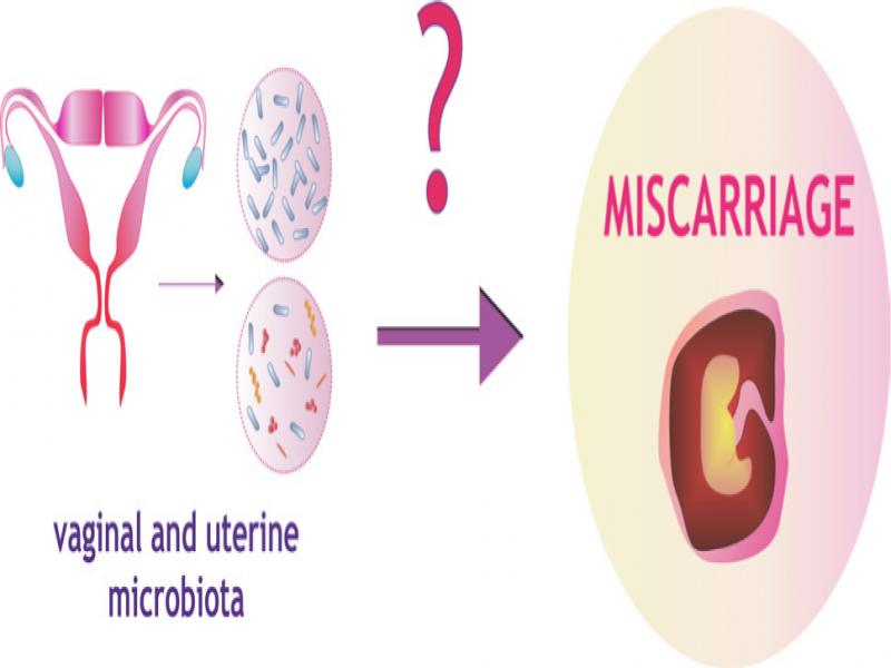
Major causes of miscarriage include genetic (Franssen et al. 2006; Branch et al. 2010) and epigenetic disorders of the embryo (Daher et al. 2012; Yin et al. 2012), immunological (Holers et al. 2002; Calleja-Agius et al. 2012), and endocrine factors (Cocksedge et al. 2009), uterine malformations (Chan et al. 2011), improper embryo selection (Salker et al. 2010), and lifestyle (Larsen et al. 2013) (Fig. 1). Perhaps a hitherto underappreciated cause of miscarriage may be an abnormal microbiota composition of the female reproductive system. Currently, the normal state of vaginal and uterine microbiota that would promote a physiological pregnancy is being sought. So far, it has been shown that a normal pregnancy is characterized by a stable vaginal bacterial composition with a dominance of Lactobacillus spp. and low diversity of other bacteria (Ravel et al. 2011; MacIntyre et al. 2015) (Fig. 2). Numerous studies show a possible relationship between preterm delivery, a decrease in Lactobacillus spp. and an increase in bacterial biodiversity in the vagina (Brown et al. 2018; Freitas et al. 2018; Al-Memar et al. 2020), bacterial vaginosis (BV), or aerobic vaginitis (AV). However, the relationship between miscarriage and the vaginal and uterine microbial composition is relatively poorly understood (Zhang et al. 2019; Al-Memar et al. 2020; Xu et al. 2020).
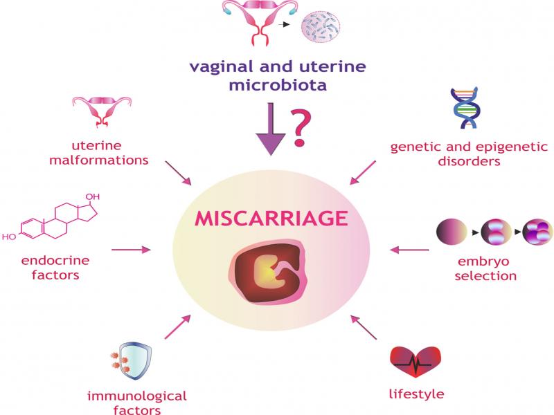
Fig. 1.
Factors affecting the risk of miscarriage.
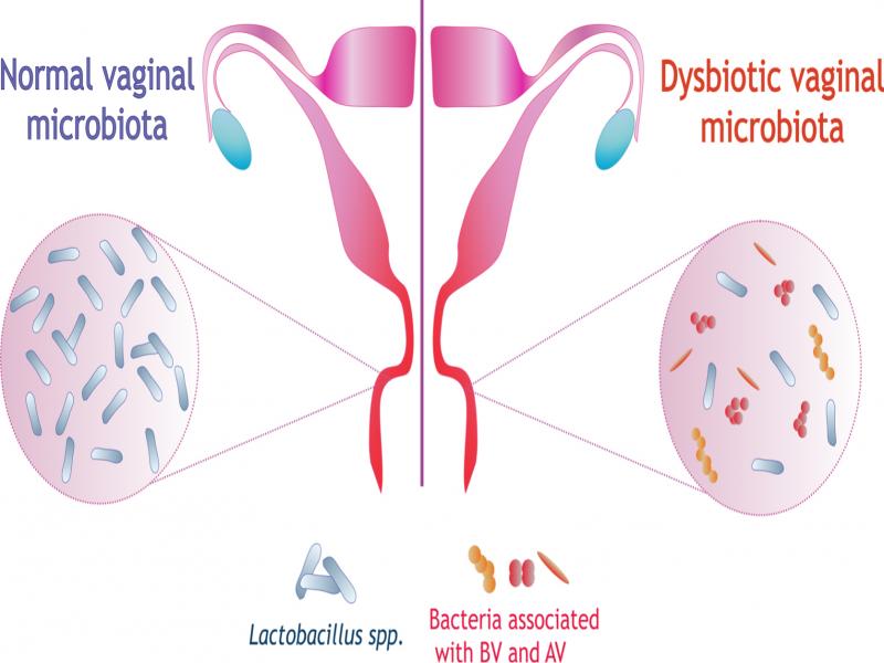
Fig. 2.
Normal vaginal microbiota and vaginal dysbiosis.
The aim of this study is an attempt to answer the question of whether, in the light of available literature, the Lactobacillus spp. can be a factor reducing the risk of miscarriage.
Protective role of Lactobacilli in the vagina
The vaginal environment is a specific ecosystem with interactions between microorganisms, the host immune system, and vaginal epithelial cells. The vaginal microbiota is a particular example of microbiota in the human body due to the definite dominance of Lactobacillus spp. (up to 99%) and low bacterial diversity (Ravel et al. 2011; Gajer et al. 2012).
There are several types of vaginal microbiota (CST, from community state types). CST-I (45.4%), CST-II (8.2%), CST-III (26.8%), and CST-V (9.3%) are successively dominated by Lactobacillus crispatus, Lactobacillus gasseri, Lactobacillus iners, and Lactobacillus jensenii (Ravel et al. 2011; Gajer et al. 2012). In the CST-IV group (10.3%), in which Lactobacillus is not a dominant genus, the following bacteria are present: Gardnerella, Prevotella, Megasphaera, Sneathia, Atopobium, Streptococcus, Dialister, Lachnospira, Anaerococcus, Peptoniphilus, Eggerthella, Finegoldia, Rhodobaca, Anaerotruncus, Ureaplasma, Mycoplasma, Aerococcus, Parvimonas, Staphylococcus, Corynebacterium, Veillonella (Ravel et al. 2011; Gajer et al. 2012; Kacerovsky et al. 2015).
The CST-I (dominated by L. crispatus) is a stable type; its transition to a pathological state has rarely been observed, provides a vaginal pH < 4.0, and may convert to the CST-III type or a microbiota consisting of different species of Lactobacillus genus. The CST-II (dominated by L. gasseri) is an unstable type; however, no transition to a pathological state has been observed, can temporarily convert to CST-I during pregnancy, provides a vaginal pH of approximately 4.4. The CST-III (dominated by L. iners) is a transitional type that facilitates transition to bacterial vaginitis (BV); this type is characterized by an increase in pro-inflammatory factors and a decrease in the level glycolysis enzymes. It is associated with an increase in vaginal pH > 4.5. Simplified metabolism of L. iners results in dependence on substances received from the host; thus, increasing sensitivity to environmental changes. In addition, the L-lactic acid produced insufficiently inhibits pathogens.
The CST-IV is the pathogenic type, dominated by anaerobic bacteria. There are CST-IVA with a small number of L. iners and CST-IVB dominated by such bacteria as Atopobium, Gardnerella, Mobiluncus, Prevotella, Sneathia. This type is most common among BV patients and healthy women of African descent. Low lactic acid levels damage to the mucin layer (which hinders Lactobacillus spp. adhesion), and the presence of a bacterial biofilm on the vaginal epithelial surface characterize the CST-IV type (Chee et al. 2020). The CST-V is a stable type (dominated by L. jensenii), is relatively poorly known, and provides a vaginal pH of approximately 4.2. There are reports that L. jensenii can affect the vaginal microenvironment by reducing lactate and succinate levels (Stafford et al. 2017). However, further studies are needed to characterize CST-V (Chee et al. 2020).
In the vagina, Lactobacilli are involved in protective functions by producing lactic acid, hydrogen peroxide (H2O2), and bacteriocins (Boskey et al. 2001; Witkin and Linhares 2017; Amabebe and Anumba 2018; Bernabeu et al. 2019).
Lactic acid is a significant protective factor of the vagina. Lactobacillus spp. produce its two isomeric types, D-lactic acid, and L-lactic acid, with D-lactic acid showing a stronger protective effect (Boskey et al. 2001). L-lactic acid acidifies vaginal secretions (to about pH < 4), thus hindering the binding of other microorganisms to vaginal epithelial cells and inducing autophagy in epithelial cells to degrade microorganisms. Lactic acid has a blocking effect on histone deacetylase, stimulating gene transcription and DNA repair (Witkin and Linhares 2017; Amabebe and Anumba 2018; Bernabeu et al. 2019). In sterile cultures, L. crispatus and L. gasseri can produce both D- and L-lactic acid, while L. iners produces only the L-isomer, whereas L. jensenii produces only the D-isomer (Witkin et al. 2013).
Another protective mechanism relies on the production of hydrogen peroxide (H2O2). This compound, which has a broad antimicrobial activity, is produced by many lactic acid bacteria isolates. It has been shown that 94–95% of L. crispatus and L. jensenii strains produce hydrogen peroxide (Vallor et al. 2001). The H2O2 shows high in vitro activity against Escherichia coli, Candida albicans, or Staphylococcus aureus ((Sgibnev and Kremleva 2015). It can inhibit the multiplication or destroy pathogenic strains of vaginal bacteria, especially those with the limited expression of hydrogen peroxide-degrading enzymes, including Prevotella, Peptostreptococcus, and Gardnerella anaerobes, among others. The lack of this compound in the vagina increases catalase-negative bacteria, associated with an increased risk of genitourinary infections, including BV and Human Immunodeficiency Virus infection (HIV) (Aroutcheva et al. 2001; Amabebe and Anumba 2018; Tachedjian et al. 2018).
Vaginal Lactobacilli also produce bacteriocins. These are proteins or protein complexes that show potent bactericidal activity. Bacteriocins kill pathogens such as Gardnerella vaginalis, Escherichia coli or Candida albicans, by inhibiting DNA synthesis. Bacteriocins resemble antibiotics in their action (Aroutcheva et al. 2001; Alpay Karaoğlu et al. 2003; Deplanche et al. 2019). Lactobacillus spp. also shows strong adhesion to the non-keratinized vaginal epithelium, displacing pathogenic microorganisms such as C. albicans, G. vaginalis, E. coli, Streptococcus agalactiae, or S. aureus from the epithelial surface. Lactobacillus spp. and G. vaginalis may bind to the same receptors on the surface of vaginal epithelial cells. However, Lactobacillus spp. has a higher affinity for vaginal epithelial cells and displaces G. vaginalis (Kovachev 2018). Studies show that the combination of L. crispatus UBLCp01, L. gasseri UBLG36 and L. johnsonii UBLJ01 may be helpful in preventing/treating vaginal dysbiosis and maintaining a healthy vaginal ecosystem when used as vaginal probiotics. Features such as adherence and antimicrobial potential, exopolysaccharide production, and biofilm-forming ability of strains are essential characteristics that influence their potential against pathogens (Ahire et al. 2021).
The composition of vaginal microbiota fluctuates under the influence of many factors such as environmental conditions (antibiotics, pre- and probiotics, contraception), lifestyle (hygiene and sexual habits), individual characteristics (genetic, immunological factors, age, hormonal status, ethnicity, socioeconomic status) and general health (Macklaim et al. 2015; Brooks et al. 2017; Kervinen et al. 2019). There are cases when certain changes in the composition of vaginal microbiota occur during pregnancy. During pregnancy, the levels of various hormones change dynamically, produced primarily by the placenta, which becomes a gland for the secretion of many biologically active substances. Such hormones include estrogen and progesterone. Estrogens increase the synthesis of glycogen in the vaginal epithelium, which provides a substrate for lactic acid bacteria (Taddei et al. 2018; Heil et al. 2019; Serrano et al. 2019). Also, the absence of menstruation or changes in cervical and vaginal secretions affects the different states of pregnancy microbiota (Walther-António et al. 2014). In addition, the precise reciprocal interaction between the microbiota and locally acting immune cells are responsible for the inhibition of pathogen growth, but also for the tolerance to paternal antigens that are present in the semen and embryo (Agostinis et al. 2019; Kervinen et al. 2019; Bardos et al. 2020; Monin et al. 2020).
Studies have also been conducted on the relationship between ethnicity and vaginal microbiota composition. Experiments conducted by Walther-António et al. (2014) on pregnant Caucasian women showed that L. crispatus was the dominant genus; for a smaller number of subjects, it was L. iners, and a small proportion of patients showed the transition from L. crispatus to L. iners. Afro-American populations have greater interindividual diversity in the vaginal microbiota than Caucasians. It is particularly important because gynecologic-obstetric complications are more common among these populations (Walther-António et al. 2014). Caucasian women have a predominantly Lactobacillus spp. dominated microbiota (approximately 90%), Asian and Hispanic women have microbiota percentages of approximately 80% and 60%, respectively, and African women only 37% (Anahtar et al. 2015). Studies show that Asian women have a different composition of the vaginal microbiota, with a higher prevalence of L. iners. The reason for these differences remains unclear. It may be related to genetic and environmental factors, including geographic location, diet, age, BMI, drug exposure, physical activity, and availability of resources such as access to medical care. However, studies show that the presence of L. iners in the vagina of Asian women does not increase the risk of abnormal pregnancy (Serrano et al. 2019; Kumar et al. 2021).
Lactobacillus iners: friend or foe? Standard culture and microscopic methods, used for many years to determine the presence of lactic acid bacteria in the vagina, could not detect L. iners in vaginal samples. Employing molecular biology methods helped to detect the presence of seven strains of L. iners in the vagina and urinary tract in 1999 (Falsen et al. 1999).
Current knowledge indicates that L. iners is the predominant genus in the vaginal microbiota among older women, pregnant women, and women of Afro-American descent (Srinivasan et al. 2010; Mls et al. 2019). The L. iners is also frequently isolated from the vagina of women diagnosed with BV, shortly after BV treatment, and during menstruation (Lopes dos Santos Santiago et al. 2011, Gajer et al. 2012). It is suggested that this genus is very flexible and can quickly adapt to changing conditions prevailing in the vaginal niche. The function analysis of proteins encoded by the L. iners genome revealed that this genus could show both commensal and pathogenic properties. The L. iners genome encodes proteins predicted to be involved in optimal adaptation to the vaginal niche, such as iron-sulfur proteins and the σ factor. Several genes have also been identified in the L. iners genome suggesting that it may be an opportunistic pathogen (Petrova et al. 2017). For example, the genome of L. iners strains encodes the toxin inerolysin (Rampersaud et al. 2011), related to the vaginolysin of G. vaginalis (Srinivasan et al. 2012). Furthermore, the genome size of L. iners is also unique among Lactobacillus genus. The AB-1 strain of L. iners has by far the smallest genome yet known among Lactobacillus spp., consisting of a single chromosome of approximately 1.3 Mbp, in contrast to other lactic acid bacteria in which the genome size is approximately 3–4 Mbp (Macklaim et al. 2011; 2013). It appears that there may be some clonal variants within the L. iners genus that show commensal properties in some cases and pathogenic properties in others (Petrova et al. 2017).
Presence of Lactobacilli in the vagina and maintenance of pregnancy
There are reports that Lactobacilli, due to their protective role, may contribute to the normal course of pregnancy (Szubert et al. 2021). Data show that their absence is observed in pre-eclampsia (Gomez et al. 2016), gestational diabetes (Dunn et al. 2019), and preterm labor (Elovitz et al. 2019), among others. Numerous studies have confirmed that women with a vaginal microbiota dominated by Lactobacillus spp. bear a lower risk of preterm birth (Ansari et al. 2020; Aslam et al. 2020; Gerson et al. 2020; Kosti et al. 2020).
Far fewer studies have examined the effects of Lactobacillus spp. on fertilization success, implantation, and early embryonic development as well as recurrent implantation failure (RIF). Based on 16S rRNA gene sequencing of the vaginal microbiota, the vaginal Lactobacillus spp. showed a significant positive correlation with the pregnancy rate and the RIF group, all of the genera were significantly increased, especially the aerobic bacteria (8.5% for the RIF group and 2.3% for the control group, p < 0.05) (Fu et al. 2020).
The relationship between miscarriage and vaginal bacterial composition has been studied by Nelson et al. (2007; 2015). They analyzed the effect of changes in the vaginal microbiota, in the first trimester of pregnancy, on the risk of miscarriage in the second trimester. It has been shown that a lack of Lactobacillus spp. in the vagina during the first trimester of pregnancy may be related to the risk of miscarriage in the second trimester (HR: 1.32; 95% CI: 1.10–1.64) (Nelson et al. 2007). Similarly, Xu et al. (2020) showed that a lack of Lactobacillus spp. may be a contributing factor for pregnancy loss. In 80% of women included in this study who had a miscarriage, the number of Lactobacillus spp. in the vagina was lower than the control group (Xu et al. 2020).
Interestingly, other reports comparing vaginal microbiota at the genus level in women with confirmed miscarriage correlated with a reduction in the number of Lactobacillus spp. in the first or second trimester of pregnancy (Al-Memar et al. 2020). It also proved that the risk of pregnancy loss in the second trimester among women with confirmed BV diagnosed in the first trimester was increased but not statistically significant. However, women with the most severe BV changes in the vaginal microbiota had a twofold increase in the risk of pregnancy loss in the second trimester compared to women with normal vaginal microbiota (HR: 2.49, 95% CI: 1.13 to 5.48). Similarly, Bretelle et al. (2015) reported a correlation between pathogenic bacteria in the vagina, including Chlamydia trachomatis, Atopobium vaginae, and G. vaginalis, and late miscarriage and high-risk pregnancies. Also, genital tract infections primarily characterized by anaerobic bacteria such as Gardnerella, Prevotella, Megastrobila, and Cyclospora increase the risk of miscarriage (Xu et al. 2020).
Interesting studies investigate the relationship between recurrent miscarriage (RM) and Lactobacillus spp. and the growth of pathogenic bacteria in the vagina. Non-pregnant women with three or more consecutive miscarriages were selected for recurrent miscarriage studies (Llahi-Camp et al. 1996; Işik et al. 2016; Kuon et al. 2017; Zhang et al. 2019). Llahi-Camp et al. (1996) microscopically evaluated Gram-stained vaginal smears of women with one miscarriage and women diagnosed with RM. Results showed that BV was significantly more common among women with a history of one second-trimester miscarriage (27/130; 21%) than among women with RM (31/370; 8%) (Llahi-Camp et al. 1996). Similarly, in a study conducted by Işik et al. (2016), the presence of BV was statistically associated with the occurrence of one miscarriage in the last six months (p < 0.05), while no significant association was found between BV and recurrent miscarriages (p > 0.05). Thus, it was concluded that there is no direct relationship between BV and RM. However, a recent study based on 16S rRNA gene sequencing of vaginal microbiota shows that women with RM have a higher genus richness (p = 0.037) in the vagina than healthy women and bacteria such as Atopobium, Prevotella and Streptococcus are identified (Zhang et al. 2019). Women with RM also showed a reduced amount of Lactobacillus spp. (Zhang et al. 2019; Fan et al. 2020). Kuon et al. (2017) found that women whose vagina is colonized by G. vaginalis, and Gram-negative anaerobes more often have RM. Almost 20% of patients with RM showed vaginal colonization by G. vaginalis and 15% by Enterobacteriaceae. In addition, vaginal Lactobacilli have been reported less frequently in women with RM (Kuon et al. 2017). Other studies also suggest that BV may contribute to chronic endometritis, which correlates with the occurrence of RM (Bardos et al. 2020). The effect of vaginal microbiota on miscarriage and recurrent miscarriage is summarized in Table I.
Table I
Effect of vaginal microbiota on miscarriage and recurrent miscarriage.
| References | Conclusions | |
|---|---|---|
| MISCARRIAGE | Nelson et al. (2007) | Lack of Lactobacillus spp. in the vagina during the first trimester of pregnancy may be associated with a risk of miscarriage in the second trimester |
| Bretelle et al. (2015) | The presence of pathogenic bacteria such as Chlamydia trachomatis, Atopobium vaginae, and Gardnella vaginalis in the vagina is associated with high-risk pregnancies and may contribute to miscarriage | |
| Nelson et al. (2015) | BV correlates with miscarriageAl-Memar et al. (2020) Decreased vaginal Lactobacillus spp. during the first or second trimester of pregnancy correlates with risk of miscarriage | |
| Chang et al. (2020) | The presence of Lactobacillus iners in the vagina increases the risk of miscarriage | |
| Xu et al. (2020) | The presence of Gardnerella, Prevotella as well as Megastrobila, and Cyclospora and the lack of Lactobacillus spp. in the vagina may contribute to pregnancy loss | |
| RECURRENT MISCARRIAGE (RM) | Llahi-Camp et al. (1996) | The BV is significantly more common among women who have had a second-trimester miscarriage than among women with RM |
| Işik et al. (2016) | The BV is associated with the occurrence of one miscarriage in the past six months. The BV does not affect the occurrence of recurrent miscarriage | |
| Kuon et al. (2017) | Lactobacillus spp. is not present in the vagina of women with RM | |
| Zhang et al. (2019) | Pathogenic bacteria in the vagina include Prevotella, Atopobium, and Streptococcus and reduced Lactobacillus spp. correlate with RM |
| References | Conclusions |
|---|---|
| Kyono et al. (2018) | Endometrium dominated by Lactobacillus spp. favors embryo implantation |
| Moreno and Simon (2018) | Dysbiotic endometrium characterized by Atopobium, Bifidobacterium, Chryseobacterium, Gardnella, Haemophilus, Klebsiella, Neisseria, Staphylococcus, and Streptococcus correlates with abnormal pregnancy |
| Leoni et al. (2019) | The presence of mixed bacterial microbiota, not always Lactobacillus spp., is associated with a normal pregnancy |
| Moreno et al. (2020) | The endometrium before miscarriage is characterized by a greater diversity of bacteria and fewer Lactobacillus spp. |
| Moreno et al. (2021) | Types of bacteria such as Enterococcus, Enterobacteriaceae, Streptococcus, Staphylococcus, Gardnerella, Mycoplasma, Ureaplasma, Chlamydia, and Neisseria are responsible for chronic endometritis and suspected to have adverse effects on implantation as well as may contribute to miscarriage |