
Table I
The characteristics of the strains used in this study.
| Strain characteristics | Pseudomonas aeruginosa 6-1B | Rhodococcus erythropolisT7-2 | Pseudomonas aeruginosa PAO1 |
|---|---|---|---|
| Optimum temperature (°C) | 42 | 30 | 37 |
| Fermentation product2 | Rhamnolipid | Saccharides, protein, lipid | ND |
| Emulsification index (EI24) | 100% | 100% | ND |
| Cell surface hydrophobicity (CSH%)1 | 38% | 85% | 16% |
| Degradation range of n-alkenes | C8-C20 | C12-C36 | ND |
| Degradation rate of crude oil3 | 60.09% | 75.43% | ND |
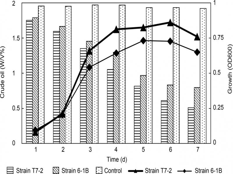
Fig. 1.
Growth and degradation of crude oil by P. aeruginosa 6-1B and R. erythropolis T7-2 strains.
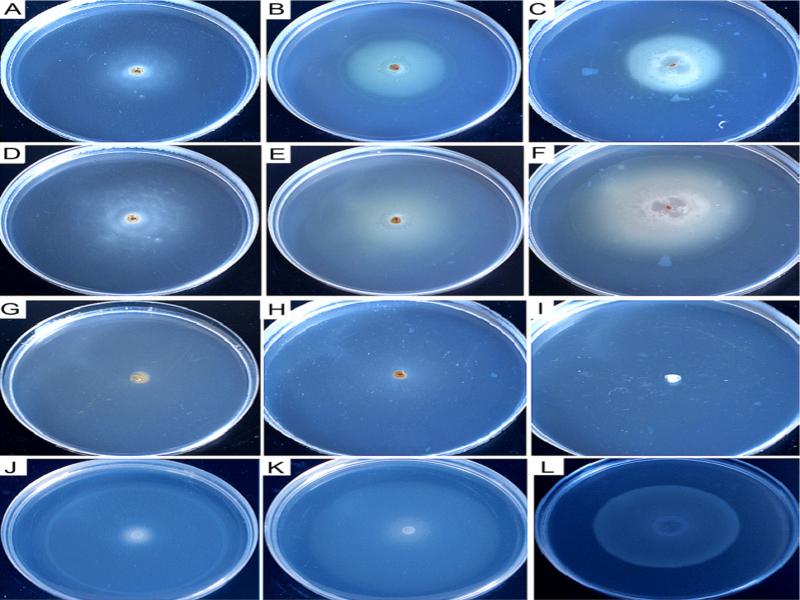
Fig. 2.
Chemotaxis of the strain 6-1B towards tridecane, liquid paraffin, and crude oil in the swarm plate assay.
The chemotaxis of the strain 6-1B towards 0.01% of tridecane (A) and (D), liquid paraffin (B) and (E), crude oil (C) and (F), and the chemotaxis buffer without attractant (G) were determined, respectively. The chemotactic responses of the control strain R. erythropolis T7-2, and P. aeruginosa PAO-1 to crude oil are shown in (H) and (I). Among them, Fig. 2A, 2B, and 2C were photographed after 24 hours of cultivation; Fig. 2D, 2E, 2F, 2G, 2H, and 2I were taken after 48 hours of incubation. The chemotactic responses of the strain 6-1B towards sucrose, glycine, glycerol after 24 hours of cultivation are also shown in Fig. 2J, 2K, and 2L separately.
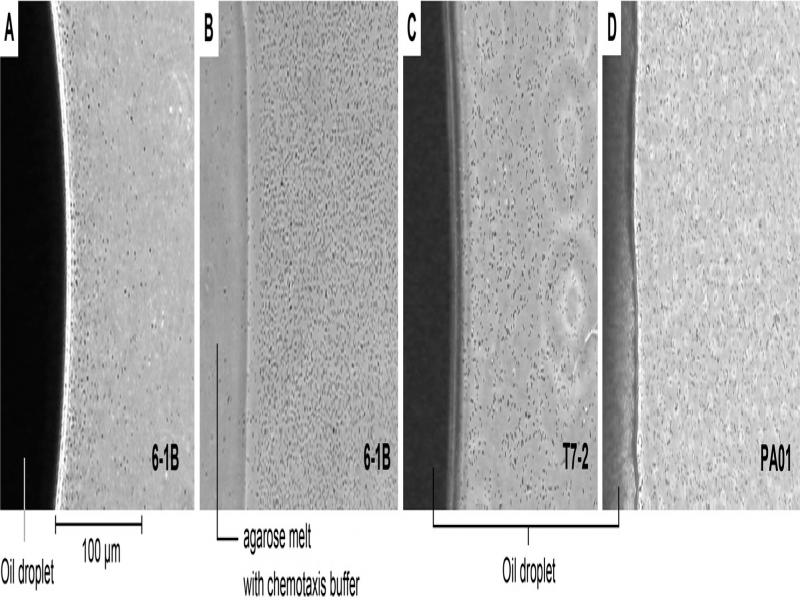
Fig. 3.
The chemotactic responses of the strain 6-1B (A), T7-2 (C), and PAO1 (D) towards crude oil via modified agarose plug assay. The chemotaxis of the strain 6-1B towards the chemotaxis buffer served as the negative control (B).
Table II
Chemotaxis responses of the different strains to various components of the Daqing crude oil and their respective relative
degradation rates.
| Attractants1 | Pseudomonas aeruginosa 6-1B | Rhodococcus erythropolis T7-2 | Pseudomonas aeruginosa PAO-1 | |||
|---|---|---|---|---|---|---|
| Chemotaxis response2 | Oil degrading rate (%)3 | Chemotaxis response2 | Oil degrading rate (%)3 | Chemotaxis response2 | Oil degrading rate (%)3 | |
| Dodecane | + | 63.22 | – | 78.17 | – | ND |
| Tridecane | + | 56.18 | – | 75.62 | – | ND |
| Tetradecane | + | 54.28 | – | 67.57 | – | ND |
| Pentadecane | + | 57.97 | – | 62.84 | – | ND |
| Hexadecane | + | 55.54 | – | 59.73 | – | ND |
| Liquid paraffin | + | 58.13 | – | 65.11 | – | ND |
| Crude oil | + | 60.09 | – | 75.43 | – | ND |
| Naphthalene | – | ND | – | ND | – | ND |
| Diphenyl | – | ND | – | ND | – | ND |
| Sulfur | – | ND | – | ND | – | ND |
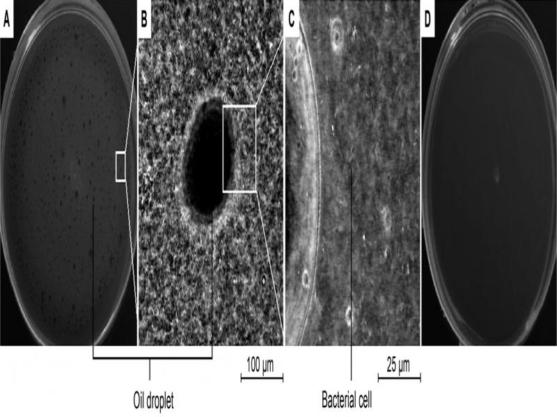
Fig. 4.
The visualization of chemotaxis rings in the swarm plate using microscopy.
The chemotaxis of the strain 6-1B toward crude oil (0.1%) visualized using a phase-contrast microscope (Olympus BH2 microscope, Japan) with magnifications of 0× (A), 125× (B) and 500× (C), respectively; the control swarm plate which contained the same composition except for crude oil, visualized at a magnification of 0× (D).
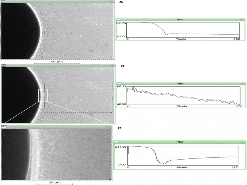
Fig. 5.
P. aeruginosa 6-1B cells’ distribution around the crude oil droplet.
The strain 6-1B movement towards the oil droplets was analyzed under the phase-contrast microscope (Olympus BH2 microscope, Japan) using the Scion Image 3b Software (Scion, Frederick, MD). A) The chemotactic trend of the strain 6-1B toward crude oil (125× magnification); B) the chemotactic trend of the dashed area; C) the chemotactic trend of the strain 6-1B toward crude oil of the area of the white line in Fig. 5B (400× magnification).
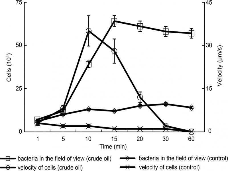
Fig. 6.
The number of chemotactic cells (left y-axis) and chemotactic velocity curves (right y-axis) of P. aeruginosa 6-1B toward the crude oil.
The control chamber was treated without crude oil, which served as the chemoattractant in the other setups. A chosen 50 μm × 50 μm area was magnified 500 times for the determination of cell velocity. The chemotaxis videos were divided into images, and an average velocity was determined from observed cell movements that had relatively straight trajectories. Data represent the averages and the standard deviations of five independent experiments.