Introduction
Microbial enhanced oil recovery (MEOR) is an environment-friendly process that may be useful to petroleum recovery. MEOR may be an efficient and inexpensive alternative method to enhance the physicochemical recovery of oil (EOR) (Kryachko 2018). Microorganisms can be used to reduce the paraffin build-up in producing wells, produce solvents or polymers above ground, and for pumping into the oil-bearing formation, as in EOR (Brown 2010). It is widely accepted that microorganisms can enhance oil recovery by their ability to produce some metabolic products, including biosurfactants. Biosurfactants are one of the most important microbial metabolic products that can reduce surface tension in oil and facilitate the emulsification of oil in with water. The latter increases the bioavailability of the residual crude oil and enhances its biodegradability (Batista et al. 2006). Furthermore, the properties of the microbes themselves, such as high cell surface hydrophobicity and motility, help them to attach to the interface and the surface of oil droplets in sandstones and carbonate strata (Rocha et al. 2020). The degradation of crude oil generally occurs at oil-water points of contact. The oil-water contact provides conditions that are the most conducive to microbial activity. The transport of hydrocarbons from the oil droplets will provide a plentiful supply of electron donors needed for metabolism, whereas inorganic nutrients required for microbial growth can be transported by water flow or diffusion to the biosphere on the oil-water contact (Head et al. 2003).
Flagellum-dependent chemotaxis is an important advantage of motile bacteria. These bacteria move through a fluid medium by rotating one or more flagella (Nakamura and Minamino 2019). Both metabolism-dependent and independent chemotaxis of Pseudomonas sp. toward aromatic compounds had been studied (Sampedro et al. 2015). Chemotactic bacteria, such as Escherichia coli, Pseudomonas putida, Pseudomonas aeruginosa, Rhodococcus erythropolis, and others can adapt to the chemical environment by detecting changes in concentrations of certain chemicals and by changing their movement patterns essentially based on the chemical gradient present (Waite et al. 2018). The bacterial chemotaxis ability provides more opportunities for bacterial cells to move into an area with high concentrations of the necessary chemical attractants (Ni et al. 2020; Yang et al. 2020). Some microorganisms have evolved to use chemotaxis to resist degradation and survive conditions that lead them to utilize poisonous carbon sources, such as non-aqueous phase liquids (NAPLs), especially when there are no usable growth substrates available (Parales and Harwood 2002). Bacterial chemotaxis here is an important prelude to metabolism, as it can increase the degradation rate of NAPL-associated hydrophobic compounds (Law and Aitken 2003). Marx and Aitken (2000) have reported that chemotaxis to naphthalene by Pseudomonas putida G7 increased the rate of naphthalene degradation in an aqueous system in which a concentration gradient of naphthalene was imposed.
Crude oil contains NAPL-associated compounds, which are mainly composed of hydrocarbons, and aromatic compounds. It has been reported that hydrocarbon-degrading microbes migrate toward pure alkanes by chemotaxis, which may enhance the alkanes’ biodegradation rate by facilitating microbial contact with the substrate (Lanfranconi et al. 2003). Favorable chemotactic properties may allow oil-degrading bacteria to efficiently detect and migrate towards oil droplets, which could be utilized as a carbon source (Meng et al. 2019). However, experimental demonstrations on chemotaxis of oil-degrading microbes towards crude oil are still limited. The potential role of chemotaxis of oil-degrading microbes in EOR has not yet received much attention. Therefore, this study investigated the chemotactic characteristics of a Pseudomonas aeruginosa oil-degrading strain, 6-1B, elicited by various substrates, and the chemotactic response towards crude oil via the swarm plate assay and the modified agarose plug assay. A potential chemotaxis mechanism of the 6-1B strain toward crude oil has been proposed based on the experimental results.
Experimental
Materials and Methods
Materials and media. The crude oil used in this study was light oil obtained from the Daqing Oilfield (China). It has been sterilized using high-pressure steam before use (Table S1). Tridecane and liquid paraffin were obtained from Sinopharm Chemical Reagent Co., Ltd.
Rhodococcus erythropolis T7-2 strain was cultivated in the lab (Huang et al. 2007). The bacteria were incubated in two different kinds of mineral salt media, namely medium M1 (in g/l: KH2PO4 0.2, Na2HPO4 0.6, NaNO3 2.0, CaCl2 0.01, FeSO4 0.01, MgSO4∙7H2O 0.615, and yeast extract 0.5), and medium M2 (in g/l: Na2HPO4 1.5, KH2PO4 3.48, (NH4)2SO4 4.0, MgSO4 0.7, and yeast extract 0.01), respectively. The pH values of both media were adjusted to 7.2. The media were then autoclaved at 121°C for 30 min.
Screening of oil-degrading bacterial strains. The strain 6-1B, used in this study, was isolated from the oil-water of Daqing Oilfield. The strain can degrade crude oil efficiently with the production of biosurfactants. The isolation method was as follows: 10% of oil-water (w/v) was added into the mineral medium M1 supplemented with 0.2% sucrose (w/v) and 2.0% liquid paraffin (w/v) as the carbon sources and was shaken with 150 rpm at 42°C for seven days. After subculturing twice in this medium, 5% seed culture (v/v) was transferred into medium M2 supplemented with 2% liquid paraffin (w/v) as the sole carbon source. The bacteria were incubated for five days at 42°C and were subcultured for more than five times to ensure the selected strains’ activity. After enriching the bacteria in the M2 medium, aliquots were diluted and smeared on Luria-Bertani (LB) agar plates to screen for single colonies. The selected colonies on the LB plate were then respectively cultured in the M2 medium at 42°C, supplemented with 2% crude oil (w/v) as the sole carbon source, and these enriched cultures were collected for subsequent testing. The degradation rate of crude oil was analyzed by a standard test method for oil and grease and petroleum hydrocarbons in water (ASTM D3921-85.1990), commonly known as the IR method. Absorbance readings were then taken by following the manufacturer’s instructions, using a fixed wavelength model DM600 IR analyzer (AilunGroup, CHN). The measurement range of the IR is 0.1–10,000 ppm.
Bacterial characteristics. The biosurfactant formed by the strain 6-1B from liquid paraffin, which was the sole carbon source needed for production, was analyzed as previously reported (Patowary et al. 2017). The fermentation products of strains 6-1B and T7-2, which could emulsify diesel oil with an Emulsification Index (EI24) value of 100%, were also characterized according to respective methods well-described previously (Gandhimathi et al. 2009). In order to assess cell surface hydrophobicity (CSH), the bacterial adherence to hydrocarbon (BATH) assay was performed as described previously (Gomes et al. 2013). The taxonomy of the isolated strain 6-1B was identified according to the BLAST result of the 16S rDNA sequence with the GenBank database. The 16s rDNA was extracted from isolated colonies of the strain 6-1B and amplified using universal primers 27F (5’-AGAGTTTGATCCTGGCTCAG-3’) and 1492R (5’-GGTTACCTTGTTACGACTT-3’). The 16S rDNA sequence was then deposited in NCBI with an accession number of JQ012217.
Chemotaxis assays. Swarm plate assay. To characterize the chemotactic behavior of the strain 6-1B toward crude oil, the swarm plate assay was performed as previously described (Ha et al. 2014). Briefly, cells grown on oil were collected by centrifugation at 8,000 × g for 5 min and washed twice with a chemotaxis buffer, which contained 25 mM Na2HPO4, 25 mM KH2PO4, and 0.01% yeast extract. Cells were then resuspended in the chemotaxis buffer at a final concentration of 1×10⁹ CFU/ml. 0.01 ml of suspension of the strain 6-1B in the chemotaxis buffer was gently poured on the centre of the swarm medium agar plate, which contained (g/l): Na2HPO41.5, KH2PO4 3.48, (NH4) 2SO4 4.0, MgSO4 0.7, 0.01% of Triton X-100, 0.01% yeast extract (v/v), and 0.01% of attractants (crude oil, liquid paraffin, and tridecane, v/v). The 0.01% of Triton X-100 (v/v) and the yeast extract were added to the swarm agar plate to improve the oil solubility and keep the mobility of cells, respectively. Overnight cultures of P. aeruginosa PAO-1 and R. erythropolis T7-2 in LB broth were used as controls. The plates were incubated at 42°C, 30°C, and 37°C, respectively, and were observed every six hours.
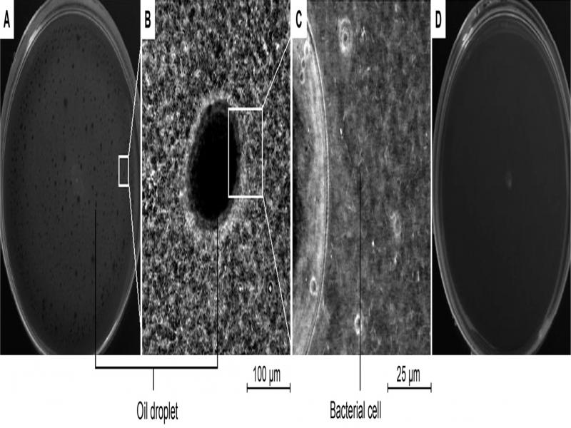
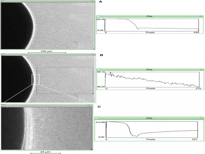
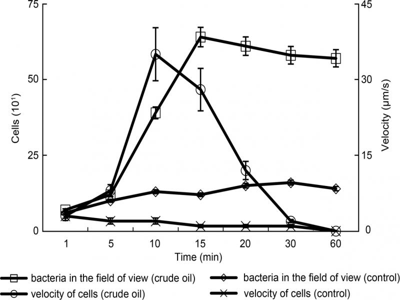
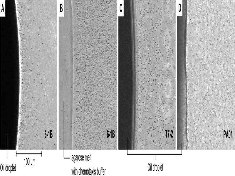
Modified agarose plug assay. The agarose plug assay was performed as described previously (Roggo et al. 2018), with a slight modification, by adding a coverslip on top of a concave slide to form a chamber. The cells used in these assays were harvested in the mid-logarithmic phase, washed, and resuspended with the chemotaxis buffer (with a density of about 1 ×10⁷ CFU/ml). Plugs with crude oil sample, or melted agarose in the chemotaxis buffer (negative control), were dropped in the chamber’s center. Then, 50 μl of the freshly harvested bacterial culture was then infused around the crude oil droplets. A glass coverslip was placed on top of the chamber, which was then sealed with petroleum jelly (Vaseline) to ensure that there were no air bubbles in the chamber. The movement of cells of the strain 6-1B towards the oil droplets was analyzed under the phase-contrast microscope (Olympus BH2 microscope, Japan) using the Scion Image 3b Software (Scion, Frederick, MD). The chemotaxis of the strain 6-1B was determined based on a relative velocity. A chosen 50 μm × 50 μm area was magnified 500 times to determine the relative velocity of the strain 6-1B. The chemotaxis videos were divided into frames, and an average relative velocity (derived from the average of n = 5 independent experiments) was determined from observed cell movements that had relatively straight trajectories. The area magnified and visualized was the interface between the oil and water; the number of cells in the field of view was counted. The changes in the pixel intensity (ranging from 0–255 PPI) reflected the bacterial density in the oil droplet’s vicinity.
Morphology and image analysis. The morphology of the bacteria used in this study was observed through transmission electron microscopy (TEM, Philips EM400-ST, Japan). Chemotactic responses were observed at respective magnifications of 100×, 125×, 400×, and 500×, using a phase-contrast microscope (Olympus BH2 microscope, Japan) equipped with a CCD camera (Hitachi KP-D50 Colour Digital, Tokyo, Japan), and Axio-Vision software. The sizes of the chemotactic rings were determined using Axio-Vision software. Videos were analyzed by the method previously described (Boudko et al. 2003). The Scion Image 3b software was used to make line scan image plots.
Results and Discussion
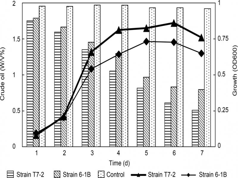
The isolation of the strain chemotactic towards crude oil. The strain 6-1B was isolated from an oil/water sample collected in the Daqing Oilfield and can degrade crude oil upon producing a rhamnolipid biosurfactant. A 16S rRNA sequence-based phylogenetic analysis revealed that the stain 6-1B represented Pseudomonas aeruginosa species and was named P. aeruginosa 6-1B. The biosurfactant produced by the strain 6-1B was detected in the mineral salts medium M1 culture, which was supplemented with 2.0% (w/v) liquid paraffin as a carbon source. Strain 6-1B can also grow with n-alkanes (C8 to C20) as its sole carbon and energy source. The oil degradation rate of strain 6-1B was up to 60% (Table I).
Table I
The characteristics of the strains used in this study.
| Strain characteristics | Pseudomonas aeruginosa 6-1B | Rhodococcus erythropolisT7-2 | Pseudomonas aeruginosa PAO1 | |||
|---|---|---|---|---|---|---|
| Optimum temperature (°C) | 42 | 30 | 37 | |||
| Fermentation product2 | Rhamnolipid | Saccharides, protein, lipid | ND | |||
| Emulsification index (EI24) | 100% | 100% | ND | |||
| Cell surface hydrophobicity (CSH%)1 | 38% | 85% | 16% | |||
| Degradation range of n-alkenes | C8-C20 | C12-C36 | ND | |||
| Degradation rate of crude oil3 | 60.09% | 75.43% | ND | |||
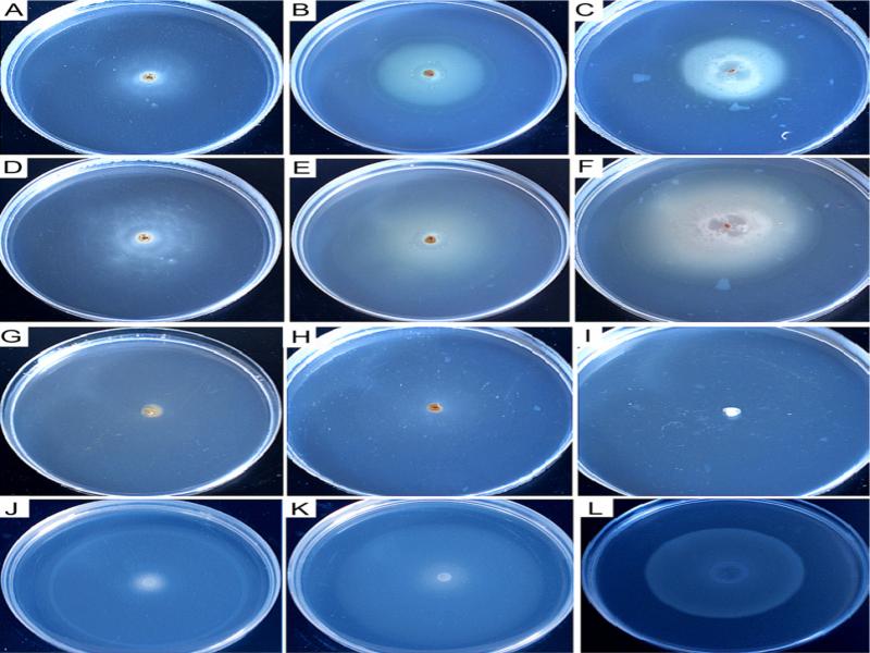
| Attractants1 | Pseudomonas aeruginosa 6-1B | Rhodococcus erythropolis T7-2 | Pseudomonas aeruginosa PAO-1 | |||
|---|---|---|---|---|---|---|
| Chemotaxis response2 | Oil degrading rate (%)3 | Chemotaxis response2 | Oil degrading rate (%)3 | Chemotaxis response2 | Oil degrading rate (%)3 | |
| Dodecane | + | 63.22 | – | 78.17 | – | ND |
| Tridecane | + | 56.18 | – | 75.62 | – | ND |
| Tetradecane | + | 54.28 | – | 67.57 | – | ND |
| Pentadecane | + | 57.97 | – | 62.84 | – | ND |
| Hexadecane | + | 55.54 | – | 59.73 | – | ND |
| Liquid paraffin | + | 58.13 | – | 65.11 | – | ND |
| Crude oil | + | 60.09 | – | 75.43 | – | ND |
| Naphthalene | – | ND | – | ND | – | ND |
| Diphenyl | – | ND | – | ND | – | ND |
| Sulfur | – | ND | – | ND | – | ND |