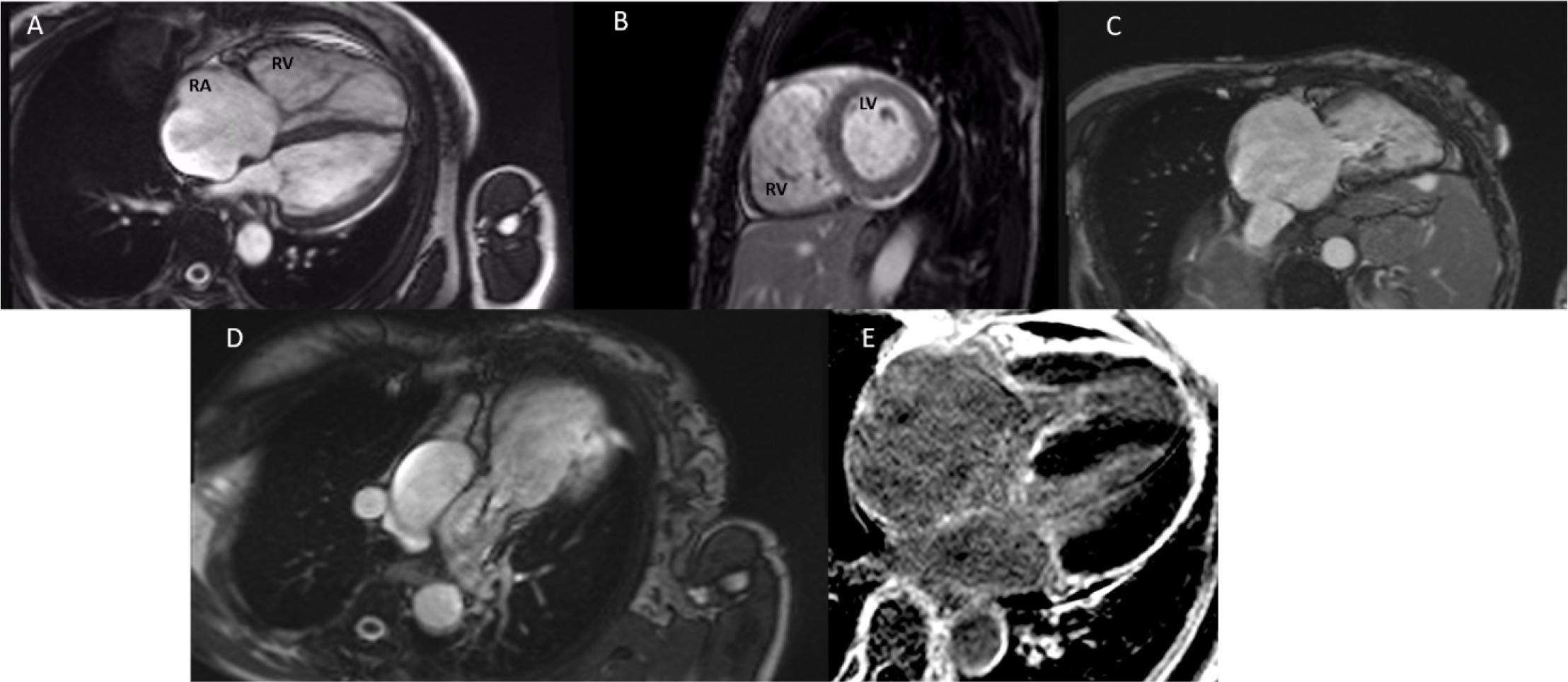
Figure 1
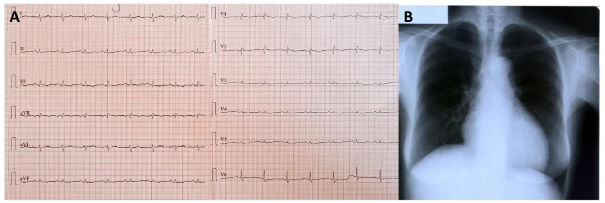
Figure 2
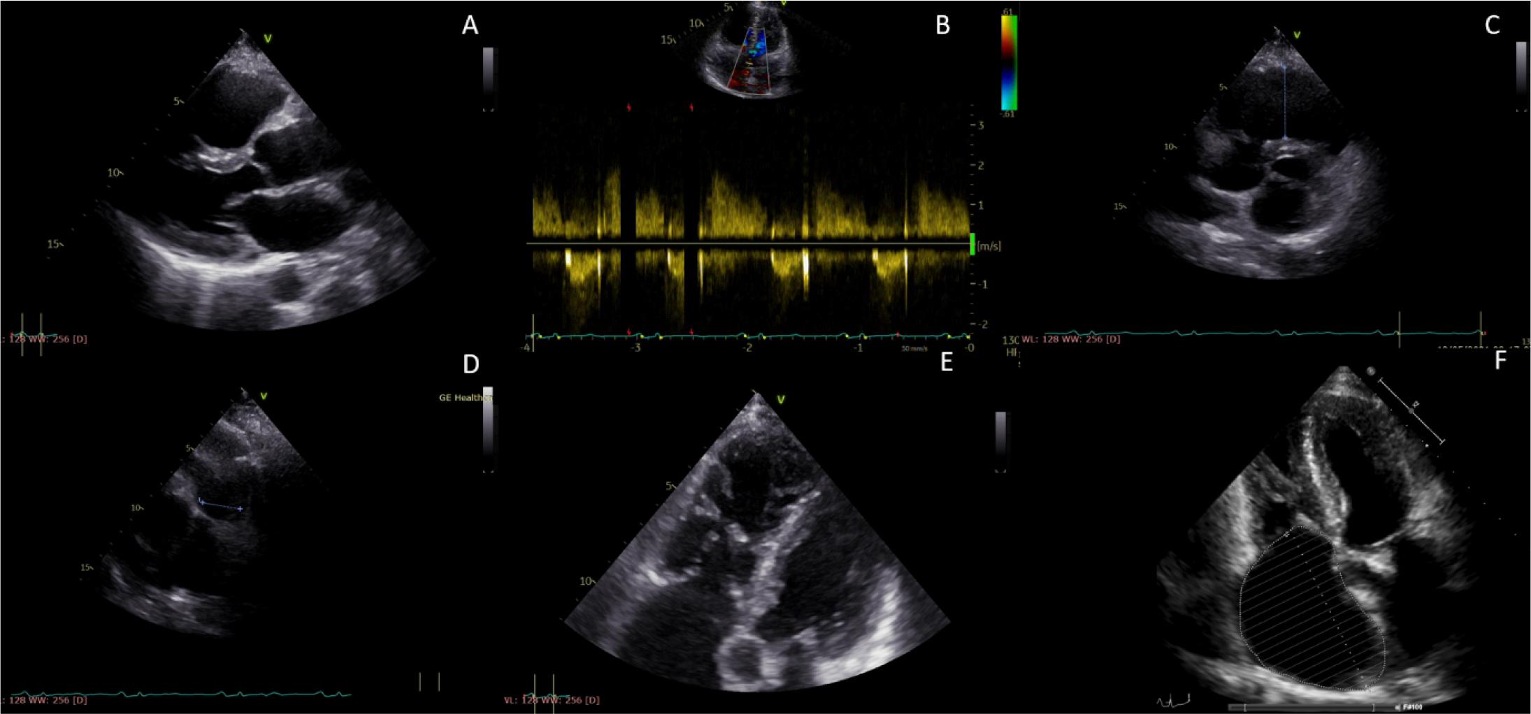
Figure 3
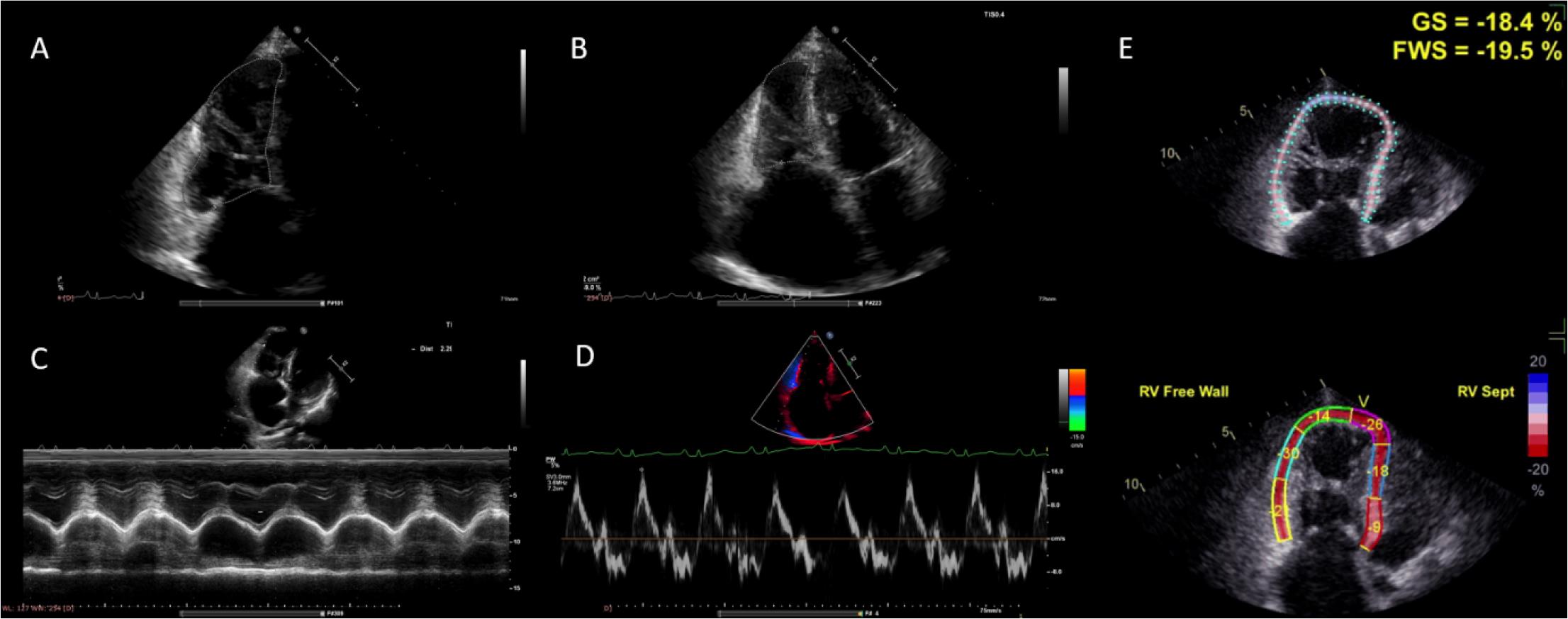
Figure 4
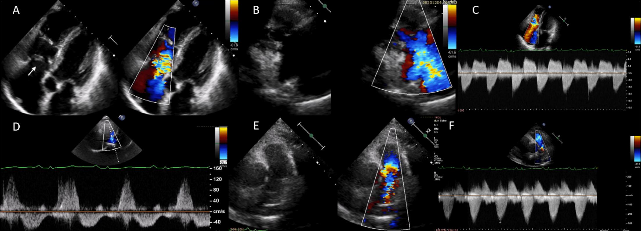
Figure 5
© 2025 Laura Cătălina Benchea, Larisa Anghel, Radu Andy Sascău, Cristian Stătescu, published by Romanian Society of Cardiology
This work is licensed under the Creative Commons Attribution-NonCommercial-NoDerivatives 4.0 License.