
Introduction
Phagocytosis is a process of innate immunity and consists of the engulfment and internalisation of large particles, microorganisms and apoptotic cells by several cell types (8). The most efficient phagocytic cells are short-lived neutrophils, playing a crucial role in antibacterial defence (11, 12). The B-1 subset of B-lymphocyte cells also has a phagocytic property (4, 9). Macrophages, residing in lungs (25) and lymphoid organs (13), are another important cell compartment, removing microorganisms and apoptotic cells from the organism. Pinocytosis is a form of endocytosis in which cells internalise extracellular fluid and dissolved solutes through the formation of membrane-bound vesicles. The function of pinocytosis may be also fulfilled by other cells than macrophages, such as dendritic cells by formation of active membrane protrusions (20, 24).
Cultures of RAW 264.7 macrophages represent a convenient model to study various aspects of phagocytosis. Studies have shown that the endocytic function in this model is stimulated by glycoproteins (40) and polysaccharides (6, 18, 28, 30, 33) as Toll-like receptor 4 (TLR4) ligands (26). The metabolic events resulting from interaction of the stimulatory ligands include enhanced phagocytosis (5, 18) associated with polarisation of macrophages to the M1 phenotype (40), stimulation of pro-inflammatory cytokines and inducible nitric oxide (NO) (7, 18), prostaglandin E2 and cyclooxygenase 2 (COX 2) (7) production. However, at the same time, superoxide dismutase (SOD) may be produced, which protects against oxidative damage (18).
Yolkin is a recently discovered egg yolk–derived glycoprotein with distinct immunoregulatory properties which render it suitable for application in prevention and therapy as a nutraceutical (35). Very recent studies by the present authors revealed protective actions of yolkin in mouse endotoxaemia (38) and in a homologous model of viral infection (unpublished). In targeting bone marrow–derived macrophages via TLR2/4, the protein activates mitogen-activated protein (MAP) kinases and production of TNF-α and type I interferons (IFNs) (14). In the model of RAW 264.7 mouse macrophages, yolkin induced expression of MAP kinases and COX 2 at both the mRNA and protein levels (37).
The accumulated data on the protective properties of yolkin in inflammation and infection prompted an investigation of the potential actions of yolkin on the pinocytic and phagocytic activity of macrophages, which play a key role in antimicrobial defence. The RAW 264.7 mouse macrophage model was chosen for the investigation as it is frequently used to study mechanisms of phagocytosis. More specifically, an evaluation was made of the capability of the RAW 264.7 cell to endocytose neutral red (NR) and NR-labelled zymosan particles in culture with yolkin. The accompanying production of TNF-α, IL-6, and IL-1 was also determined.
Material and Methods
Reagents
Neutral red (NR, N4638), 3-(4,5-dimethylthiazol-2-yl)-2,5-diphenyltetrazolium bromide (MTT), zymosan A (Z4250), and dimethyl sulfoxide (DMSO) were purchased from Sigma-Aldrich (St. Louis, MO, USA) and PBS, DMEM High Glucose (L0103-500), penicillin, streptomycin, and foetal calf serum (FCS) were from Biowest (Nuaillé, France). A TNF-α kit (cat. No. 430904), IL-6 kit (cat. No. 431304), and IL-10 kit (cat. No. 431414) were from BioLegend (San Diego, CA, USA).
Yolkin
Yolkin was isolated from egg yolk as originally described (22). It was prepared as three protein fractions with molecular weight below 45 kDa. Detailed characteristics of the protein were recently presented (21).
RAW 264.7 cell line
Cultures of RAW 264.7 mouse macrophages were conducted in high-glucose DMEM supplemented with 10% FCS and a penicillin/streptomycin solution. Upon cell transfer, the supernatants were removed, the cells in the flasks were washed with the DMEM and the flasks again filled with the medium. The adherent cells were then harvested from the flasks with a plastic cell scraper. The cell suspension was adjusted to an appropriate density (1 : 3), transferred to another flask and incubated at 37°C in an 8% CO2 atmosphere.
Determination of RAW 264.7 viability
Volumes of 100 μL of RAW 264.7 cells with cell density of 3 × 10⁴/well were pipetted into 96-well plates and incubated for 24 h at 37°C in an 8% CO2 atmosphere. Then, yolkin at 20, 10, 5, 2, 1, 0.5, 0.25 and 0.125 μg/mL concentrations was distributed onto a monolayer cell culture, and the culture was incubated for 48 h at 37°C in an 8% CO2 atmosphere. The control culture contained no yolkin. The quantitative and morphological changes were evaluated in an inverted microscope. The minimal concentration of yolkin causing degeneration of 50% of cells was defined as the 50% tissue culture cytotoxic dose. The analysed parameters of growth, cell morphology, and cell viability were determined by a colorimetric MTT test (10). In short, 25 μL from the stock solution (5 mg/mL) was added to each well at the end of the cell incubation time, and the plates were incubated for an additional 2 h in a cell culture incubator. Then, 85 μL of the extraction buffer (20% SDS with 50% dimethylformamide, pH 4.7) was added. After overnight incubation, the OD was measured at 550 nm with the reference wavelength of 630 nm (550/630 nm) in a spectrophotometer (Bio-Tek Instruments Inc., Winooski, VT, USA). The experiment was performed in two replicates.
Induction of cytokine production by yolkin
Cultures of RAW 264.7 cells (2 × 10⁴/well in DMEM with 5% FCS and antibiotics) were incubated overnight, after which yolkin was added to make final concentrations of 10–0.5 μg/mL. After 48 h incubation, the supernatants were collected and frozen at −80°C. Each experiment was performed in 6 or 7 replicates within a single assay (supernatants were pooled). The number of cells was estimated separately for each study group in a Bürker haemocytometer. The levels of TNF-α, IL-6 and IL-10 were determined using the appropriate ELISA kits as noted above. The lowest standard concentrations were 7.8 pg/mL for TNF-α and IL-6 and 31.3 pg/mL for IL-10. When the detected cytokine concentration was below the indicated value, the sample was classified as non-detectable and assigned a value of zero. The data were calculated using four-parameter logistic regression with Assayfit Pro (Assay Cloud, Nijmegen, the Netherlands).
Pinocytosis
The studies were performed according to protocols in the literature (2, 3, 23) with some modifications. Neutral red was initially dissolved in DMSO (10 mg/mL) and subsequently diluted in DMEM containing 5% FCS to a concentration of 50 μg/mL. Cultures of RAW 264.7 cells were incubated with NR for 30 min at 37°C in an incubator. The control cultures included only NR in the medium and the other only the cells without NR. The cells were washed 4× with PBS. The cell pellet was re-suspended in 0.1 mL of acidic ethanol (1% v/v of glacial acetic acid and 50% ethanol in distilled water), and the culture plate was shaken for 10 min at 400 rpm. The absorbance was determined spectrophotometrically at 550/630 nm using the microtitre plate reader (Bio-Tek Instruments). The concentration of NR in each well was calculated based on a standard curve, and the reported NR values were normalised in relation to 106 cells. Each experiment was performed in seven replicates. The number of cells was estimated separately for each experiment group in the protocol with a preincubation step. In the experiment without preincubation, the cell numbers were determined only for the control group. The cell enumerations were performed in a Bürker haemocytometer in triplicate using three additional samples prepared exclusively for this purpose. Each summary graph presents a percentage in relation to the control value in the given experiment.
Phagocytosis of NR-labelled zymosan (NR-zymosan)
These studies were also conducted according to protocols in the literature (2, 27, 29) with minor modifications. Neutral red was initially dissolved in PBS (0.2 mg/mL), and 3 mL was added to 3 mL of zymosan A in PBS (20 mg/3 mL). The suspension was subsequently incubated for 30 min at room temperature with occasional agitation. Next, the suspension was centrifuged at 1,000 × g for 10 min and washed with PBS, and the pellet was re-suspended in PBS. The NR-zymosan suspension was added to the RAW 264.7 culture at a density of 20 × 106 zymosan particles/well. The cells were incubated at 37°C for 3 h. The control cultures were also included as described above. The cultures were washed 5× with PBS. Further steps, including measurement of absorbance, the cell enumerations, and data presentation, are as described above, except that the plates were shaken for 30 min instead of 10 min.
Statistical analysis
The results are presented as mean values ±SD. The Brown–Forsyth test was used to determine the homogeneity of the variance between groups. When the variance was homogenous, one-way ANOVA was applied, followed by post-hoc comparisons with Tukey’s test to evaluate the significance of the difference between groups. Nonparametric data were evaluated with the Kruskal–Wallis analysis of variance. Significance was defined as P-value < 0.05. Statistical analysis was performed using Statistica 7.0 for Windows (StatSoft, Tulsa, OK, USA).
Results
The effect of yolkin on cell viability
The yolkin concentration of 20 μg/mL resulted in a decrease in cell metabolism (Fig. 1), accompanied by distinct morphological changes such as spreading on the plastic surface (data not shown). Therefore, for evaluating the functional activities of RAW 264.7 cells, the highest concentration of yolkin was set at 10 μg/mL.
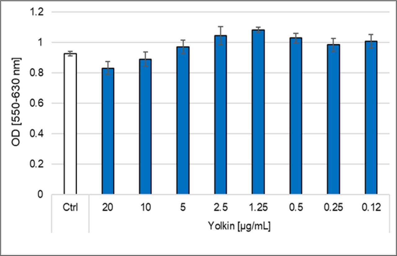
Fig. 1.
The effects of yolkin on viability of RAW 264.7 cells. The results of one of the two separate experiments conducted are presented as the mean OD at 550/630 nm ±SD from five determinations for each concentration. Ctrl – control incubation of RAW 264.7 cells without yolkin
The effect of yolkin on NR pinocytosis
The effects of yolkin on NR pinocytosis are presented in Fig. 2. In the first protocol, RAW 264.7 cells (2 × 10⁵/well) were cultured overnight, the cultures were treated with yolkin and NR, and no prior preincubation step of cells with yolkin was included. As shown, the applied experimental protocol did not reveal any effects of yolkin on NR pinocytosis (Fig. 2A). In the second protocol, yolkin was added to RAW 264.7 cells (2 × 10⁴/well) which had been cultured overnight, the treated cells were preincubated for 48 h, and NR was added subsequently. The results show a significant stimulation of the pinocytic capacity of the cells with a maximum yolkin concentration of 10 μg/mL (Fig. 2B).
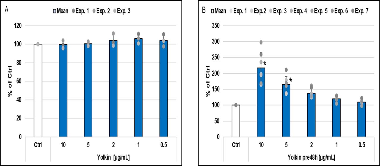
Fig. 2.
The effects of yolkin on the capability of RAW 264.7 cells to internalise neutral red (NR). (A) Addition of yolkin and NR simultaneously to the culture (no preincubation step). The means ±SD from three replicates of the experiment are presented. (B) Addition of NR after preincubation with yolkin for 48 h. The means ±SD from seven replicates of the experiment are presented. * – Statistical significance (P-value < 0.05) compared to the control (RAW 264.7 cells in culture medium only); Ctrl – control incubation; pre48h – incubation with yolkin for 48 h
The effect of yolkin on phagocytosis of NR-zymosan particles
The effects of yolkin on phagocytosis of NR-zymosan particles are presented in Fig. 3. In the first protocol, RAW 264.7 cells (2 × 10⁵ cells/well) were cultured overnight, after which yolkin and NR-zymosan were added. The results show no effect of yolkin on the phagocytosis (Fig. 3A). On the other hand, when the cells (2 × 10⁴/well) were preincubated for 48 h with yolkin prior to NR-zymosan addition, a very significant stimulation of the phagocytosis with a maximal effect at 10 μg/mL of yolkin could be demonstrated (Fig. 3B).
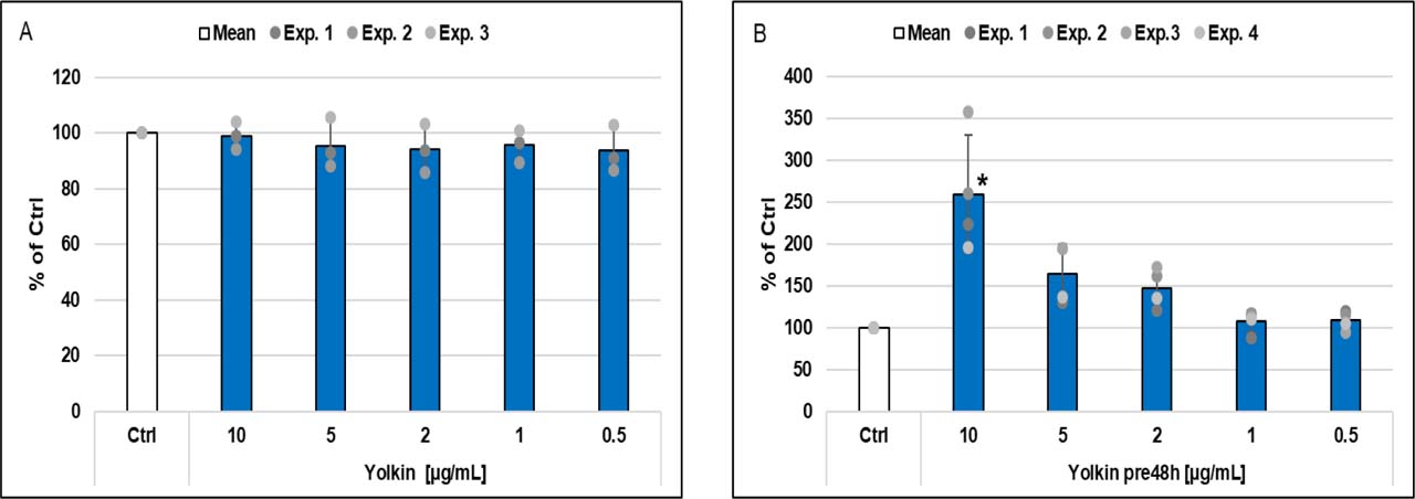
Fig. 3.
The effects of yolkin on the capability of RAW 264.7 cells to engulf neutral red (NR)-stained zymosan particles. (A) Yolkin and NR-zymosan were added simultaneously to the culture (no preincubation step). The means ±SD from three replicates of the experiment are presented. (B) Addition of NR-zymosan after preincubation with yolkin for 48 h. The means ±SD from four replicates of the experiment are presented. * – statistical significance (P-value < 0.05) compared to the control (RAW 264.7 cells in culture medium only); Ctrl – control incubation; pre48h – incubation with yolkin for 48 h
The effect of yolkin on cytokine production by RAW 264.7 cells
These cells, incubated with polysaccharides or glycoproteins, were shown to produce cytokines with a prevailing Th1 profile (18, 40). For the present investigation, TNF-α, IL-6, and IL-10 were selected to evaluate production of Th1-profile cytokines by the cells following incubation with yolkin (Fig. 4). The results show a strong induction of TNF-α production after 48 h incubation with yolkin in a dose-dependent manner (Fig. 4A). The production of IL-6 (Fig. 4B) and IL-10 (Fig. 4C) was rather modest in comparison with that of TNF-α. Of importance is that these results revealed the immunological status of the macrophages just before exposure of the cells to the phagocytic particles.
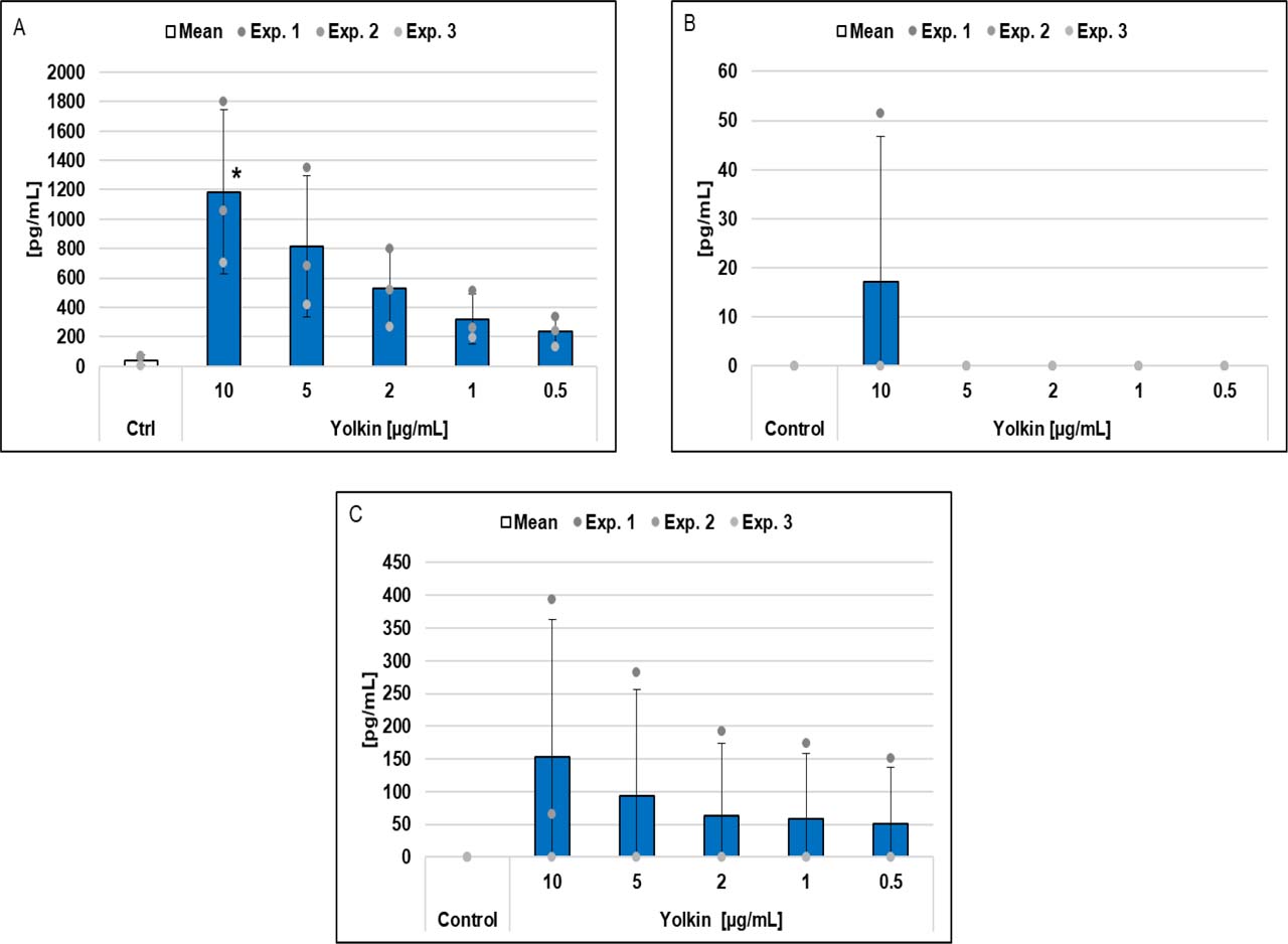
Fig. 4.
The effect of incubation of RAW 264.7 cells for 48 h with yolkin on cytokine production. (A) Production of TNF α. (B) Production of IL-6. (C) Production of IL-10. A culture of RAW 264.7 cells was incubated overnight, yolkin was added and the cells incubated for 48 h. The results are presented as the mean ±SD from three independent experiments. Statistical significance is indicated by asterisks (* – P-value < 0.05) compared to the control (RAW 264.7 cells in culture medium only). Ctrl – control incubation
Discussion
In this work, we provided evidence that yolkin promotes the pinocytic and phagocytic functions of RAW 264.7 mouse macrophages. The stimulatory effects on pinocytosis and phagocytosis could resemble the action of yolkin on bone marrow–derived macrophages via TLR2 and TLR4 (15). Further, yolkin was a strong activator of pro-inflammatory cytokine production in mouse splenocyte cultures (37) and in bone marrow–derived macrophages (15). These events were correlated with a large increase in NFκB expression in RAW 264.7 cell cultures and upregulation of extracellular signal-regulated kinases 1/2 and c-Jun N-terminal kinase expression. In addition, yolkin strongly induced COX 2 expression and production in RAW 264.7 cultures (37).
The presented profiles of the secreted cytokines support the suggestion made by other authors (14) that yolkin directs polarisation of macrophages to the M1 phenotype. In this study, we demonstrated that following culture with yolkin, RAW 264.7 cells produce more TNF-α than IL-10. The TNF-α : IL-10 ratio was 7.8:1 after 48 h incubation. Following incubation with yolkin, the cytokine profile corresponded to the M1 macrophage phenotype capable of efficient pinocytosis and phagocytosis and characterised by that efficiency (36).
Additionally, the results indicate that activation of macrophages by yolkin could engage TLRs, as demonstrated for bone marrow–derived macrophages (15), since acquisition of the pinocytic ability was partially inhibited by anti-TLR2 and -TLR4 antibodies (data not shown). Taking into account the very complex glycosylation pattern of yolkin and the significant content of mannose (35), yolkin may also target RAW 264.7 cells via the mannose receptor (19) and sialoadhesin (31). More research is required to identify other yolkin receptors on relevant cells. Of note is that another key egg yolk protein – phosvitin – demonstrated similar actions in the RAW 264.7 model by enhancing phagocytosis and expression of pro-inflammatory mediators (17). The structure of its oligosaccharide also predisposes the protein to interact with these cells (32).
The activation of RAW 264.7 cells via TLR4, leading to the M1 phenotype and evidenced by the production of IL-2, TNF-α, and IFN, was also induced by a polysaccharide (18). Redirection of RAW 264.7 cells to the M1 phenotype was also induced by a glycoprotein, which stimulated pinocytic and phagocytic activity, and increased NO, TNF-α, IL-6, and chemokine production (36). Taken together, these alterations in the inflammatory mediators in macrophages induced by yolkin resemble those by which RAW 264.7 macrophage pinocytosis and phagocytosis are elicited by other glycoproteins and glycans (6, 7, 18, 36).
A finding of importance is that the registered stimulatory effects of yolkin on macrophage phagocytic function were accompanied by production of cytokines known to support antibacterial defence (11). Macrophages were also implicated in antibacterial defence in mice by another naturally occurring glycoprotein, lactoferrin (34), which exhibited a mechanism of action closely resembling that of yolkin (16). The protein was also shown to act in concert with specific bacteriophages in response to lethal bacterial infection (38) and to exhibit an adjuvant property when administered with BCG immunisation (39).
The ability of yolkin to enhance phagocytosis may also explain its strong stimulatory effect on the humoral immune response to sheep erythrocytes (21). In the mouse model used in that experiment, yolkin and an antigen (sheep red blood cells) were introduced by the same intraperitoneal route. Under these conditions, professional antigen-presenting cells, such as peritoneal macrophages and B-1 cells may be activated for enhanced phagocytosis, antigen degradation and presentation. We also speculate that the ability of yolkin to enhance phagocytosis may be applied to improve vaccine efficiency (1).
Conclusion
We demonstrated that yolkin stimulated the capability of cells to perform pinocytosis of NR and phagocytosis of NR-zymosan in a dose dependent manner, and to induce cytokine production of the Th1 profile. The results supplement the current knowledge on the immunotropic properties of yolkin, which may also play antibacterial and antiviral roles in developing embryos. Going forward, yolkin and other egg yolk–derived proteins may find use as nutraceuticals and ingredients of functional foods for boosting immunity to various infectious agents.
Notes
[1] Conflicts of interest Conflict of Interests Statement:
The authors declare that there is no conflict of interests regarding the publication of this article.
[2] Financial disclosure Financial Disclosure Statement:
This research was funded by National Science Centre, Poland, No. DEC-2021/41/B/NZ6/01167. For the purpose of open access, the author has applied a CC-BY public copyright license to any Author Accepted Manuscript (AAM) version arising from this submission.
[4] Contributed by CRediT Authorship Contribution Statement:
Michał Zimecki: research concept and design, collection and assembly of data, data analysis and interpretation, writing the article, critical revision of the article, final approval of the article. Jolanta Artym: critical revision of the article, final approval of the article. Maja Kocięba: collection and assembly of data, data analysis and interpretation, critical revision of the article, final approval of the article. Ewa Zaczyńska: collection and assembly of data, data analysis and interpretation, critical revision of the article, final approval of the article. Aleksandra Zambrowicz: collection and assembly of data, final approval of the article. Łukasz Bobak: collection and assembly of data, final approval of the article.