
Cyst nematodes of the subfamily Heteroderinae (Tylenchida: Heteroderidae) are obligate plant parasites that cause substantial yield losses in a wide range of agricultural and horticultural crops across temperate, subtropical, and tropical regions worldwide (Subbotin et al., 2010). The subfamily currently comprises approximately seven valid genera, including Heterodera, Globodera, Punctodera, Cactodera, Dolichodera, Betulodera, and Paradolichodera (Subbotin et al., 2010). These genera are distinguished primarily by morphological features of cysts, such as the type of vulval fenestration, presence or absence of an underbridge and bullae, and characteristics of second-stage juveniles (J2) including stylet length, tail morphology, and hyaline tail terminus.
The genus Cactodera was erected by Krall and Krall (1978) and currently comprises 20 valid species, many of which are adapted to hosts in the families Cactaceae, Amaranthaceae, Chenopodiaceae, and Polygonaceae (Ni et al., 2024a, 2024b; Prado Vera et al., 2024; Subbotin et al., 2010). Among them, the cactus cyst nematode, C. cacti, is the most widespread and representative species, having been reported from numerous countries across the Americas, Europe, and Asia, mainly on plants of family Cactaceae grown in glasshouses as ornamentals (Subbotin et al., 2010). Infected plants typically exhibit root branching and proliferation of rootlets, and heavy infestations may eventually lead to plant death.
Dragon fruit (Selenicereus undatus) is a perennial climbing vine from the Selenicereus genus of the Cactaceae family (Korotkova et al., 2017). It has emerged as a commercially important fruit crop worldwide, driven by its high nutritional value, visual appeal, climate resilience, and growing global demand (Luu et al., 2021; Hossain et al., 2021). Originating from the tropical and subtropical forest regions of Mexico, Central America, and South America, dragon fruit is now extensively cultivated in Vietnam, China, and other Southeast Asian countries (Sisir & Prodyot, 2024). In China, the cultivation area has reached approximately 67,000 ha, ranking first in the world (Sisir & Prodyot, 2024; Zhao, 2023). The crop is primarily grown in several southern provinces, including Hainan, Guangdong, Guangxi, and Fujian. Among these, tropical Hainan province – with its abundant sunshine and year-round frost-free climate – has experienced rapid expansion of dragon fruit cultivation in recent years, now accounting for approximately one-fifth of China’s total production area.
To date, C. cacti associated with dragon fruit has been reported from Taiwan Province of China, and Vietnam, but its occurrence in mainland China (specifically Hainan Province) has not been documented (Chan et al., 2016; Thanh Thi Vo et al., 2026). In February 2025, a cyst nematode was discovered in soil and root samples collected from a dragon fruit (Selenicereus undatus) orchard in Danzhou County, Hainan Province of China, where affected plants exhibited poor growth (Figure 1). The objective of this study was to provide morphological and molecular characterization of this nematode, identified herein as Cactodera cacti. This report constitutes the first record of C. cacti infecting dragon fruit in Hainan Province of China.
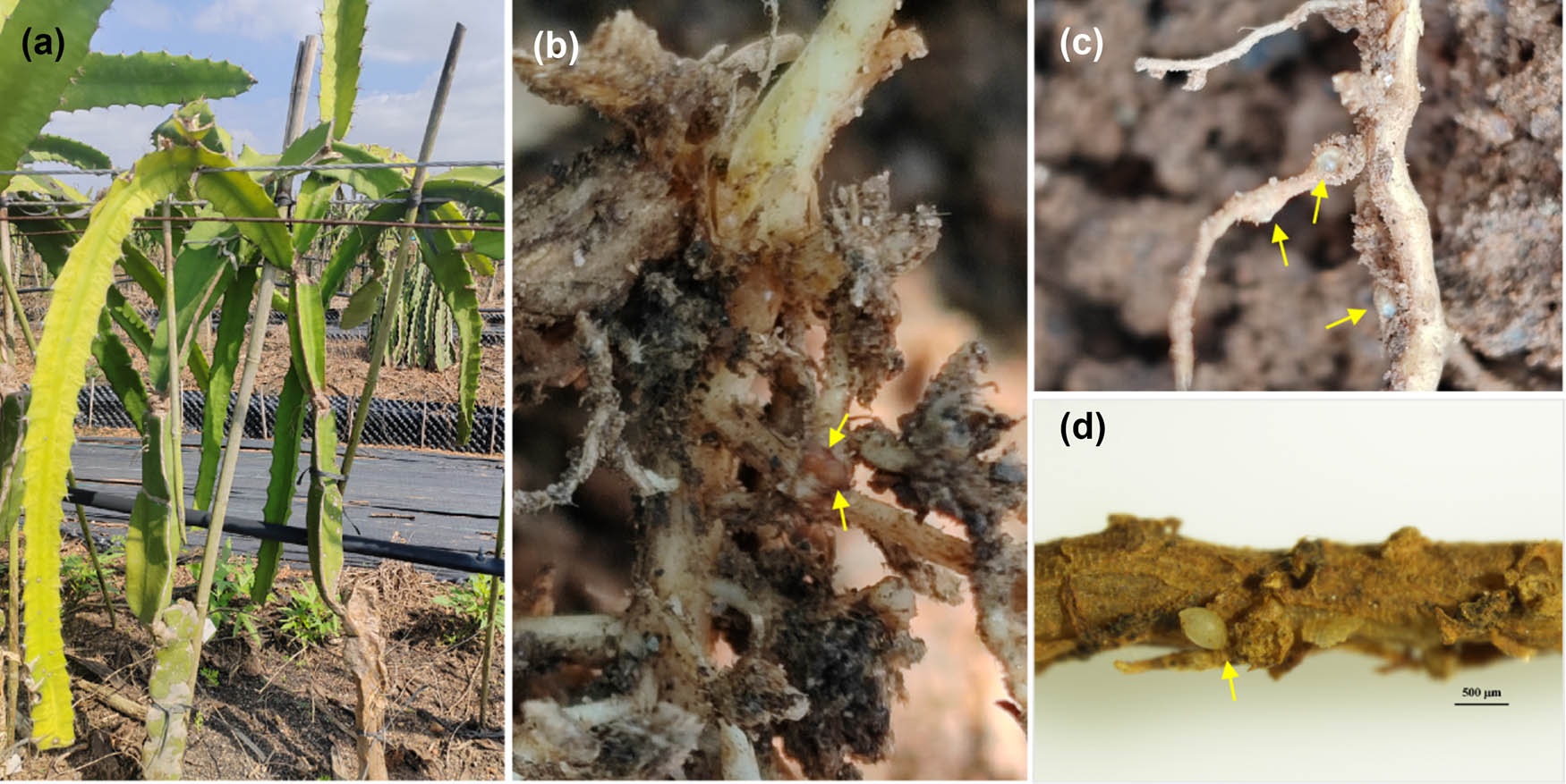
Figure 1
Symptoms of dragon fruit infected by Cactodera cacti. Stunted and yellowing plant (a); roots showing cyst nematode (indicated by arrows) (b–d).
1. Materials and methods
1.1. Nematode sampling and extraction
Soil and root samples were collected from the rhizosphere of dragon fruit (Selenicereus undatus) plants in Danzhou County, Hainan Province, China (19.485901°N; 109.489160°E). Approximately 2 kg of soil was obtained from each sampling site using the five-spot sampling method. A total of 10 soil and root samples were collected to laboratory for further analysis. Each sample was thoroughly mixed, then 200 g soil was used for nematode extracted. Cysts were extracted from soil samples using the standard centrifugal flotation method (Jenkins, 1964). 200 g of the mixed soil was weighed and transferred into a 2,000 mL triangular bottle, followed by the addition of 1,800 mL water. After 1–5 min, the floating objects were filtered through a 250 µm sieve. The cysts were then collected on this sieve. The cysts picked from the root samples were observed under the microscope. All these cysts were collected together for further identification. Second-stage juveniles (J2) were obtained by incubating cysts in sterile distilled water at room temperature. White females were picked directly from the roots under a dissecting microscope.
1.2. Morphological and morphometric study
Second-stage juveniles (J2) were heat-killed in 4% formalin, processed through a glycerin series, and mounted on permanent microscope slides following standard protocols (Golden, 1990; Hooper, 1970). Vulval cones of cysts were dissected, cleared of internal contents, trimmed, and mounted according to the method described by Subbotin (Subbotin et al., 2010). All specimens were examined and measured using an Olympus SZX16 stereomicroscope and an Olympus BX53 compound microscope equipped with a digital camera for photomicrography.
1.3. Molecular study
Single cysts were selected and transferred from a Petri dish to a microtube before being crushed with a pipette tip. 20 cysts were used for DNA extraction using the Proteinase K method described by Kumari and Subbotin. The extracted crude DNA was amplified in the ITS (ITS1–5.8S–ITS2) region using the primers TW81 (5′-GTTTCCGTAGGTGAACCTGC-3′) and AB28 (5′-ATATGCTTAAGTTCAGCGGGT-3′) (Joyce et al., 1994); the D2–D3 regions of 28S rRNA using the primers D2A (5′-ACAAGCTACCGTGAGGGAAAGTTG-3′) and D3B (5′-TCGGAAGGAACCAGCTACTA-3′) for 28S D2–D3 (De Ley et al., 1999); the cytochrome oxidase I(COI) genes of mtDNA using the primers JB3 (5′-TTTTTTGGGCATCCTGAGGTTTAT-3′) and JB5 (5′-AGCACCTAAACTTAAAACATAATGAAAATG-3′) (Derycke et al., 2005). The polymerase chain reactions (PCRs) were performed in 25 µL of 2× Rapid Taq Master Mix (Vazyme Biotech, Nanjing, China), 2 µL forward and reverse primers, 2 µL of template DNA, and sterile Milli-Q water up to a volume of 50 µL by a Bio-Rad T100 Thermal Cycler (Bio-Rad Laboratories, Shanghai, China). All successful PCR products were purified with the TianGEL Extraction Kit (Tiangen Biotech, Beijing, China), cloned with the pMD19-T vector (Takara Bio, Dalian, China), and then transferred into DH5α competent cells. Finally, four clones of each PCR product were sequenced by Sangon Biotech (Guangzhou, China). The resulting sequences from different cyst samples were deposited in GenBank under accession numbers PX498015 and PX498018 (ITS); PX498032 and PX498038 (28S D2–D3); and PX498440 (COI).
1.4. Phylogenetic analysis
The obtained sequences were compared against the GenBank database using the BLAST algorithm to identify closely related sequences. Relevant gene fragments from closely related species were retrieved and aligned using the online version of MAFFT version 7 (http://mafft.cbrc.jp/alignment/server/). Phylogenetic trees were constructed using MEGA 11 software with the Neighbor-Joining (NJ) method, and branch support was evaluated using bootstrap analysis with 1,000 replicates. Based on previously published data, Punctodera mulveyi (MN121006) and Rotylenchus cypriensis (KP843116) were selected as outgroups for the ITS and 28S D2–D3 datasets, respectively, while Rotylenchus eximius (KX669244) was used as the outgroup for the COI dataset.
1.5. Pathogenicity verification
Six dragon fruit (Selenicereus monacanthus cv. “Dahong”) seedlings were transplanted into perforated plastic pots containing 200 g of sterilized sandy soil, with one seedling per pot. At 7 days after transplantation, 20 well-developed cysts of C. cacti were placed into four 4 cm deep holes around the root zone, and the holes were then covered with soil. Four uninoculated plants served as controls. Plants were watered as needed and maintained in an incubator at 29°C. At 65 days after inoculation, all cysts and white females were recovered from both roots and soil. The reproduction factor (Rf) of C. cacti was calculated as Rf = final population density/initial population density (Pi = 20 cysts per pot).
2. Result
2.1. Morphological and morphometric characteristics
A total of 15 J2s were used for morphometric measurements and compared with populations from China (Taiwan Province and Liaoning Province), the Netherlands, and Vietnam. The body length (L) of J2s from Hainan Province ranged from 434.6 to 549.1 μm. The body width (W) ranged from 17.8 to 24.1 μm. The stylet length ranged from 18.9 to 26.0 μm. The tail length ranged from 39.2 to 59.9 μm. The tail hyaline region ranged from 16.9 to 25.4 μm (Table 1). J2s were vermiform (Figure 2a), the stylet was well developed and bore semi-rounded basal knobs that were convex both anteriorly and posteriorly (Figure 2b, c). The tail was tapering, with a U- or V-shaped hyaline region (Figure 2d, e).
Table 1
Morphometric measurements of Cactodera cacti from Hainan Province and four reference populations from China, Taiwan, Vietnam, and the Netherlands
| Character | The Netherlands (Adam, 1932) | China (Duan et al., 2012) | Taiwan (Chan et al., 2016) | Vietnam (Thanh Thi Vo et al., 2026) | Hainan China |
|---|---|---|---|---|---|
| n | 20 | 20 | 20 | 30 | 15 |
| Body length | 486 (428–584) | 513.8 (467.3–542.5) | 479.7 (426.5–578.6) | 447.6 ± 38 (341–485) | 503.1 (434.6–549.1) |
| Body width | — | — | 23.98 (19.7–28.2) | 20.37 ± 1.16 (17–23) | 20.1 (17.8–24.1) |
| Stylet length | 25.2 (22–28.2) | 24.6 (23.0–25.8) | 24.74 (22.6–27.4) | 27.63 ± 1.3 (25–31) | 22.6 (18.9–26.0) |
| Tail length | — | 52.2 (45.9–59.5) | 53.11 (45.6–59.3) | 52.8 ± 4.89 (39–61) | 51.4 (39.2–59.9) |
| Hyaline tail | — | 19.1 (16.3–23.2) | 20.18 (16.1–24.7) | 19.37 ± 2.28 (13–25) | 20.7 (16.9–25.4) |
| DGO | — | _ | 4.32 (2.4–6.7) | — | — |
All measurements of J2s are in μm.
DGO: Distance from Dorsal Gland Opening to top of median bulb.
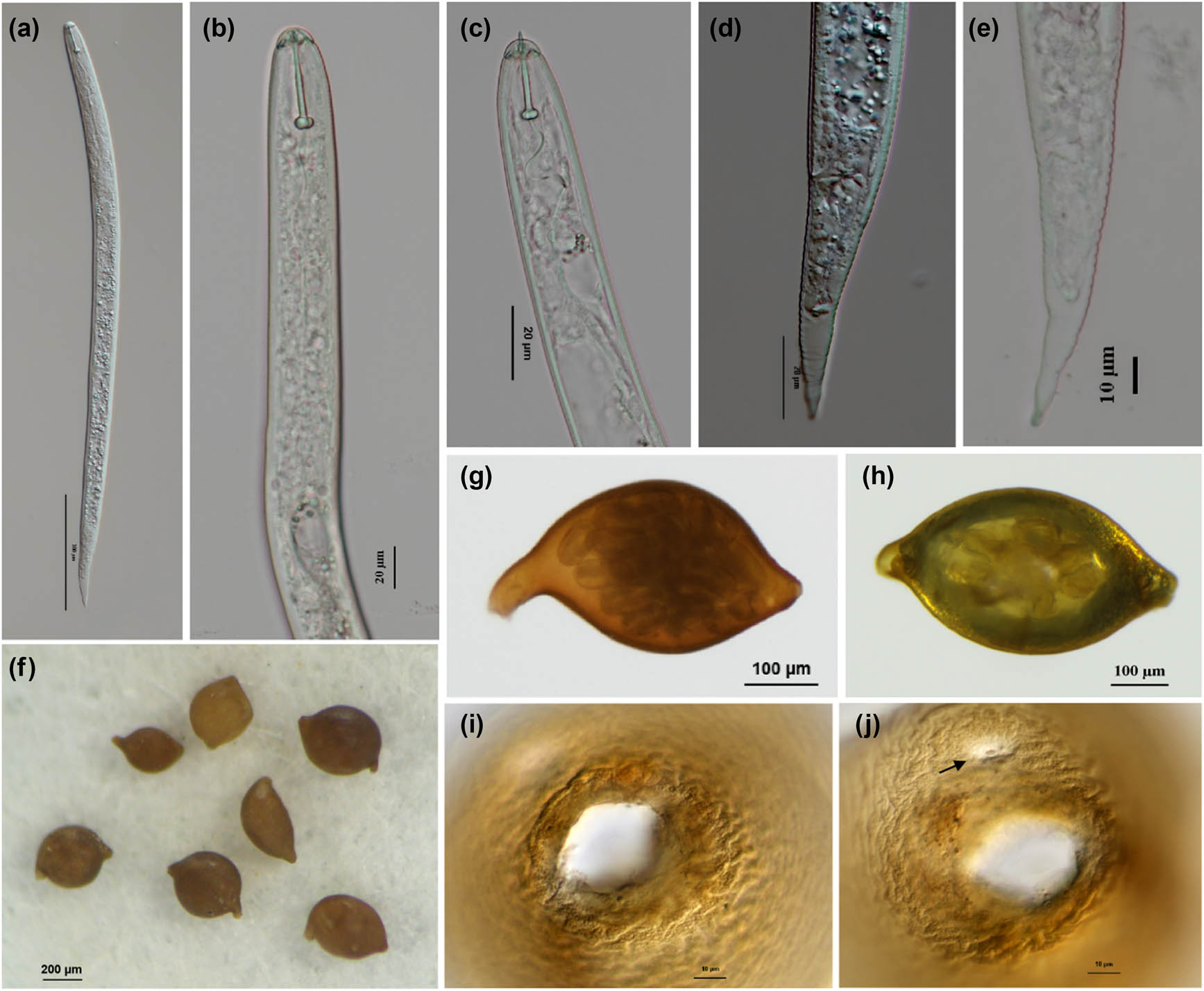
Figure 2
Morphological characteristics of second-stage juveniles (J2), cysts, and vulval cone of C. cacti. Entire J2 body (a); anterior end of J2 (b and c); posterior end of J2 (d and e); lemon shaped cyst (f–h); vulva (i and j). Arrow shows the anus (j). Scale bar: 200 μm (f); 100 μm (a, g, h); 20 μm (c, d); 10 μm (e, i, j).
15 cysts were measured (Table 2). Body length ranged from 299.4 to 453.6 μm (mean value = 391.3 μm), body width ranged from 239.3 to 306.3 μm (mean value = 274.0 μm). The L/W ratio ranged from 1.0 to 1.9. The fenestral diagram had the average of 21.4 μm (19–23 μm). The measurements of Hainan cysts were compared with the other populations from Netherland, Italy, Iran, Taiwan, and Vietnam and found to be similar (Table 2).
Table 2
Morphometrics of Cactodera cacti cyst from Hainan Province population and five reference populations from the Netherlands, Italy, Iran, Taiwan, and Vietnam
| Population characters | The Netherlands (Adam, 1932) | Italy (Ambrogioni, 1969) | Iran (Maafi, 2003) | Taiwan (Chan et al., 2016) | Vietnam (Thanh Thi Vo et al., 2026 | Hainan China |
|---|---|---|---|---|---|---|
| n | 50 | 25 | 21 | 20 | 10 | 15 |
| Length | 497 (328–617) | 536 (384–780) | 490 (380–600) | 457 (358–563) | 411 ± 102 (284–647) | 391 (299–454) |
| Width | 447 (309–598) | 378 (240–588) | 382 (280–490) | 359 (296–475) | 332 ± 88 (200–491) | 274 (239–330) |
| L/W | 1.1 | 1.4 (1.1–2.0) | _ | 1.28 (1.1–1.6) | 1.26 ± 0.17 (1.07–1.66) | 1.44 (1.03–1.89) |
| Fenestral diameter | _ | (26–28) | 28 ± 3.3 (23–35) | 23.2 (20–29) | _ | 21.4 (19–23) |
All measurements are in μm.
Cysts were typically lemon-shaped with a distinct neck and vulva (Figure 2). The vulval cone was circumfenestrate, lacking an underbridge and bullae, but generally bearing vulval denticles (Figure 2i). The anus was prominent, and punctations were often present in the terminal area of the cyst (Figure 2j).
2.2. Molecular analysis
Amplification of the ITS1–5.8S–ITS2 rDNA region yielded two fragments of 940 bp (PX498018) and 943 bp (PX498015). These two fragments were from two different cyst samples. The two sequences shared 99.7% identity, with three gaps (3/943 bp). Homology search comparison analysis showed that the sequences were 99.3–100% identical to those of C. Cacti from the USA (MH477533, MH477535); Vietnam (PV242139); and Taiwan and China (JX051344, JQ341140). In the phylogenetic tree, the Hainan isolates formed a strongly supported clade with 14 other C. cacti sequences, with Punctodera mulveyi (MN121006) used as the outgroup (Figure 3).
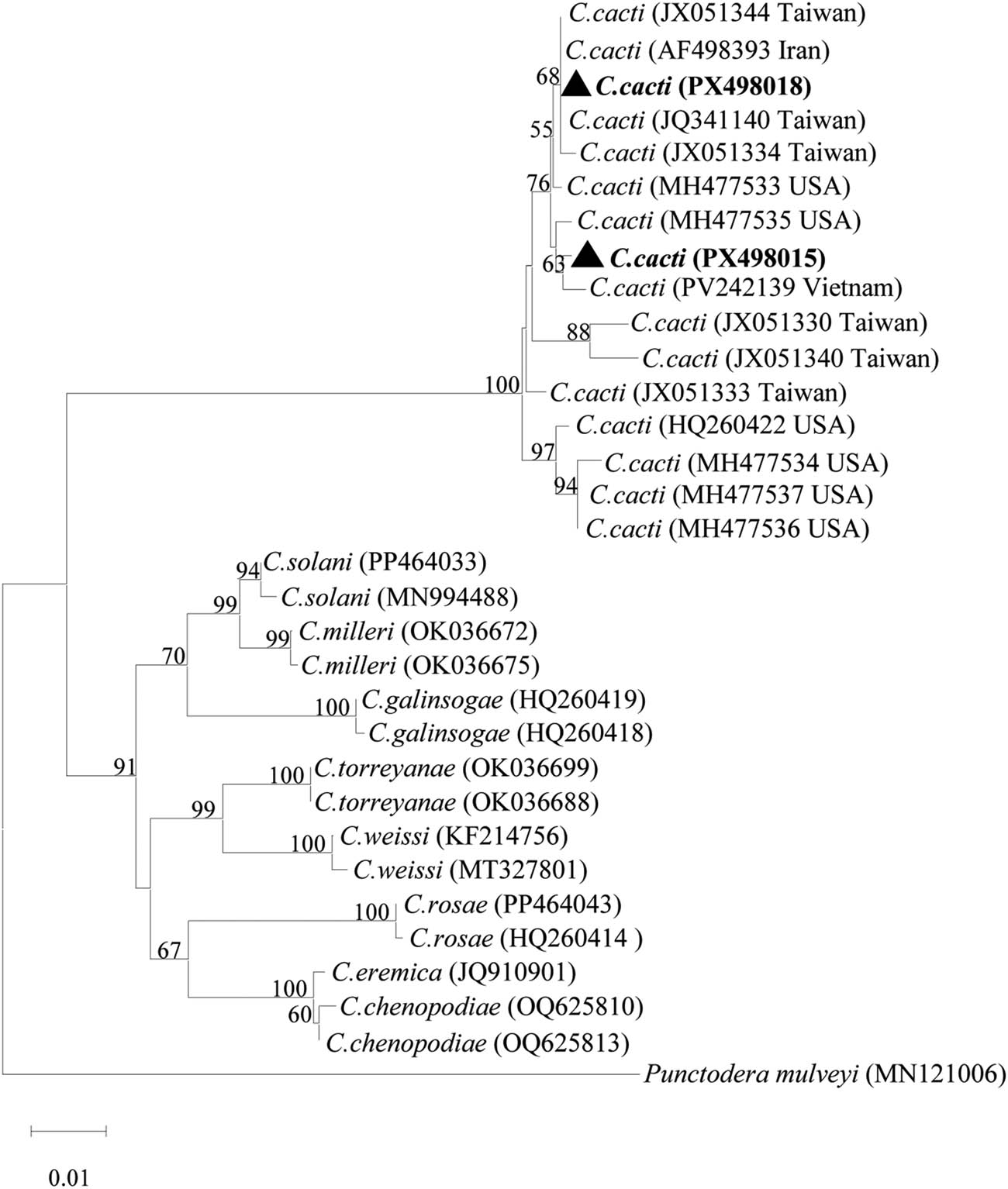
Figure 3
Phylogenetic tree based on ITS rDNA sequences of the genus Cactodera. The tree was constructed using the NJ method with bootstrap support from 1,000 replicates. Bootstrap values are shown at each node. Sequences obtained in this study are marked in bold (PX498015 and PX498018). Sequences of Cactodera cacti used in the phylogenetic analysis are labeled with accession numbers and geographic origin. Sequences originating from Taiwan Province of China are labeled as “Taiwan” in the tree to maintain consistency with GenBank records.
Amplification of the 28S D2–D3 region yielded two fragments, both was 739 bp (PX498032 and PX498038). This two accession numbers were from two different cyst nematode samples. The two sequences shared 99.73% identity with 2 bp difference (2/739 bp). Homology search comparison analysis showed that the sequences were 99.3–100% identical to those of C. Cacti from China (PX602974), the USA (MH478572, MH478573, MH478574), and Brazil (MW562830). In the phylogenetic tree, the two isolates of Hainan formed a well-supported clade with five other C. cacti sequences, with Rotylenchus cypriensis (KP843116) used as the outgroup (Figure 4).
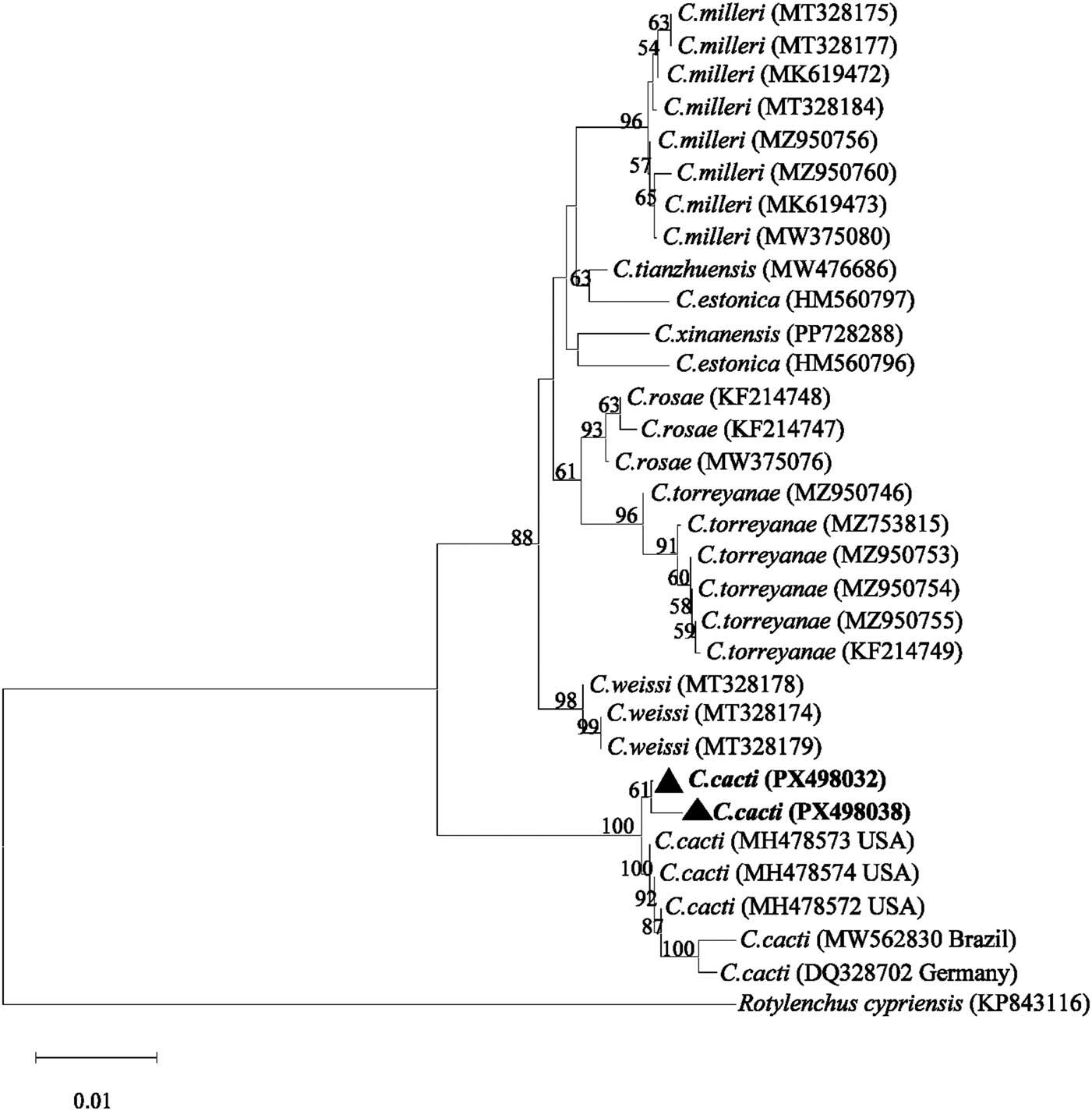
Figure 4
Phylogenetic tree based on D2–D3 28S rDNA sequences of the genus Cactodera. The tree was constructed using the NJ method with bootstrap support from 1,000 replicates. Bootstrap values are shown at each node. Sequences obtained in this study are marked in bold (PX498032 and PX498038).
Amplification of the COI mtDNA region yielded a single fragment of 393 bp (PX498440). Homology search comparison analysis showed that the sequences were 99.3–100% identical to those of C. Cacti from the USA (MW018446, MT936889) and China (PX506295). In the phylogenetic tree, the Hainan isolate formed a clade with three other C. cacti sequences, with Rotylenchus eximius (KX669244) used as the outgroup (Figure 5).
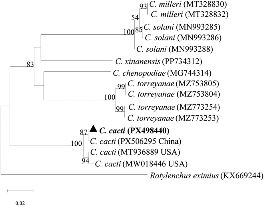
Figure 5
Phylogenetic tree based on COI mtDNA sequences of the genus Cactodera. The tree was constructed using the NJ method with bootstrap support from 1,000 replicates. Bootstrap values are shown at each node. Sequence obtained in this study are marked in bold (PX498440).
2.3. Parasitism test
The parasitism test confirmed that C. cacti was able to complete its life cycle and reproduce on dragon fruit (Selenicereus monacanthus cv. “Dahong”). At 65 days after inoculation, numerous cysts and white females were observed both on roots and in the soil (Figure 6), with an average of 116 individuals recovered per plant. The Rf of C. cacti was calculated as 11.6, confirming the pathogenicity of this nematode to dragon fruit. No cysts or females were detected in the roots or soil of non-inoculated control plants.
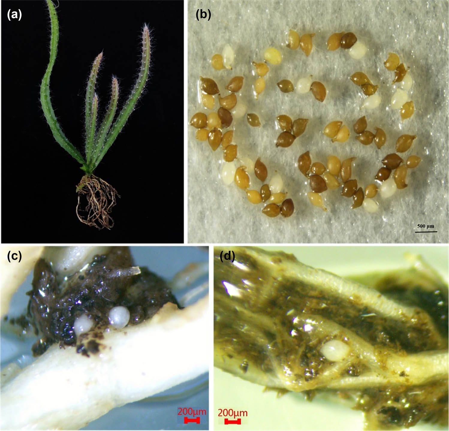
Figure 6
Pathogenicity test of C. cacti on dragon fruit. Dragon fruit seedling inoculated with C. cacti (a); cysts and white females recovered from roots and soil (b); and white females parasitizing on roots (c and d).
3. Discussion
In the present study, a cyst nematode population recovered from the rhizosphere of dragon fruit (Selenicereus undatus) in Danzhou, Hainan Province, was unequivocally identified as Cactodera cacti through integrative morphological and molecular approaches. The morphological features of cysts and second-stage juveniles were consistent with previous descriptions of C. cacti from China, Brazil, and the USA (Café et al., 2021; Duan et al., 2012; Skantar et al., 2019). Molecular characterization based on ITS rDNA, 28S D2–D3, and mitochondrial COI sequences further corroborated the morphological identification, with phylogenetic analyses placing the Hainan isolates in well-supported clades with other C. cacti populations, clearly distinguishing them from congeners such as C. solani, C. milleri, and C. torreyanae.
Cactodera cacti has a broad but primarily host-restricted distribution, having been reported mainly on ornamental cacti and occasionally on non-cactaceous hosts such as celery (Esser, 1992). The association between C. cacti and dragon fruit was first documented in Taiwan (Chan et al., 2016). More recently, a survey across 60 dragon fruit orchards in the Mekong Delta, Vietnam, detected C. cacti in 100% of soil samples and 70% of root samples (Thanh Thi Vo et al., 2026). Notably, C. cacti was the most consistently detected species among ten genera found in soil and five in roots, while Meloidogyne spp. were absent from all surveyed orchards, but were reported as major nematode pests in Brazil and China (Long et al., 2022; Moura de Souza et al., 2022) –were absent from all surveyed orchards (Thanh Thi Vo et al., 2026). This contrast suggests that C. cacti may surpass root-knot nematodes in prevalence and economic importance in the Mekong Delta. Collectively, these records indicate that C. cacti is expanding its host range from ornamental cacti to commercially important dragon fruit and widening its geographical distribution across tropical and subtropical Asia.
The pathogenicity test demonstrated that C. cacti can complete its life cycle and reproduce effectively on dragon fruit. The high Rf (11.6) observed in this study indicates that dragon fruit is a favorable host for C. cacti. To the best of our knowledge, this is the first report of C. cacti infecting dragon fruit in Hainan, China. Given the expanding dragon fruit cultivation in Hainan, the presence of this highly pathogenic nematode underscores the need for continued surveillance and the development of integrated management strategies to prevent potential outbreaks. Future research should focus on the distribution, host range, and population dynamics of C. cacti in Hainan, as well as the evaluation of resistant cultivars and sustainable control measures.
Funding information
Central Public-interest Scientific Institution Basal Research Fund for Chinese Academy of Tropical Agricultural Sciences (No.1630042022008) and Hainan Province Science and Technology Special Fund (No. ZDYF2023XDNY076).
Author contributions
Yanfang Sun: Writing-original draft, Visualization, Methodology, Conceptualization. Yueling Pei: Resources, Review & editing; Yuan Chen: Resources, Review, Data curation. Tuizi Feng: Review & editing. Haibo Long: Visualization, Methodology, Investigation, Conceptualization.
Conflict of interest statement
Authors state no conflict of interest.
Data availability statement
Data will be made available on request.