
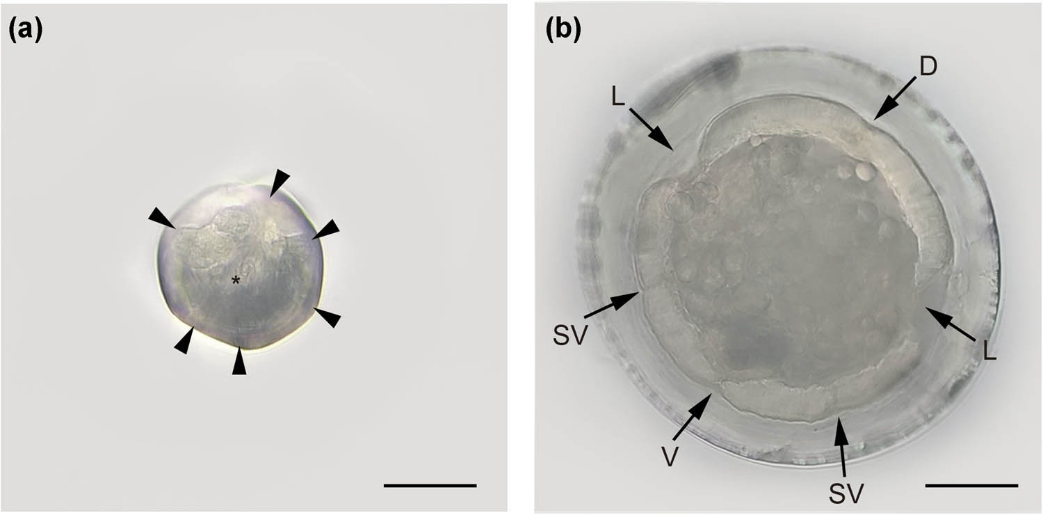
Figure 1
Hexamermis philaeni n. sp. (a) Head, en face; arrowheads showing the cephalic papillae and asterisk marking the mouth. (b) Cross section, midbody; dorsal chord (D), lateral chord (L), sub-ventral chord (SV) and ventral chord (V). Scale bars: A and B: 100 μm.
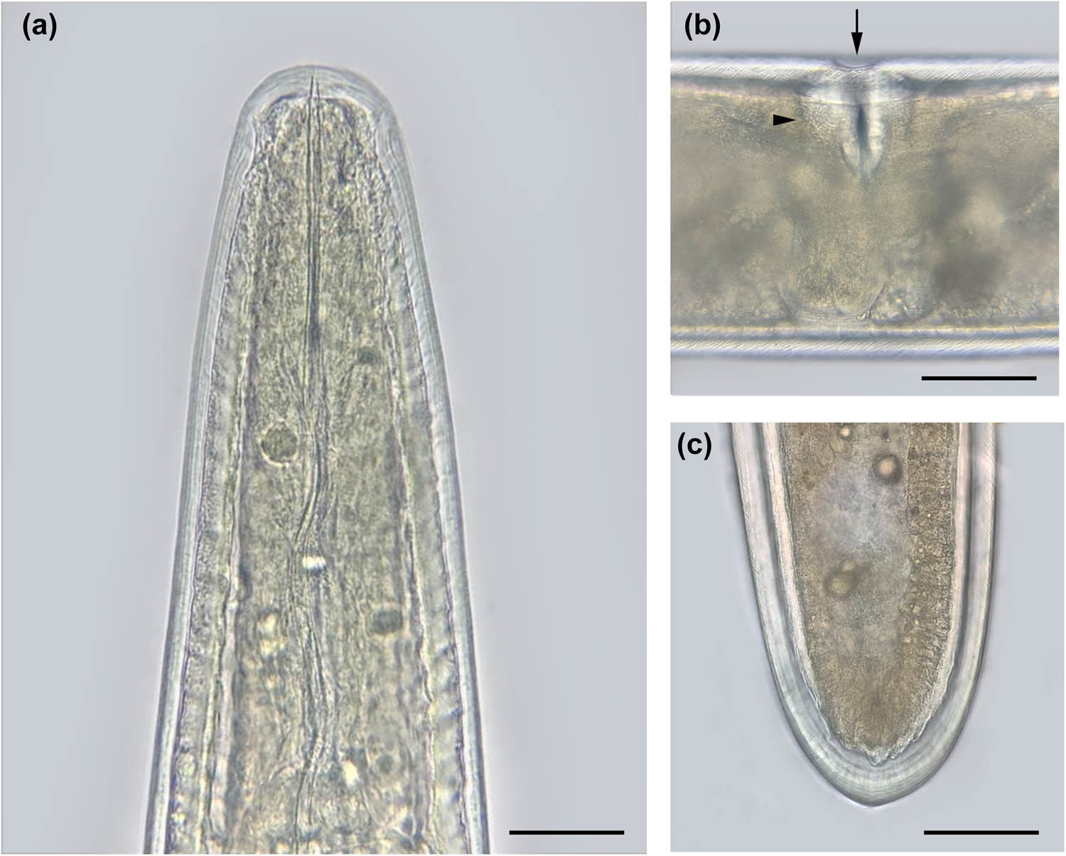
Figure 2
Hexamermis philaeni n. sp. Female. (a) Head. (b) Vulvar opening (arrow) and vulvar cone (arrowhead). (c) Posterior region. Scale bars: A, B, and C: 100 μm.
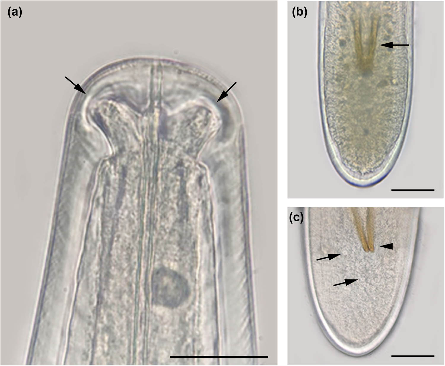
Figure 3
Hexamermis pilaeni n. sp. Male. (a) Head showing the cephalic papillae (arrows). (b) Tail showing the spicules (arrow). (c) Tail with cloaca (arrowhead) and genital papillae (arrows). Scale bars: A: 40 μm, B and C: 100 μm.
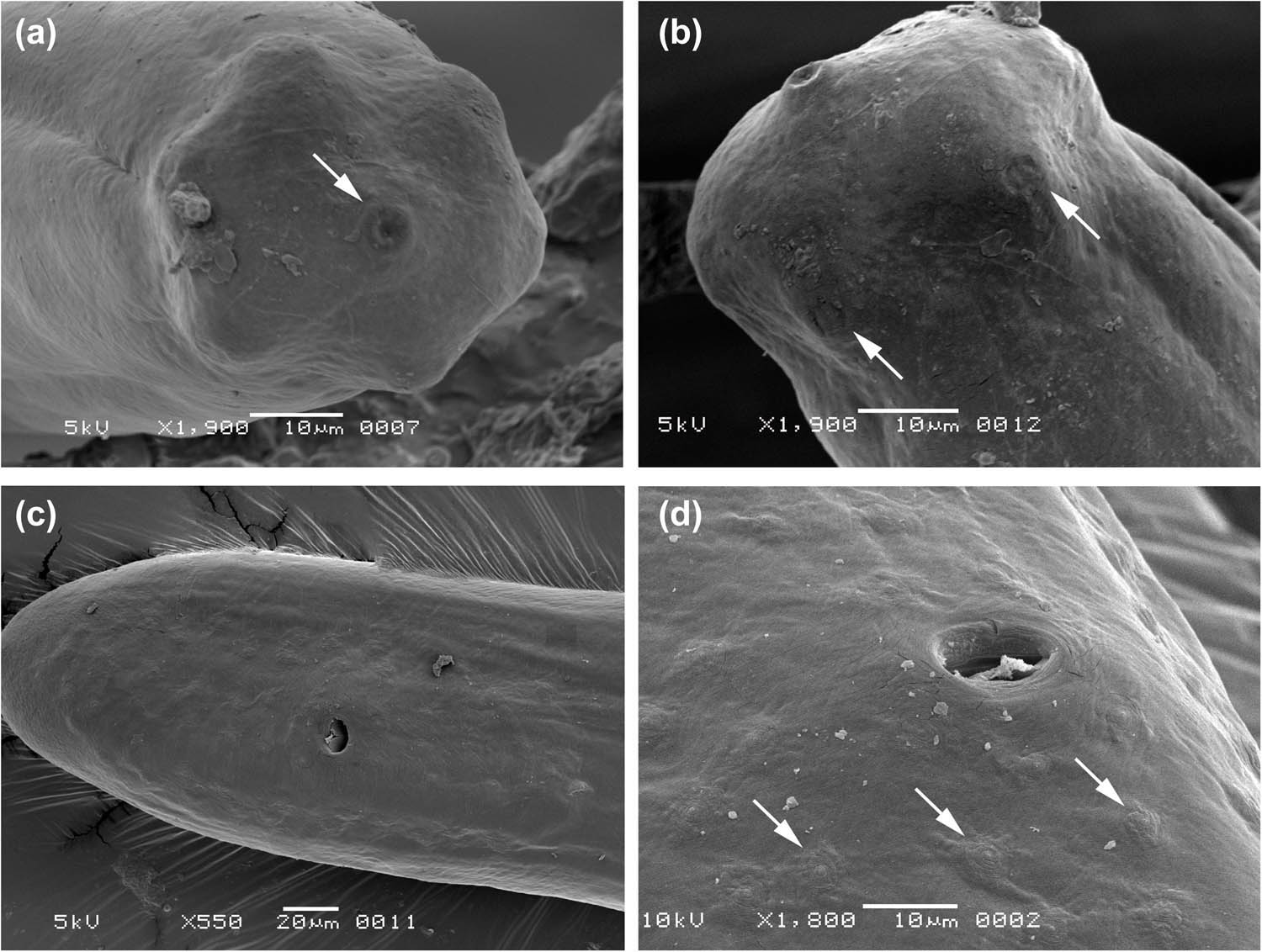
Figure 4
Hexamermis philaeni n. sp. Male. (a) Head showing the mouth (arrow). (b) Head showing two cephalic papillae (arrows). (c) Genital papillae in the tail. (d) Detail of genital papillae (arrows).
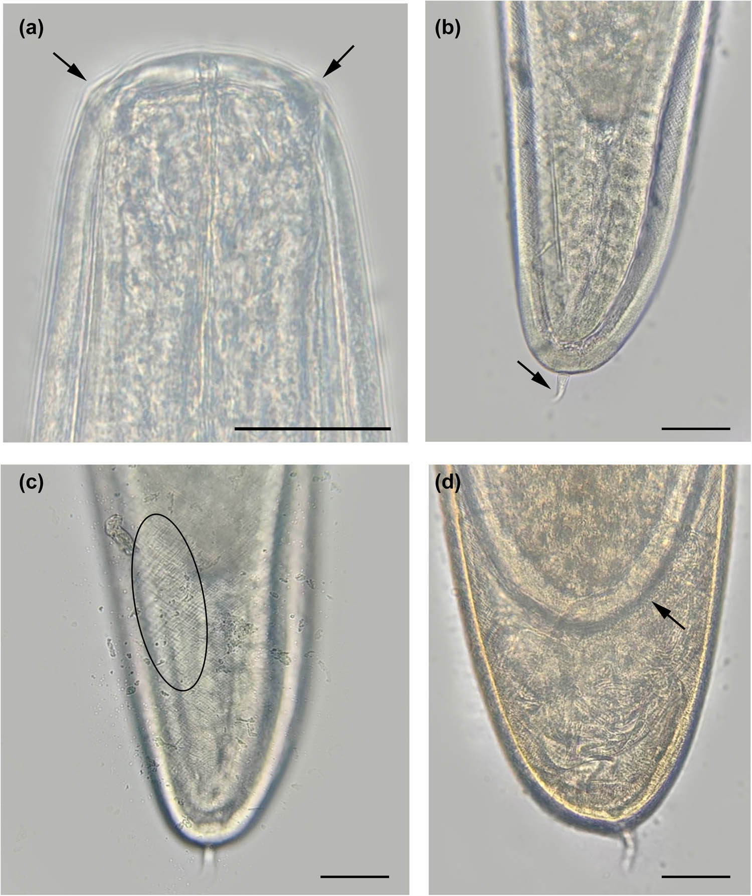
Figure 5
Hexamermis philaeni n. sp. Postparasitic juvenile. (a) Head showing the cephalic papillae (arrows). (b) Tail with curved tail appendage (arrow). (c) Cuticle with cross-fibers (oval). (d) Molt, arrow showing a female inside. Scale bars: A: 40 μm, B, C, and D: 100 μm.
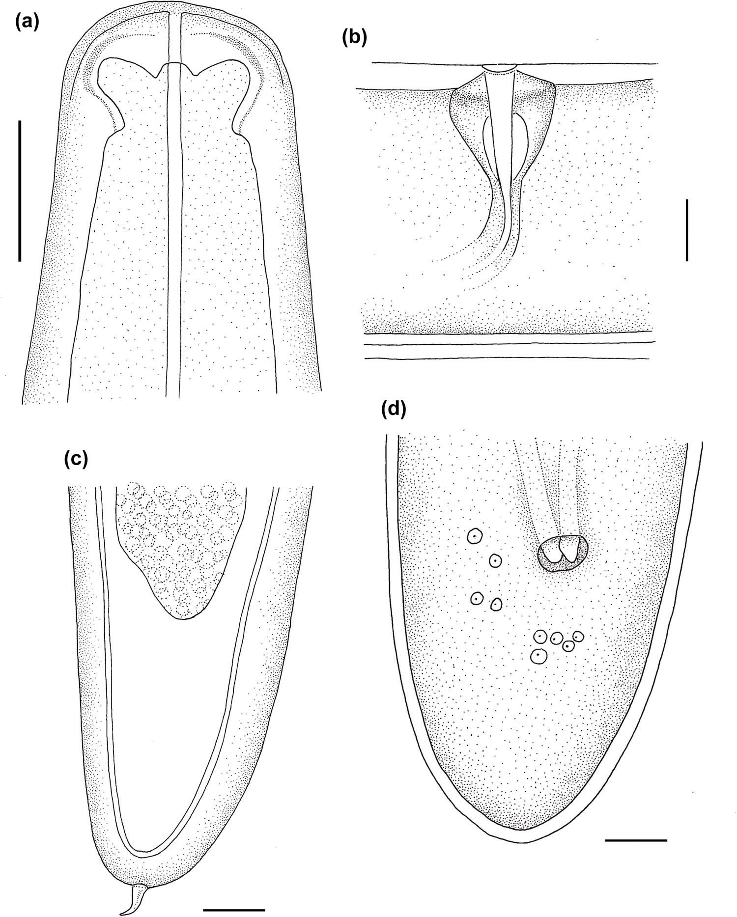
Figure 6
Line drawings of Hexamermis philaeni n. sp. (a) Male head. (b) Vulval region. (c) Postparasitic juvenile tail. (d) Male tail.
Table 1
Morphometrics of Hexamermis philaeni n. sp. males, females, and postparasitic juveniles
| Characters | Male (n = 6) | Female (n = 3) | Post-parasitic juvenile (n = 3) |
|---|---|---|---|
| Length (L) | 4.6 ± 0.8 (3.2–5.6) cm | 5.5 ± 0.5 (4.9–5.8) cm | 5.1 ± 1.5 (4.2–6.8) cm |
| GW | 205 ± 16.5 (181–223) | 267 ± 31.5 (234–297) | 172 ± 11.1 (162–184) |
| DNR | 281 ± 17.8 (259–302) | 285 ± 34.6 (261–310) | 247 ± 15.5 (230–260) |
| DEP | 265 ± 25.4 (247–283) | ||
| % Vulva (V) | 57 ± 2.1 (56–59) | ||
| LVC | 95 ± 2.8 (93–97) | ||
| WVC | 93 ± 2.1 (91–94) | ||
| SL | 157 ± 31.1 (109–160) | ||
| SW | 13 ± 1.6 (12–16) | ||
| WLC | 195 ± 21.2 (176–237) | ||
| WPE | 216 ± 21.9 (202–241) | 142 ± 9.2 (132–150) | |
| TL | 204 ± 22.5 (165–220) | 141 ± 32.6 (103–160) | |
| LTA | 70 ± 10.6 (58–78) |
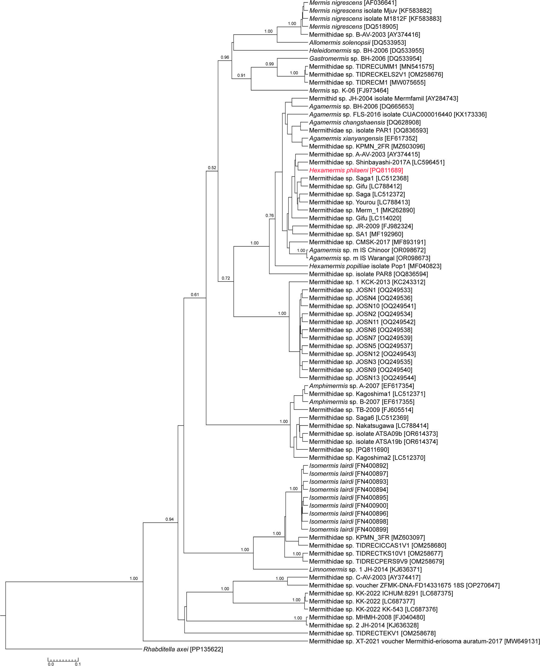
Figure 7
Phylogeny of the Mermithidae family based on 18S rDNA data including the new species, Hexamermis philaeni. Bayesian inference tree is shown. Posterior probability values are annotated near the respective nodes.
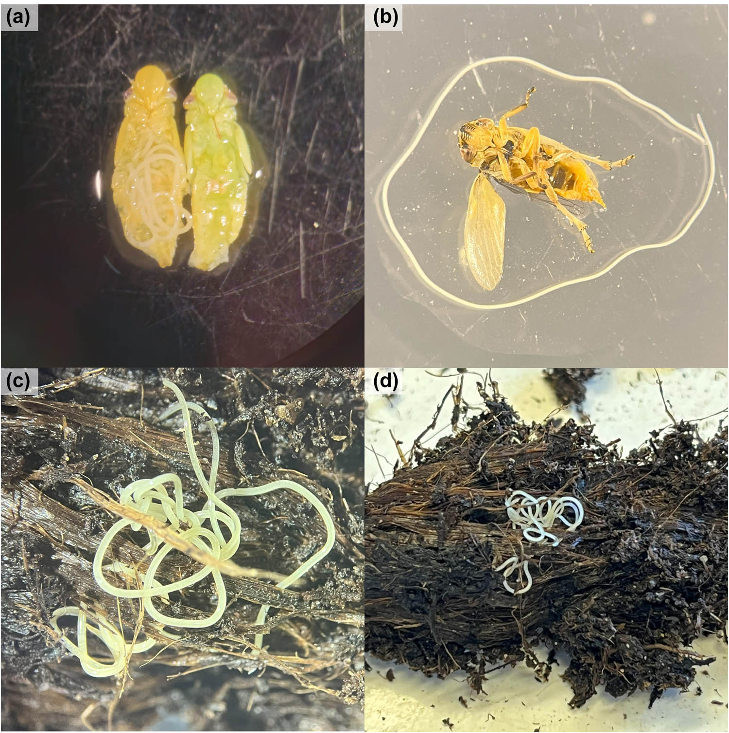
Figure 8
(a) Mermithid nematode inside the abdomen of a fifth-stage nymphs of Philaenus spumarius; (b, c) postparasitic juvenile (J4) emerging from an adult male of P. spumarius; (d) adult mermithid developed inside universal potting soil and retrieved after manual sifting.