
The genus Hexamermis (Steiner, 1924) (Mermithidae) comprises long white nematodes (length: 15–450 mm) that are obligate parasites of several invertebrate taxa, including insects, harvestman, and mollusks (Kaiser, 1991). Taxonomic identification of Hexamermis species is challenging since this genus seems to be particularly rich in species that are not very different from each other. For example, the type species Hexamermis albicans (von Siebold, 1848) is currently considered a complex of similar species that could be separated through the investigation of their specific hosts and their reproductive biology (Kaiser, 1991; Mazza et al., 2017). However, species belonging to the genus Hexamermis can be distinguished from those of other genera by the crisscross fibers in the cuticle, terminal mouth opening, six cephalic papillae, males with short spicula, and females with horn-shaped vagina and well-developed, cuticularized vulval cones. Moreover, postparasitic juveniles typically show finger-like tail appendages. Hexamermis species are reported as parasites of orthopteran, lepidopteran, dipteran, coleopteran, and hemipteran insects (Welch, 1963; Poinar et al., 1981; Poinar & Linares, 1985; Hernández-Crespo & Santiago-Alvarez, 1997; Achinelly & Camino, 2008; Mazza et al., 2017).
Recently, two unidentified mermithid species were found parasitizing the meadow spittlebug Philaenus spumarius L. in Central Italy (Nencioni et al., 2025). Phylogenetic analysis performed by the authors suggested that one of these species could be ascribed to the genus Hexamermis. However, the lack of mermithid adult specimens has hinder the complete morphological identification. Consequently, the aim of the present study was to collect P. spumarius individuals in the field to obtain and rear putative Hexamermis mermithids to the adult stage, enabling their identification to the species level. As a result, the description of a new species, Hexamermis philaeni n. sp., was provided.
1. Materials and methods
1.1. Insect sampling and nematode rearing
The following description of Hexamermis n. sp. is based on specimens obtained from P. spumarius individuals. Spittlebugs were collected in two organic olive groves, named as Site A and Site B, located in Central Italy and previously described in the study by Nencioni et al. (2025). P. spumarius nymphs (fourth and five instar) and adults were collected in the field between May and June and reared in Bugdorm® cages on potted herbaceous plants (Fragaria sp., Achillea millefolium, Artemisia dracunculus, Rumex sp., Ocimum basilicum). The cages were kept in an unheated greenhouse at ambient conditions throughout the experiment. These settings enable the full development of both P. spumarius immatures and possible parasitic nematodes. Moreover, any stress for spittlebugs was reduced, avoiding forced emergence of mermithids, that could negatively affect their survival. The universal potting soil (Radicom, Vigor Plant Italia srl) provided with potted plants was used as growth substrate for mermithids that emerged from spittlebugs. Cages were checked daily, and mermithids were left free to deepen into the soil and develop until the adult stage. Potted plants were watered as needed to ensure a good level of moisture for nematodes. After 40 days, potted soil was manually sifted through, and mermithids were removed and fixed in T.A.F. (2% triethanolamine and 7.5% formaldehyde in distilled water) for morphological identification.
Apart from rearing attempts, five postparasitic juveniles were collected soon after their emergence and stored in 1.5 mL tubes filled with 180 µL ATL Buffer supplied with QIAmp DNA Extraction Kit (Qiagen) for molecular analyses. Moreover, three postparasitic juveniles (one for Site A and 2 for Site B) were fixed in T.A.F. for taxonomic identification.
1.2. Morphological and morphometrical analyses
Nematodes were measured using an ocular micrometer in a Leica DM 500 microscope. All measurements were in micrometers unless otherwise specified and were presented as the mean followed by the range in parentheses. Photographs were taken with an Olympus DP-71 camera and a Leica DM 500 microscope, and drawings were made using an Olympus CX-31 with a drawing tube. The different stages were taken into consideration the following morphological and morphometric characters: length (L), greatest width (GW), distance from head to nerve ring (DNR), distance from anterior end to excretory pore (DEP), % vulva (V), length of vulvar cone (LVC), width of vulvar cone (WVC), length of vulva (LV), width of vulva (WV), width at posterior end (WPE), tail length (TL), and length of tail appendage (LTA). In the case of males, the following morphometric characters were also considered: spicule length (SL), spicule width (SW), and width at the level of the cloaca (WLC).
1.3. Scanning electron microscopy
Fixed nematodes were rinsed in distilled water three times and dehydrated at 15-min intervals through 30, 50, 70, 80, 90, and 100% ethanol. The nematodes were critical point dried with carbon dioxide, then were mounted on metal stubs with silver paste, coated with gold, and examined in a Philips 505 scanning electron microscope equipped with a digital imaging program (Philips Electron Optics BV, Eindhoven, Netherlands).
1.4. Molecular and phylogenetic analyses
Molecular analyses were performed to verify if the identity of mermithid collected in site A and B was in accordance with the findings of Nencioni et al. (2025). Nematodes were homogenized in a 1.5 mL microcentrifuge tube using a hand pestle. Subsequently, 20 µL of Proteinase K 20 mg/mL was added, and samples were incubated at 55°C for 3 h. The following DNA extraction steps were performed according to QIAmp DNA Extraction Kit (Qiagen) specifications, and the final elution step was performed in 50 µL of AE buffer supplied with the kit. The concentration of eluted DNA was measured using a Qubit photometer (ThermoFisher).
Amplification of 18S-ITS and D2-D3 rDNA loci were performed through assembly of amplicons obtained from four amplifications. Each polymerase chain reaction was performed in a total volume of 50 µL made with HotStart DreamTaq Master Mix 1X, (ThermoFisher), 0.6 µM each primer, and 5.0 µL of template DNA. The thermal program was 94°C for 3 min followed by 45 cycles of denaturation at 94°C for 30 s, primer annealing at Ta (see Table S1 in supplementary materials for primer list and annealing temperature) for 30 s, extension at 72°C for 60 s, and a final elongation step of 10 min at 72°C. Amplicons were checked on Tapestation 4200 (Agilent). Amplicons were sequenced “in house” using SeqStudio (Applied BioSystem), and the resulting sequences were assembled using Geneious Prime build 2025 (GraphPad Software LLC): The final assemblies were submitted to GenBank.
Phylogenetical analysis was performed on 18S locus. Ortologous sequences (n = 84 including outgroup) were mined from NCBI nucleotide database and aligned using Geneious Prime build 2025 (GraphPad Software LLC). Ambiguous alignment regions were removed using Gblocks v0.91b (Talavera et al., 2007). Phylogenetic inference was obtained from Bayesian statistics on molecular data using BEAST v2.7.8 (Bouckaert et al., 2014), with TPM2 + G + I assumed as the nucleotide substitution matrix and the Yule speciation model used. The operators were fine-tuned to explore the entire available space; the model was tested using a Markov–Monte Carlo chain with 10,000,000 iterations, with the first 10% discarded as burn-in.
2. Results
Family: Mermithidae Braun, 1883
Genus: Hexamermis Steiner, 1924
Hexamermis philaeni Rusconi, n. sp. (Figures 1–6)
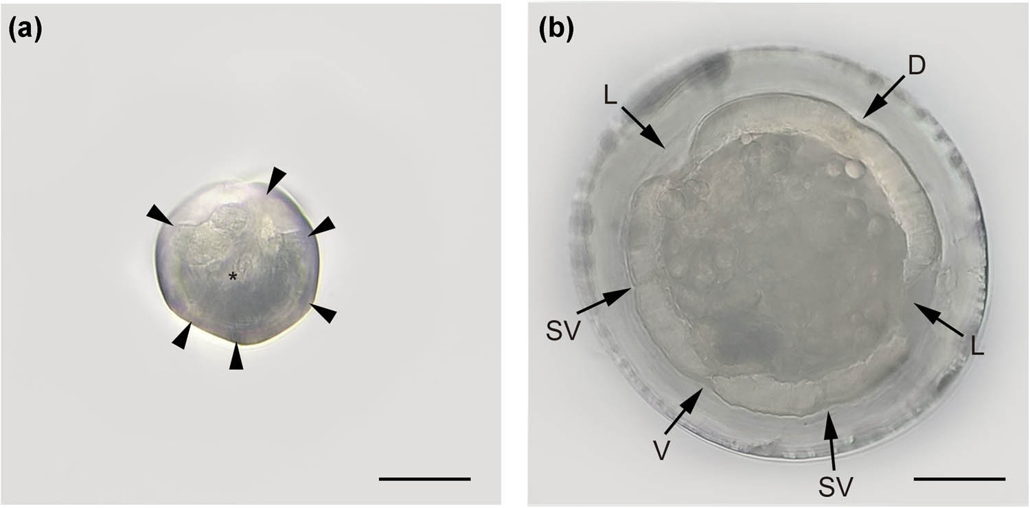
Figure 1
Hexamermis philaeni n. sp. (a) Head, en face; arrowheads showing the cephalic papillae and asterisk marking the mouth. (b) Cross section, midbody; dorsal chord (D), lateral chord (L), sub-ventral chord (SV) and ventral chord (V). Scale bars: A and B: 100 μm.
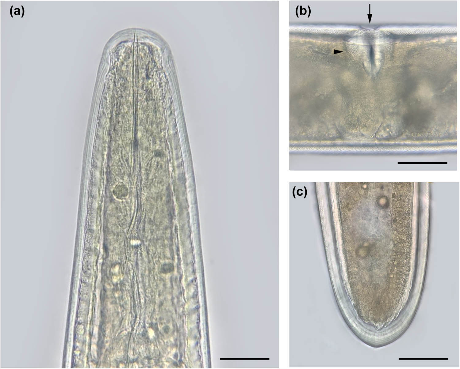
Figure 2
Hexamermis philaeni n. sp. Female. (a) Head. (b) Vulvar opening (arrow) and vulvar cone (arrowhead). (c) Posterior region. Scale bars: A, B, and C: 100 μm.
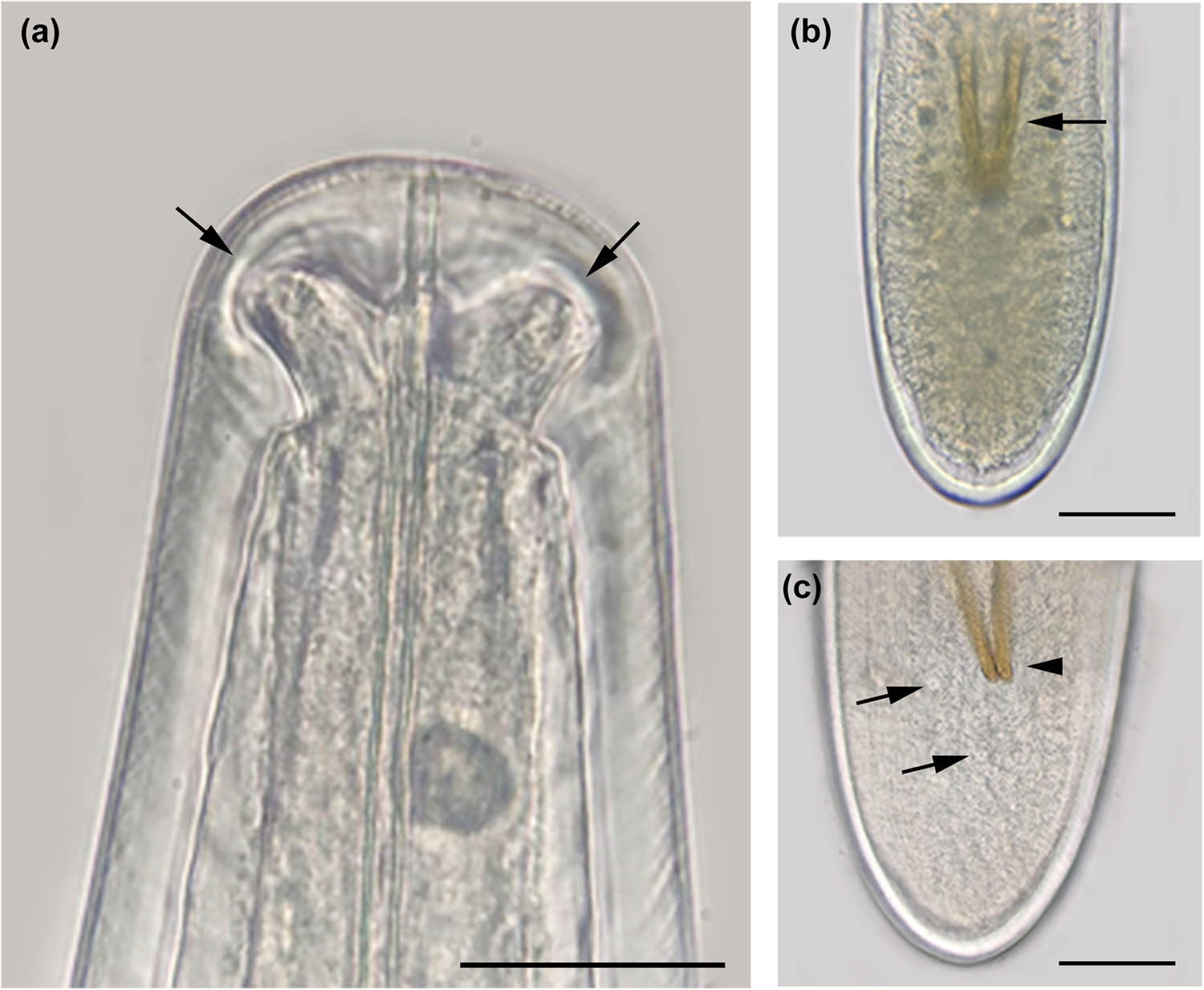
Figure 3
Hexamermis pilaeni n. sp. Male. (a) Head showing the cephalic papillae (arrows). (b) Tail showing the spicules (arrow). (c) Tail with cloaca (arrowhead) and genital papillae (arrows). Scale bars: A: 40 μm, B and C: 100 μm.
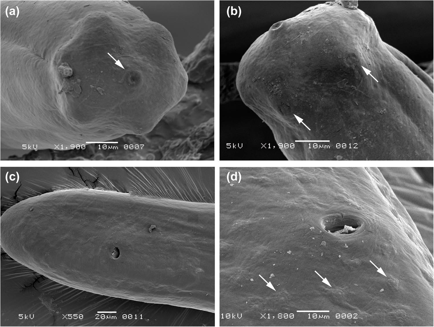
Figure 4
Hexamermis philaeni n. sp. Male. (a) Head showing the mouth (arrow). (b) Head showing two cephalic papillae (arrows). (c) Genital papillae in the tail. (d) Detail of genital papillae (arrows).
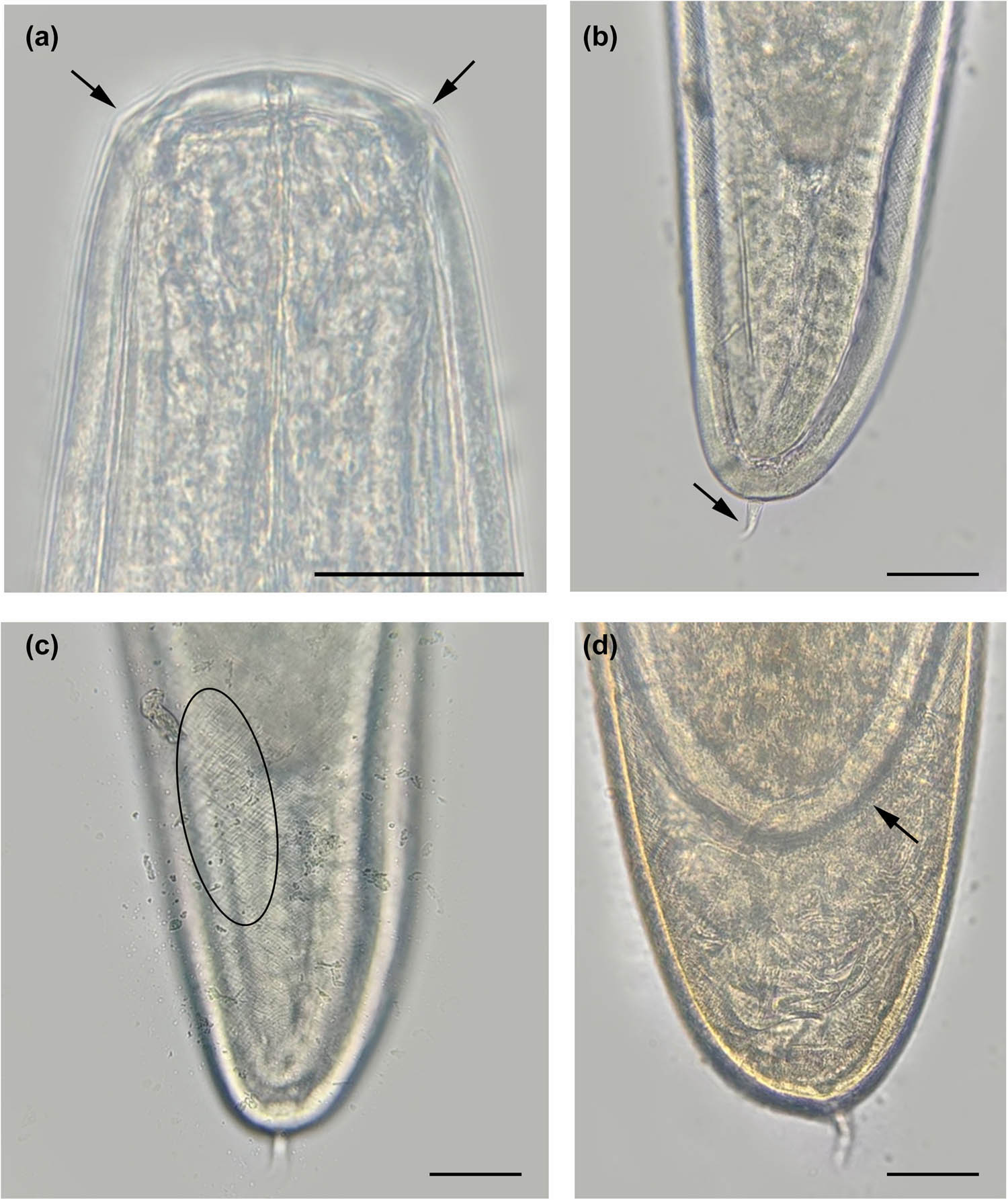
Figure 5
Hexamermis philaeni n. sp. Postparasitic juvenile. (a) Head showing the cephalic papillae (arrows). (b) Tail with curved tail appendage (arrow). (c) Cuticle with cross-fibers (oval). (d) Molt, arrow showing a female inside. Scale bars: A: 40 μm, B, C, and D: 100 μm.
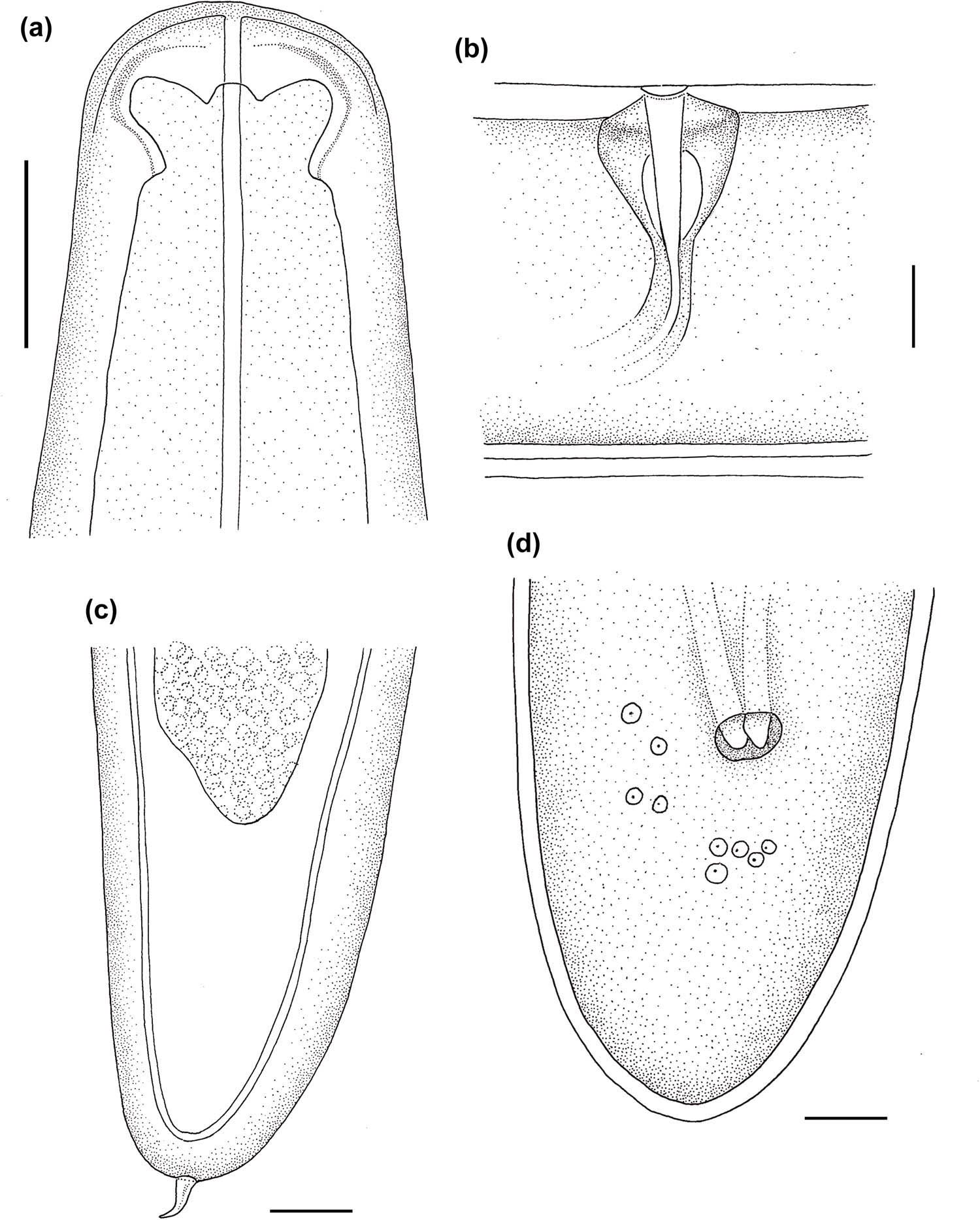
Figure 6
Line drawings of Hexamermis philaeni n. sp. (a) Male head. (b) Vulval region. (c) Postparasitic juvenile tail. (d) Male tail.
Measurements (see Table 1)
Table 1
Morphometrics of Hexamermis philaeni n. sp. males, females, and postparasitic juveniles
| Characters | Male (n = 6) | Female (n = 3) | Post-parasitic juvenile (n = 3) |
|---|---|---|---|
| Length (L) | 4.6 ± 0.8 (3.2–5.6) cm | 5.5 ± 0.5 (4.9–5.8) cm | 5.1 ± 1.5 (4.2–6.8) cm |
| GW | 205 ± 16.5 (181–223) | 267 ± 31.5 (234–297) | 172 ± 11.1 (162–184) |
| DNR | 281 ± 17.8 (259–302) | 285 ± 34.6 (261–310) | 247 ± 15.5 (230–260) |
| DEP | 265 ± 25.4 (247–283) | ||
| % Vulva (V) | 57 ± 2.1 (56–59) | ||
| LVC | 95 ± 2.8 (93–97) | ||
| WVC | 93 ± 2.1 (91–94) | ||
| SL | 157 ± 31.1 (109–160) | ||
| SW | 13 ± 1.6 (12–16) | ||
| WLC | 195 ± 21.2 (176–237) | ||
| WPE | 216 ± 21.9 (202–241) | 142 ± 9.2 (132–150) | |
| TL | 204 ± 22.5 (165–220) | 141 ± 32.6 (103–160) | |
| LTA | 70 ± 10.6 (58–78) |
Description
2.1. General
Medium-sized nematodes, the body usually narrowing in the head region; color white-yellow, cuticle with evident cross-fibers in adults and juveniles; mouth opening terminal and central; six cephalic (head) papillae arranged in one plane (Figures 1a, 3a); lip papillae absent; six hypodermal cords at midbody (Figure 1b); amphids not visible; excretory pore only visible in the male, anterior to nerve ring.
2.2. Female (n = 3)
The vulvar opening is wide and visible, without lips; vulvar cone developed (Figure 2b); vagina in equatorial position, perpendicular to long axis of the body, bend at connection with the uterus, muscular and initially straight, curved when reaching the uterus; uterus without eggs, tail bluntly rounded.
2.3. Male (n = 6)
The spicules are paired, well developed, with rounded tips, separated and equal in size, slightly curved near the distal end and shorter than the width of the body at the level of the cloaca (Figures 3c and 6d); cloaca very evident; genital papillae surrounding the cloaca; 79 genital papillae, genital papillae surpassing the base of the spicules and arranged as follows: 24 pre-cloacal, 5 ad-cloacal and 50 postcloacal; tail bluntly rounded.
2.4. Postparasitic juvenile (n = 3)
Similar in size to the adult. Head rounded. Cuticle with visible cross-fibers, attached to the adult while molting; tail rounded with curved tail appendage; length of tail appendage 58 − 78 ± 10.6 (70).
2.5. Molecular and phylogenetic analyses
Mermithid specimens obtained from P. spumarius collected in the field belong to the species previously found in the same sampling sites by Nencioni et al. (2025) and tentatively named as “Species 1.” The sequences of the 18S-ITS loci and the D2-D3 region belonging to the 28S gene were submitted to GenBank under accession numbers PQ811689 and PQ811687, respectively. However, the D2−D3 region of 28S gene has the maximum ID% with unidentified mermithid samples from Japan (96.7 %ID and coverage 100% with LC88415 and LC88415) and from South Africa (96.4 %ID and coverage 100% with MF192965), while ITS locus does not produce significant results due to lack of sequences in GenBank; the maximum %ID was found with a basidiomycetes (Pluteus septocystidiatus 27.0 %ID and 6.15% coverage).
The Bayesian reconstruction of phylogenetic relationships within the family Mermithidae, and obtained using a fragment of the 18S gene reflects general known relationships between mermithid groups. The computed tree showed a polyphyletic origin for some genera such as Mermis, Agamermis, and Hexamermis (Figure 7). In particular, the species H. philaeni is found within a well-supported clade that includes the genera Hexamermis and Agamermis (posterior probability = 1.00). However, the reconstruction of the internal structure of the clade does not reach acceptable posterior probability values to topologically resolve the two genera.
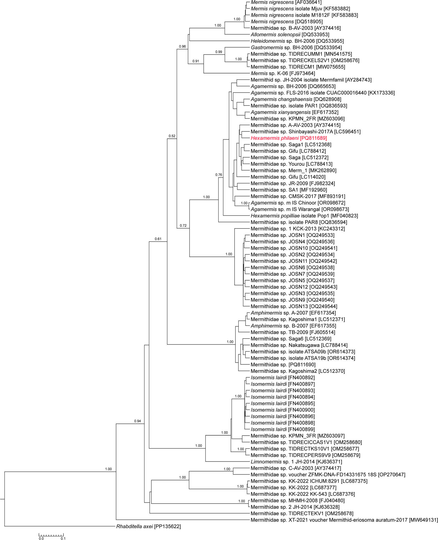
Figure 7
Phylogeny of the Mermithidae family based on 18S rDNA data including the new species, Hexamermis philaeni. Bayesian inference tree is shown. Posterior probability values are annotated near the respective nodes.
2.6. Type host and locality
The type host is represented by nymphs and adults of P. spumarius (Hemiptera: Aphrophoridae). Spittlebugs were collected at Site A, Vaiano (43.938753 N, 11.141433E), Prato province, Tuscany (Central Italy) and Site B, Carmignano (43.811145 N, 11.031118E), Prato province, Tuscany (Central Italy).
2.7. Type material
All types have been deposited in the Colección Helmintológica del Museo de Ciencias Naturales de La Plata with the following accession number: MLP-He 8347 (1 female, 1 male and 1 postparasitic juvenile).
2.8. Biology and ecology
Juvenile mermithids develop inside insects occupying the thoracic and abdominal regions (Figure 8a). In parasitized fourth- and fifth-instar of P. spumarius, the body color appears yellowish, while healthy individuals are green. On the other hand, the only exterior sign of mermithid parasitization in P. spumarius adults is the abdomen swelling. Mermithids inside the insect body cannot be externally seen through the insect’s integument. When the postparasitic juvenile emerges from the host, it breaks the insect integument in the region comprised between the VII and X tergits (Figure 8c), causing the host death in a few minutes.
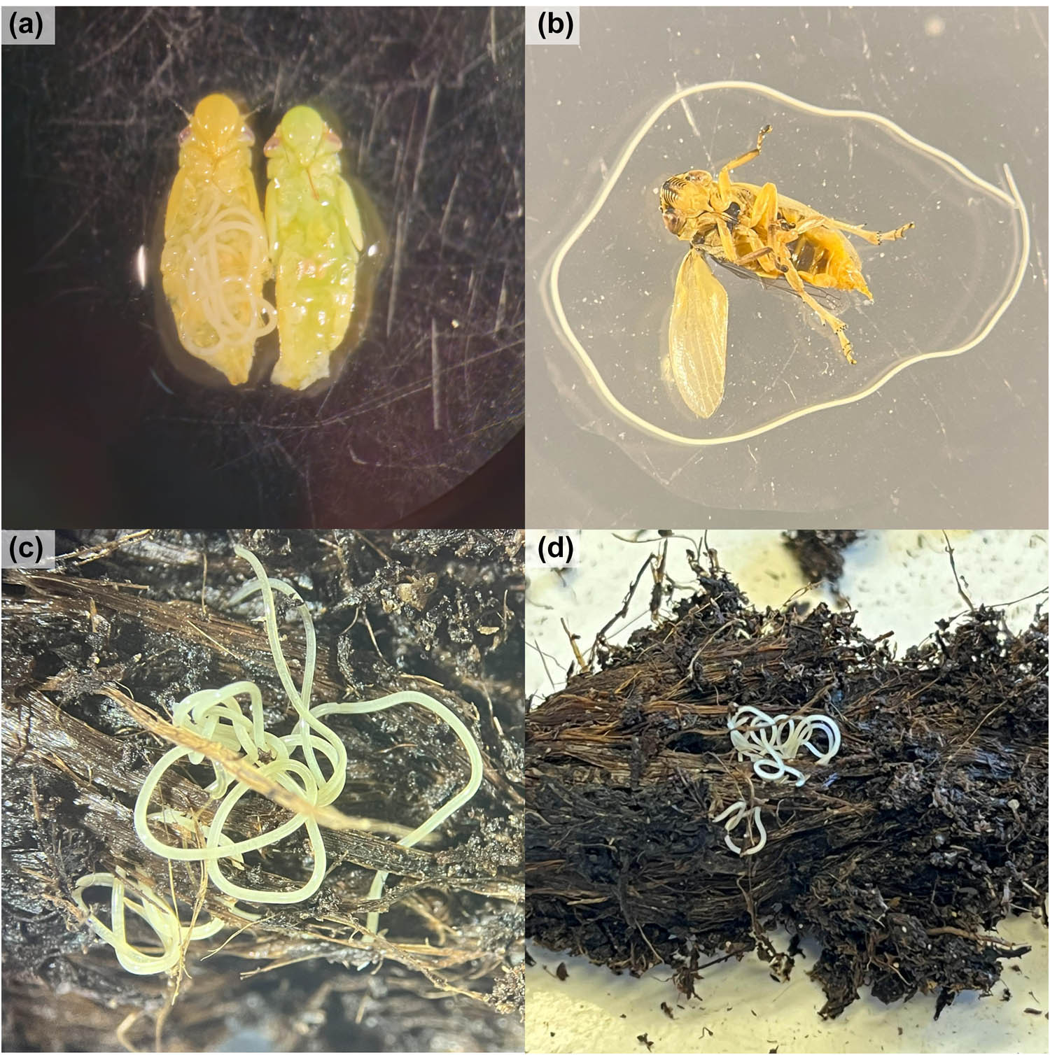
Figure 8
(a) Mermithid nematode inside the abdomen of a fifth-stage nymphs of Philaenus spumarius; (b, c) postparasitic juvenile (J4) emerging from an adult male of P. spumarius; (d) adult mermithid developed inside universal potting soil and retrieved after manual sifting.
2.9. Etymology
The name of the species refers to the host P. spumarius.
2.10. Differential diagnosis and remarks
Hexamermis philaeni n. sp. Rusconi is placed in the genus Hexamermis due to the cross fibers in the cuticle, the short, paired, and equal in size spicules in the male; the vulval cone in the female; and the presence of a tail appendage in the postparasitic juvenile.
Hexamermis philaeni n. sp. is characterized by 1) mouth opening terminal; 2) six cephalic papillae; 3) six hypodermal cords at midbody; 4) vulvar opening wide and visible, without lips and a vulvar cone developed; 5) vagina bend at connection with the uterus; 6) spicules paired, developed, separated and equal in size, slightly curved near the distal end and shorter than the width of the body at the level of the cloaca; and 7) genital papillae surrounding the cloaca, surpassing the base of the spicules and arranged as follows: 24 pre-cloacal, 5 ad-cloacal, and 50 postcloacal.
The new species was obtained from nymphs and adults of the spittlebug Philaenus spumarius in Tuscany, Central Italy. To the best of our knowledge, only three mermithid species were recorded in the aforementioned country: Hexamermis albicans von Siebold, reported in a forest in Apulia inside the larvae of two lepidoptera, Archis crataegana Hb. and Aleimma loeflingiana L. (Manfredi et. al., 1995), Hexamermis popilliae Mazza et al., 2017 found in Popillia japonica Newman (Coleoptera: Scarabaeidae) in Oleggio (Novara, Piedmont), and a Hexamermis sp. in Rhaphigaster nebulosa Poda (Hemiptera: Pentatomidae) in a hazel wood in Benevallo (Cuneo) (Manachini and Landi, 2003).
Establishing a comparison between these two species and ours, we can observe that: a) Hexamermis albicans can be differentiated from H. philaeni n. sp. in the length of the adult body (22 vs. 5.5 cm); b) Hexamermis popilliae differs from H. philaeni n. sp. in the shape of the head of the female (pointed vs. rounded), the length of the body (1.3 vs. 4.6 cm), the number of genital papillae (47 vs. 79), and the length of the spicules (115 vs. 157 µm) in the male.
3. Discussion
The present study describes H. philaeni n. sp., a parasite of the meadow spittlebug P. spumarius, found in Central Italy. P. spumarius is a Palearctic species, belonging to the family Aphrophoridae, widely distributed in almost all continents. It is a highly polyphagous species, living in most terrestrial habitats. So far, mermithid parasitization in P. spumarius was sporadically reported in the United States, where some spittlebugs were probably parasitized by Agamermis decaudata (Cobb, Steiner & Christie, 1923; Weaver & King, 1954). However, the identity of these nematodes is uncertain (Weaver & King, 1954 instead of Weaver and King, 1954).
Mermithids belonging to the genus Hexamermis are reported as parasites of terrestrial insects, including Diptera, Lepidoptera, Coleoptera, Orthoptera, and Hemiptera (Achinelly & Camino, 2008). Hexamermis dactylocercus is the only species found to be associated with a member of the Cercopoidea superfamily – the froghopper Aenolamia varia (Fabricius, 1787) – prior to this study. Thus, the observation of H. philaeni parasitizing P. spumarius expands the knowledge on Hexamermis host range and represents the first record of a mermithid of this genus associated with the meadow spittlebug worldwide.
Phylogenetic reconstruction assigned the newly described species to a clade containing the genera Hexamermis and Agamermis. However, the 18S locus alone is insufficient to resolve the deeper clade structure. A multiloci approach is also limited by the lack of a complete dataset for a significant number of mermithids in GenBank, as well as the large number of unidentified samples. In this study, we have deposited the complete 18S-ITS sequence and the D2−D3 region of the 28S gene in the databases, which will be useful for further studies. The Mermithidae family is widespread but understudied, so dedicated sampling is challenging, and complete data available in databases is a valuable resource.
According to the observation of Nencioni et al. (2025), the life history of H. philaeni appear to be similar to that of other terrestrial mermithids. The infection may occur in spring, when the preparasitic juvenile can intercept P. spumarius nymphs crawling in the soil or climbing on short herbs, where spittlebug nymphs feed. Then, the mermithid penetrates the host and develops inside its body cavity, meanwhile allowing the spittlebug to develop to the adult stage. The postparasitic juvenile exits from young P. spumarius adults, causing their death shortly after the completion of the preimaginal development. Finally, the postparasitic juvenile enters the soil to molt to the adult stage and then mate and reproduce (Kaiser, 1991; Poinar, 2010). Further studies are necessary to elucidate H. philaeni life cycle, as well as to assess a possible synchronization with the host phenology or the association with other host species.
Philaenus spumarius is considered a serious pest in Europe since it has been assessed as the primary vector of the plant pathogenic bacterium Xylella fastidiosa (Wells et al., 1987). In recent years, the need for environmentally friendly control strategies has stimulated the investigation of P. spumarius natural enemies that could be eventually used for the integrated control of this spittlebug. In this regard, the present work contributes to expanding the knowledge on P. spumarius parasites that are naturally present in Italian olive agroecosystems. However, the use of mermithids for the biological control of insect pests is generally considered unfeasible, mainly due to difficulties in their mass rearing (Petersen, 1985). Further research on the biology and ecology of H. philaeni will be crucial to understand its role in controlling P. spumarius populations, as well as the impact of agricultural practices on the occurrence of this mermithid species in olive groves. This information could be particularly useful in the evaluation of a conservative biological control approach for the management of P. spumarius in different agroecosystems.
Acknowledgements
The Authors would like to thank the farms “Siro Petracchi” and “Società Agricola Fabio” for their collaboration during the field sampling.
Funding information
This work was supported by the project: “Contenimento insetti vettori di Xylella fastidiosa con metodi a basso impatto ambientale – COVEXY, D.M. 0664980, 29/12/2022,” funded by Ministero dell’agricoltura, della sovranità alimentare e delle foreste (MASAF) (Italy).
Author contributions
AN, EG, and IC: conceived and designed research. AN, EG, GB, AS, PS and IC: conducted experiments in fields and laboratory. JMR: made taxonomic identification and morphological description. AS: performed molecular analysis, sequencing and phylogenetic analysis. AN, JMR: wrote the original draft. All authors revised the original draft and approved the manuscript. EG provided funds for research.
Conflict of interest statement
Authors state no conflict of interest.