
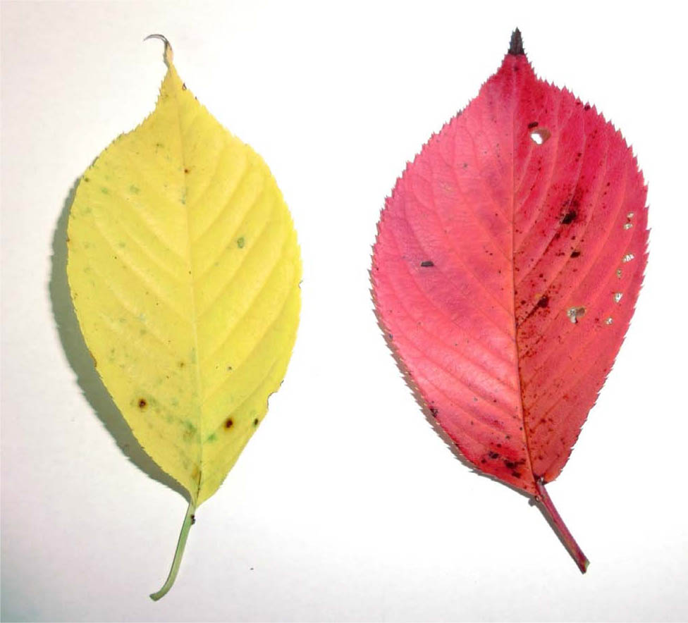
Figure 1.
Yellowing leaf (left) and red leaf (right) of a Japanese cherry (Cerasus × yedoensis ‘Someiyoshino’). Photograph: J. Ueda.
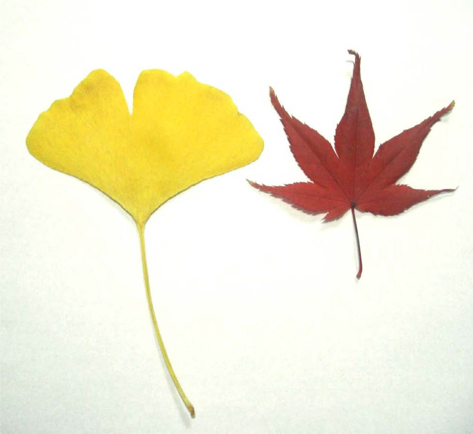
Figure 2.
Yellowing ginkgo (Ginkgo biloba) leaf (left) and red maple (Acer palmatum) leaf (right). Photograph: J. Ueda.
Yellowing: In plants like the Ginkgo, the degradation of chlorophyll reveals the underlying carotenoids (yellow pigments) that were present but masked by green.
Reddening: In plants like the Maple, sugars produced by photosynthesis accumulate in the leaf. Under the influence of ultraviolet or blue light, these sugars are used to synthesize anthocyanins (red pigments).
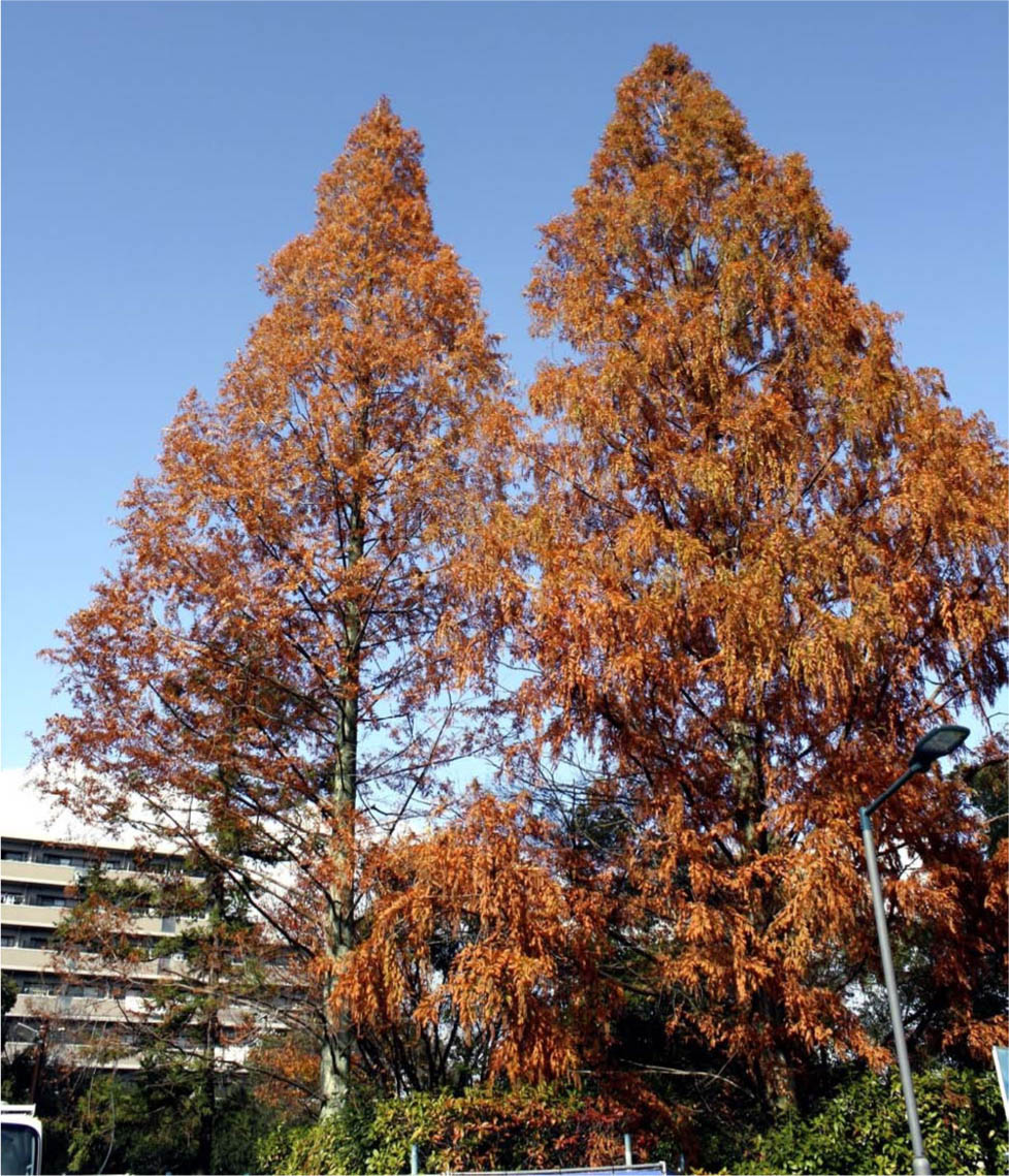
Figure 3.
Metasequoia glyptostroboides from late autumn to winter. Photograph: J. Ueda.
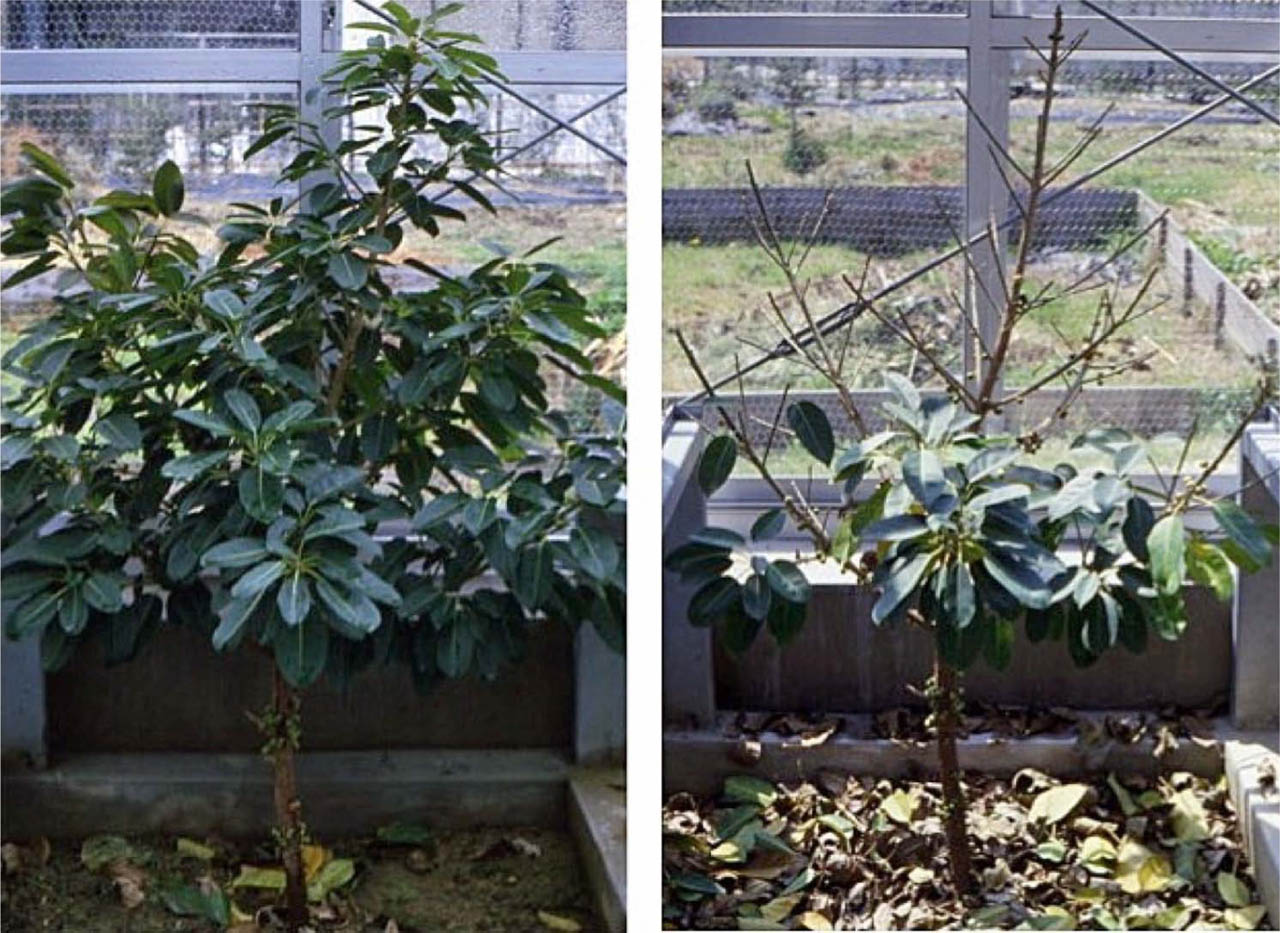
Figure 4.
Vernal leaf abscission of Ficus superba var. japonica. Photographs: J. Ueda.
Left: Overwintering with green leaves. Right: Simultaneous shedding in early spring before new budding.
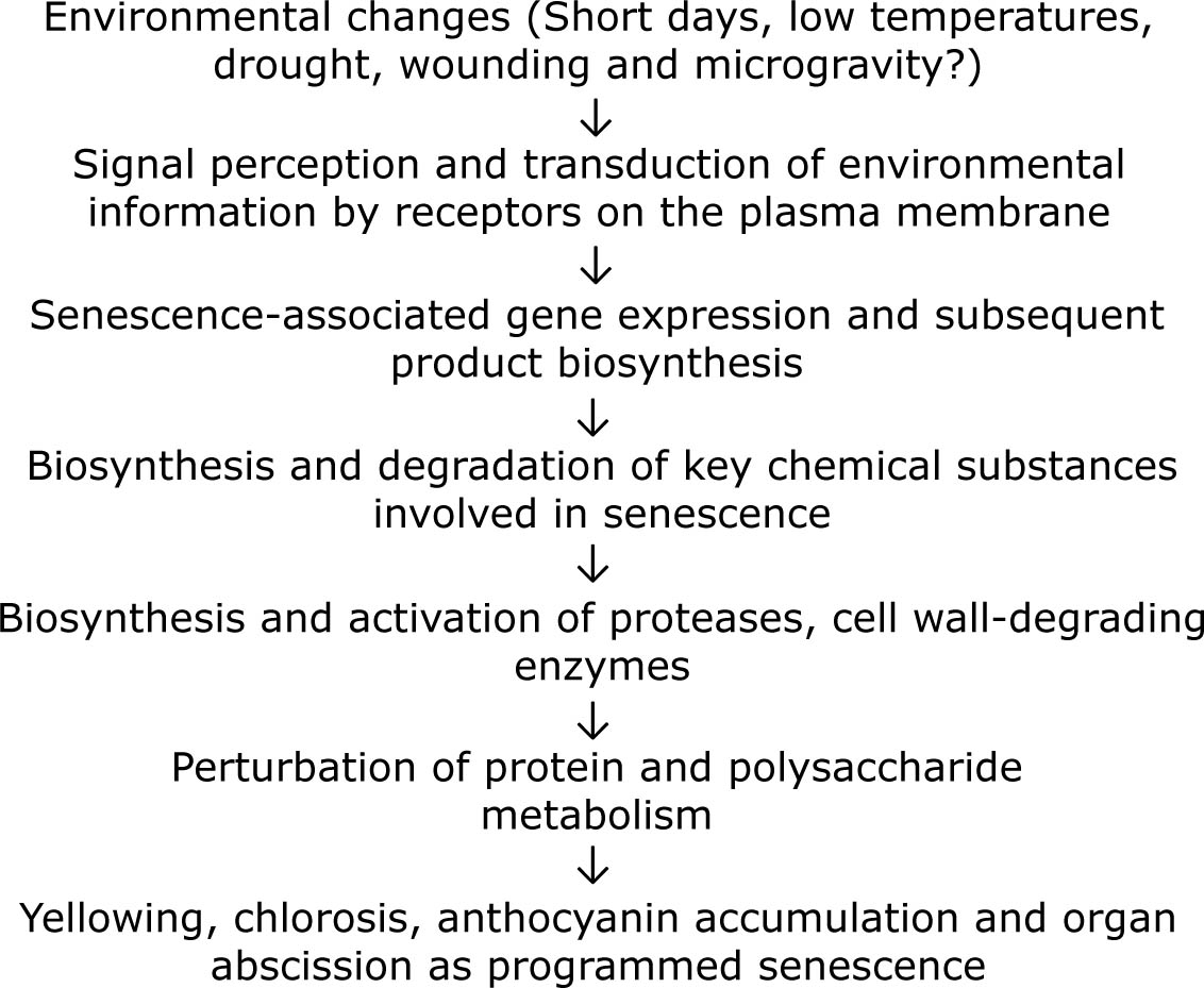
Figure 5.
Signal transduction and sequence of physiochemical changes occurring during plant senescence.
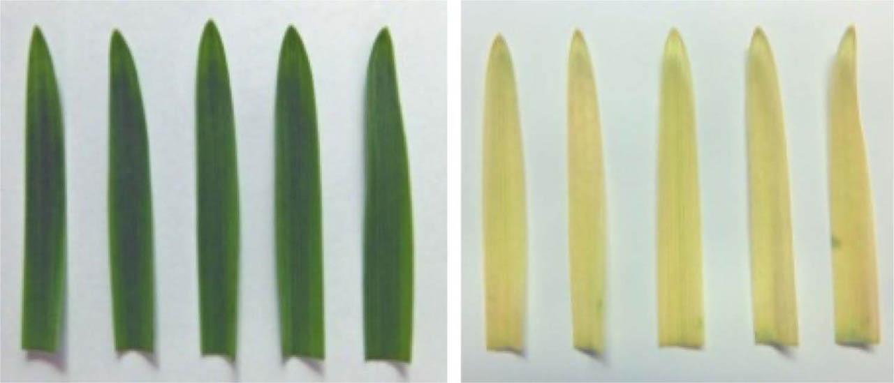
Figure 6.
Comparison of oat (Avena sativa) leaf segments kept in light vs. darkness. Photographs: J. Ueda.
Segments 3 cm in length were cut from the tips of the first leaves of healthy oat plants. These segments were placed leaf-surface up in Petri dishes lined with moistened filter paper. Senescence begins to appear when the chlorophyll contents, measured by absorbance at 665 nm, decrease to approximately 50%. After four days of incubation, the segments kept in light (left) remained relatively stable, whereas the segments kept in darkness (right) completely changed from green to yellow, indicating advanced senescence.
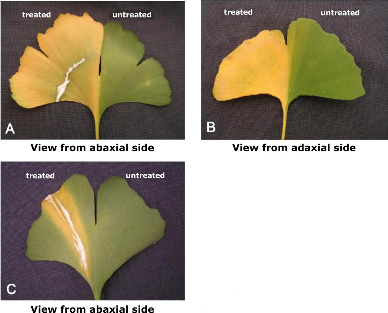
Figure 7.
Methyl jasmonate-induced senescence of Ginkgo biloba leaves.
Methyl jasmonate was applied across (A and B) or along (C) to the vein of leaf blade of abaxial side of a half leaf in 12 years old Ginkgo biloba tree. Treatment with methyl jasmonate was carried out in the abaxial side of half leaf blade (left); opposite half leaf blade was of without treatment as control (right). Photographs were taken 3 weeks after the treatment.
(denoted by Saniewski et al., 2020 with modifications)
Table 1.
Brief history of plant senescence researches focusing on key chemical compound.
| Year | Author | Achievement |
|---|---|---|
| 1924 | Denny F.E. | Reported the promotion of degreening (yellowing) in lemon fruits by ethylene |
| 1929 | Molisch H. | Published “The Longevity of Plants” (Die Lebensdauer der Pflanze) |
| 1933a, b | Laibach F. | Reported auxin production in Orchid pollen, and it inhibited petiole abscission |
| 1935 | Yemm E.W. | Studied metabolism in starved barley leaves |
| 1937 | Vickery H.B. et al. | Researched chemical changes in tobacco leaves kept in light and darkness |
| 1949 | Hemberg T. | Studied growth-inhibiting substances in the cortical tissue of potato tubers |
| 1953 | Bennet-Clark T.A. and Kefford N.P. | Named a growth inhibitor found in many plants as “inhibitor-β” |
| 1954 | Chibnall A.C. | Hypothesized the existence of a new plant hormone supplied from roots to leaves |
| 1957 | Richmond A.E. and Lang A. | Discovered that kinetin (a cytokinin) inhibits plant senescence, supporting Chibnall's hypothesis. |
| 1958 | Phillips I.D.J. and Wareing P.E. | Studied growth inhibitors in the terminal buds and leaves of Acer pseudoplatanus (Sycamore Maple) |
| 1961 | Carns H.R. et al. | Reported the promotion of abscission in cotton by gibberellin |
| 1963 | Eagles C.E. and Wareing P.E. | Named a dormancy-inducing substance “dormin” (later identified as abscisic acid) |
| 1963 | Ohkuma K. et al. | Isolated abscisin II (later identified as abscisic acid) |
| 1965 | Ohkuma K. et al. | Determined the chemical structure of abscisin II (abscisic acid) |
| 1966 | Fletcher R.A. and Osborne D.J. | Reported that gibberellin inhibits chlorophyll degradation |
| 1967 | Woolhouse H.W. | Organized the symposium “Aspects of the Biology of Aging” |
| 1970 | Shibaoka H. and Thimann K.V. | Conducted systematic research on plant senescence using leaf segments |
| 1980 | Ueda J. and Kato J. | Isolation and identification, and discovered the powerful senescence-promoting effect of methyl jasmonate |
| 1997 | Pennell R.I. and Lamb C. | Published a review on Programmed Cell Death (PCD) in plants |
| 1997 | Yamamoto R. et al. | Reported the regulation of programmed cell death by brassinosteroids |
| 2007 | Kusaba et al. | Identified the NYC1 and NOL genes using rice mutants that remain green (stay-green). |
| 2007 | Sato Y. et al. | Researched the green-cotyledon peas used by Mendel to propose the “Laws of Inheritance” (identified the STAY-GREEN gene) |
| 2016 | Shimoda Y. et al. | Proved that the STAY-GREEN gene encodes magnesium-dechelatase, an enzyme that removes magnesium from the chlorophyll |
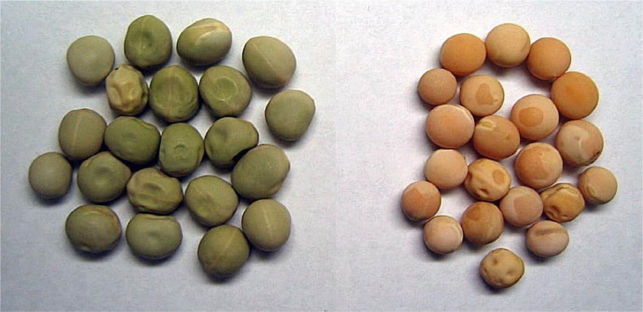
Figure 8.
Dried pea (Pisum sativum) seeds with green (left) and yellow (right) cotyledons. Photograph: J. Ueda.
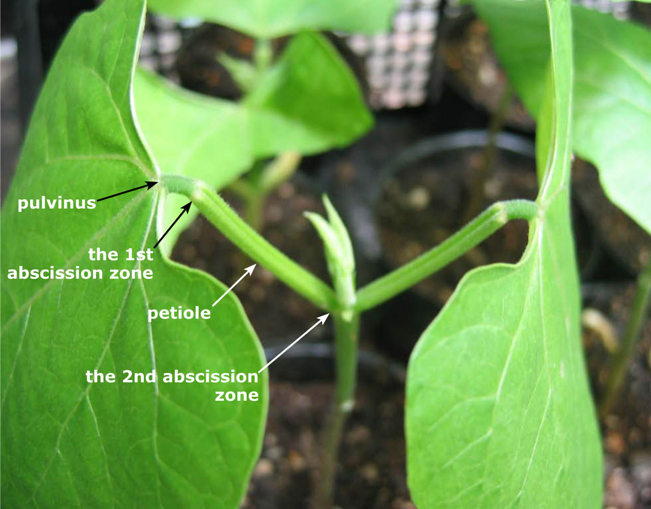
Figure 9.
Two abscission zones observed in the primary leaf of Phaseolus vulgaris seedling. Photograph: J. Ueda.
The primary abscission zone is located at the junction of the pulvinus and the petiole, and the secondary abscission zone is located at the junction of the petiole and the stem. The pulvinus refers to the thickened section at the base of a leaflet or petiole.
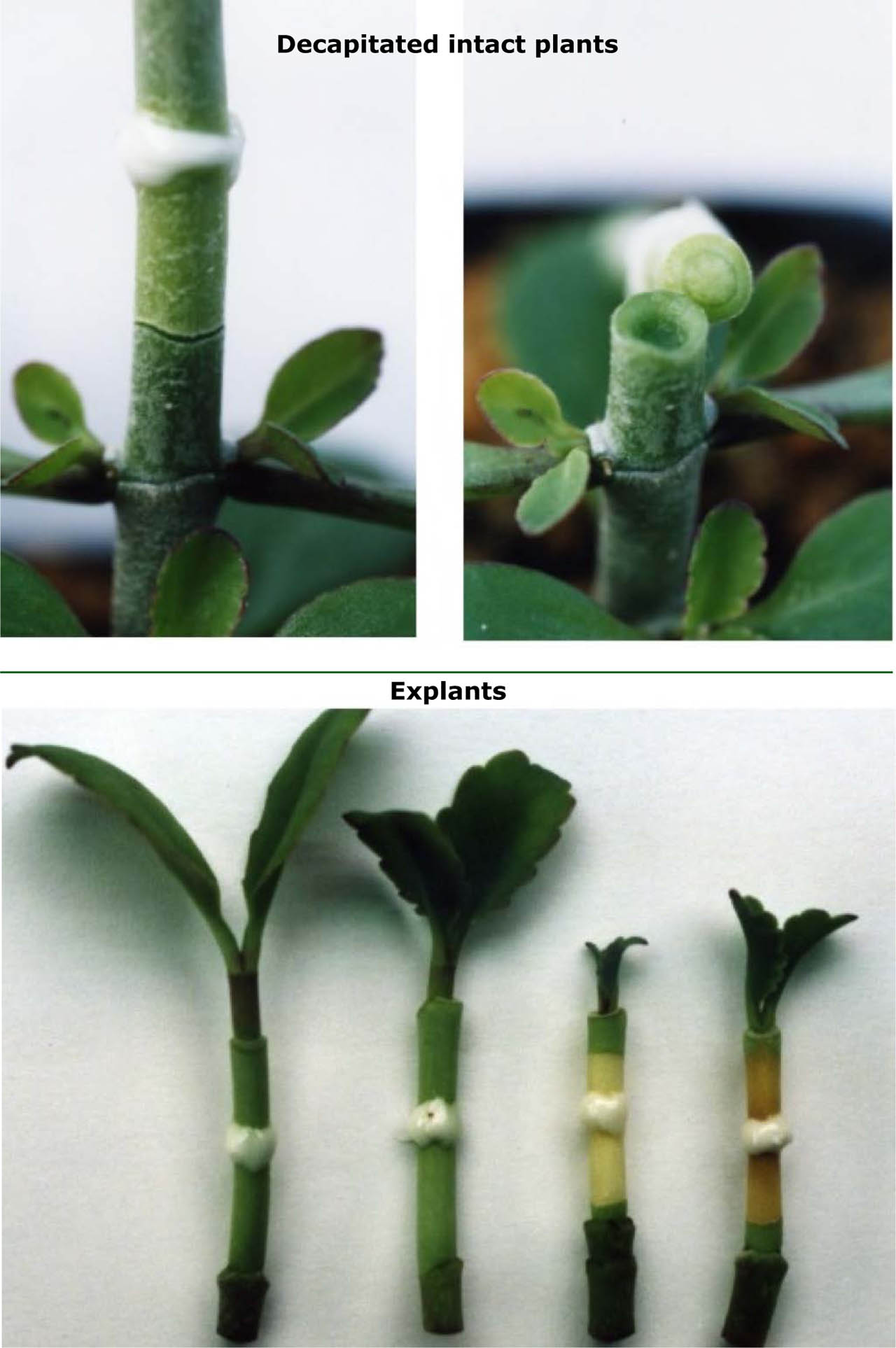
Figure 10.
Secondary abscission in Byophyllum calycinum treated with methyl jasmonate
(Upper): Decapitated intact plants (two pictures above) When methyl jasmonate in lanolin paste was applied, the secondary abscission zone was formed in the middle of the stem (left). The upper part of the stem treated methyl jasmonate was abscissed (right) (Lower): Explants
When methyl jasmonate in lanolin paste was applied to explants with small leaves, two secondary abscission zones formed in the middle of the stem. The sections between these zones turned yellow, indicating advanced senescence (the two on the right). When methyl jasmonate was applied to explants with larger leaves, no secondary abscission occurred, and the stem remained green and healthy (the two on the left); auxin produced in leaves is transported basipetally and counteracts the action of methyl jasmonate, and no two abscission zones.
(denoted by Saniewski et al., 2000)