
1. INTRODUCTION
The lifespan of organisms varies remarkably across the biological kingdom. While sea urchins and Galapagos tortoises can live for over a century, mice and guinea pigs typically perish within a few years. As an organism approaches the end of its life cycle, it undergoes a decline in physiological functions known as senescence. In plants, this process is an essential physiological phenomenon for the alternation of generations.
In the plant kingdom, we distinguish between annuals, which complete their life cycle in a single year, and perennials, such as trees, which live for multiple years. When discussing perennials, it is crucial to differentiate between aging (the chronological maturation of the whole organism) and senescence (the programmed physiological breakdown of specific organs, such as a single leaf). Recently, there has been a noticeable trend toward literature focused predominantly on the molecular mechanisms of senescence (Qiu et al., 2025; Tunc, von Wirén, 2025; Nguyen, Ha, 2026; Sun et al., 2026). However, it is equally important to look back at the historical context of this research and systematically re-organize the actual phenomena of plant senescence using concrete visual data. The conviction that such a comprehensive overview is essential for future research entering this field. This review focuses on the internal physiological processes regulated by plant hormones and other substances during plant senescence.
2. THE MECHANISMS OF LEAF YELLOWING AND REDDENING
2.1. Senescence in deciduous trees
When environmental conditions shift toward shorter photoperiods or lower temperatures – typical of autumn – green leaves undergo senescence, leading to yellowing or the development of red autumnal coloration (Figures 1 and 2). What are the underlying mechanisms of this process? Generally, environmental signals are perceived by the plant and transduced into chemical signals that regulate gene expression. The green leaves of deciduous trees turn yellow or red before eventually falling. This process, known as leaf abscission, is a vital survival strategy. By shedding leaves, trees enter a dormant state that allows them to endure the winter cold. During winter, nascent leaves are protected within tough bud scales. As temperatures rise in spring, these leaves expand and develop chlorophyll.
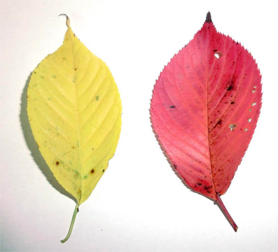
Figure 1.
Yellowing leaf (left) and red leaf (right) of a Japanese cherry (Cerasus × yedoensis ‘Someiyoshino’). Photograph: J. Ueda.
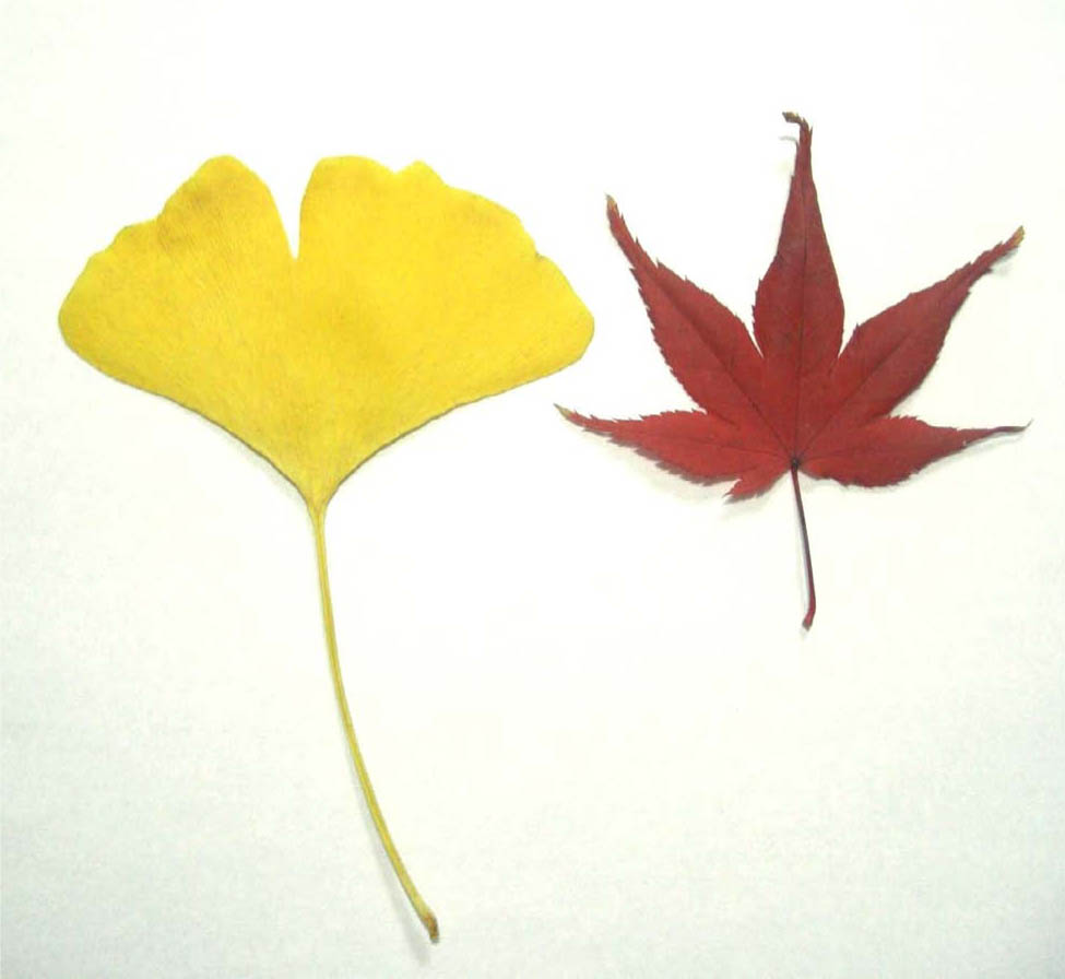
Figure 2.
Yellowing ginkgo (Ginkgo biloba) leaf (left) and red maple (Acer palmatum) leaf (right). Photograph: J. Ueda.
Yellowing: In plants like the Ginkgo, the degradation of chlorophyll reveals the underlying carotenoids (yellow pigments) that were present but masked by green.
Reddening: In plants like the Maple, sugars produced by photosynthesis accumulate in the leaf. Under the influence of ultraviolet or blue light, these sugars are used to synthesize anthocyanins (red pigments).
In areas with streetlights, such as mercury lamps, the environment remains bright even at night. In such locations, leaves on nearby branches may fail to change color, remaining a faint green even in the cold of winter. Because the streetlights simulate the long-day conditions of summer and prevent the plant from sensing the short-day conditions of autumn, the senescence of the leaves is delayed.
The reason leaves appear green to our eyes is due to chlorophyll. Chlorophyll is the substance within plant cells that captures light energy to synthesize starch (sugar) through photosynthesis. It absorbs the red and blue wavelengths of sunlight; the remaining light that is not absorbed reaches our eyes, appearing green. As we know from the seven colors of the rainbow, sunlight is composed of many different wavelengths. As a leaf ages and chlorophyll gradually decomposes, the leaf's color changes to yellow or red, depending on pigments already present or newly synthesized. In short, plant senescence can be understood as the process where chlorophyll breaks down and leaves transition from green to yellow or red. For instance, the leaves of plants like the ginkgo turn yellow before eventually falling. In contrast, plants like the maple do not just turn yellow; they turn a brilliant red (Figure 2). This autumn coloring (reddening) occurs because, alongside the decomposition of chlorophyll, sugars produced via photosynthesis accumulate in the leaf. Under the influence of ultraviolet or blue light, pigments called anthocyanins are synthesized. Depending on the species, red light may also trigger anthocyanin synthesis (Taylor et al., 2001).
2.2. Senescence in conifers such as Pinus spp. (pine) and Cedrus spp. (cedar), and that in broadleaf evergreens like Camellia japonica
The lifespan of conifer needles and broadleaf evergreens leaves is generally several years. While the new growth of the current year of conifers remain a lush green, the older needles located closer to the base of the branch turn brown in autumn.
On the other hand, senescence in broadleaf evergreens like Camellia japonica differ from conifers' one. Interestingly, redundant old leaves often turn yellow and drop from spring to early summer in Japan, precisely when new buds are beginning to sprout.
Plants do not only undergo senescence in response to autumn conditions like short days and low temperatures. For example, when barley is sown in fields after the rice harvest, the young plants overwinter and grow large in the spring. Once they flower and bear fruit in early summer, they turn yellow all at once – a season traditionally known in Japan as “Barley Autumn”. This demonstrates that plant senescence is not triggered solely by environmental changes like day length or temperature, but is also strictly controlled by the plant's own genetic information.
A senescing plant undergoes a temporary increase in respiration (the absorption of oxygen and release of carbon dioxide) before it gradually weakens toward death. Along with the change in leaf color and the decomposition of chlorophyll, various substances within the plant cells – such as proteins, sugars, and nitrogen – decrease in quantity. Finally, in most cases, this process concludes with abscission (the shedding of leaves or fruit). The specific mechanisms of how abscission occurs will be discussed in detail in the latter of this review.
2.3. Senescence in special cases: Oak or Metasequoia, and Ficus superba var. japonica
Under mild natural conditions, most deciduous trees sprout in spring, senesce as autumn deepens, and finally shed their leaves. However, if the environment changes rapidly and drastically – such as during an unexpected severe drought – immature leaves or fruits may drop while still green. In other cases, such as with certain species of oak or metasequoia, the leaves may wither completely in late autumn or winter but remain attached to the plant. These withered leaves stay on the branches through the winter and only drop one by one in the following spring when new buds begin to emerge (Figure 3). Plants like the Ficus superba var. japonica overwinter with vibrant green leaves like evergreens, but then suddenly shed all of those overwintered green leaves simultaneously in early spring (March to April, Figure 4). Immediately after, they hurriedly produce new buds to flourish with green leaves once again. This phenomenon is known as “vernal abscission” (spring leaf fall) (Ueda et al., 1991a).
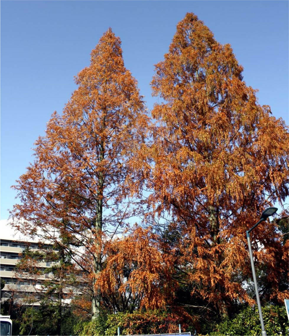
Figure 3.
Metasequoia glyptostroboides from late autumn to winter. Photograph: J. Ueda.
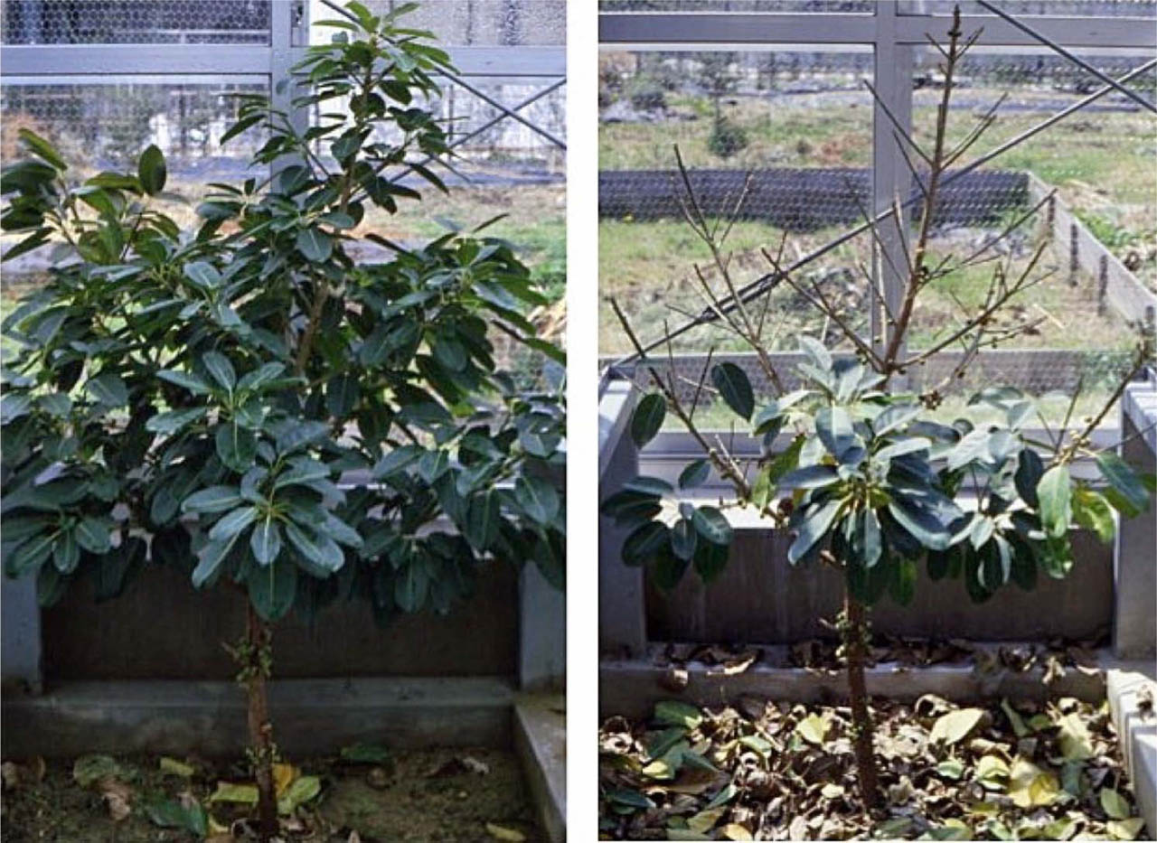
Figure 4.
Vernal leaf abscission of Ficus superba var. japonica. Photographs: J. Ueda.
Left: Overwintering with green leaves. Right: Simultaneous shedding in early spring before new budding.
The signaling pathways through which plants convert external environmental cues into internal signals are depicted in Figure 5.
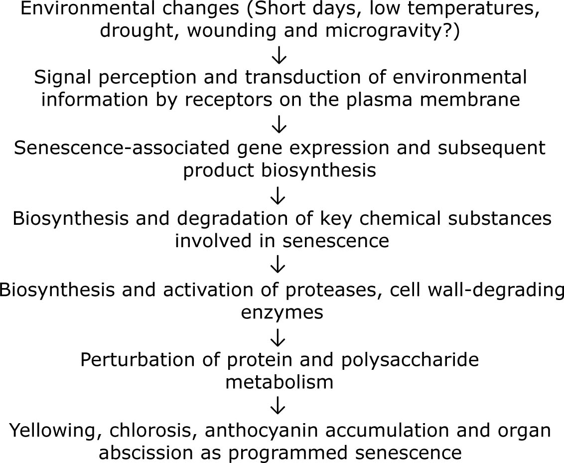
Figure 5.
Signal transduction and sequence of physiochemical changes occurring during plant senescence.
2.4. Senescence in wounding of plant organs
Plants can also undergo senescence due to human intervention. For example, if you take a healthy, vibrant leaf that is growing vigorously and cut it into sections, you can observe this process. If those leaf segments are placed in a Petri dish lined with moist filter paper for a period of time, they will begin to turn yellow (Figure 6). This indicates that senescence is occurring. Even leaves that normally would not change color until autumn will undergo a distinct aging process when triggered by the physical act of cutting, provided there is enough moisture to prevent them from drying out. Furthermore, placing these segments in total darkness significantly accelerates the rate of senescence. When a leaf segment undergoes senescence, it is believed that a signal triggered by the act of being “cut” is transmitted within the segment, leading it toward senescence and, eventually, death. It is possible that the “cutting” signal by triggering the synthesis of senescence-promoting chemicals within the plant.
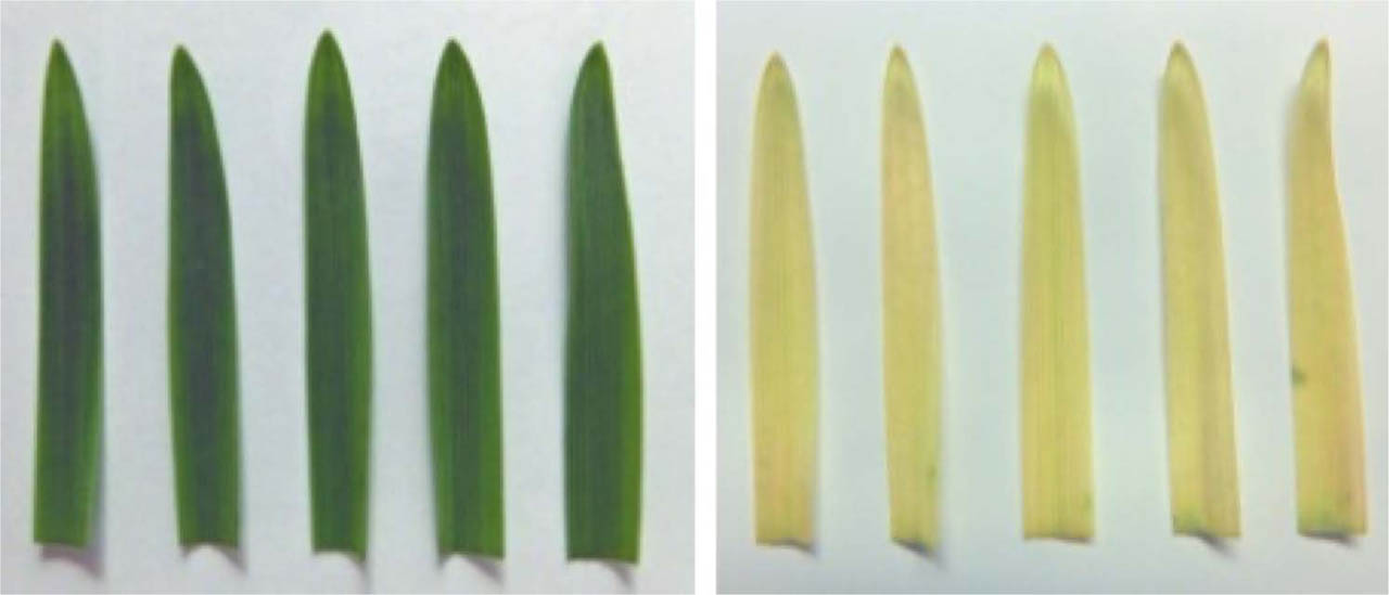
Figure 6.
Comparison of oat (Avena sativa) leaf segments kept in light vs. darkness. Photographs: J. Ueda.
Segments 3 cm in length were cut from the tips of the first leaves of healthy oat plants. These segments were placed leaf-surface up in Petri dishes lined with moistened filter paper. Senescence begins to appear when the chlorophyll contents, measured by absorbance at 665 nm, decrease to approximately 50%. After four days of incubation, the segments kept in light (left) remained relatively stable, whereas the segments kept in darkness (right) completely changed from green to yellow, indicating advanced senescence.
Wounding acts as a potent abiotic stress factor that triggers and accelerates senescence in various plant organs, most notably leaves and flower petals, suggesting it being a senescence factor. This “wound-induced senescence” is a highly regulated genetic process. Wounding induces the expression of Senescence-Associated Genes (SAGs), such as SAG12 and SAG18. Recent studies (Zhang et al., 2023) identified the MdVQ10–MdWRKY75 module as a key positive regulator in this process. Physical injury causes a rapid accumulation of Reactive Oxygen Species (ROS) which leads to rapid browning, chlorophyll loss, and massive cell death (Iakimova, Woltering, 2018). Compounds like Hydrogen Peroxide (H2 O2) or chemicals that generate superoxide radicals can cause oxidative stress, which leads to the breakdown of cellular components and triggers the senescence pathway (Ueda et al., 1991b).
3. CHEMICAL COMPOUNDS REGULATING PLANT SENESCENCE
3.1. Plant hormones
What is the carrier of signal in environmental changes? Plants, like humans and animals, contain substances called hormones that function in trace amounts but have significant effects. These are known as “plant hormones.” They belong to several chemical groups: auxins, gibberellins, cytokinins, brassinosteroids, abscisic acid, ethylene, and jasmonates. Research has shown that several of these hormones are directly involved in plant senescence. Specifically, cytokinins are effective in inhibiting senescence, while abscisic acid and ethylene significantly promote it (Taiz, Zeiger, 2006). In research on wormwood (Artemisia absinthium), it was discovered that (-)-methyl jasmonate is a potent promoter of senescence, now recognized as a key plant hormone (Ueda, Kato, 1980). This research led to methyl jasmonate, jasmonic acid, and their related compounds being recognized as members of the plant hormone family.
Methyl jasmonate applied to the abaxial side of the middle of leaf blade on the beginning of September extremely induced leaf senescence in 12 years old Ginkgo biloba tree; three weeks after the treatment the leaves disappeared chlorophylls and became yellow, while the control leaves remained still green. Contrarily methyl jasmonate applied to the adaxial side of leaves affected little to induce senescence. When methyl jasmonate was treated on the abaxial side of a half of leaf blade across to the vein, the yellowing widely took place both in acropetal and basipetal directions along vein, but no yellowing was visible in the non-treated half. On the other hand, when methyl jasmonate was applied to the abaxial side of a half of leaf blade along to the vein, yellowing was observed only in both sides of a small area along to the applied area, and little yellowing was visible in the neighboring lateral tissues. Methyl jasmonate treatment on the abaxial side of leaf blade greatly increased the levels of methyl jasmonate and jasmonic acid in leaf blade and petiole. Endogenous abscisic acid levels substantially increased during natural and methyl jasmonate-induced leaf senescence, but 1-aminocyclopropane carboxylic acid levels did not change. The contents of cytokinins, gibberellins and auxins identified changed little during natural and methyl jasmonate-induced leaf senescence (Figure 7, denoted by Saniewski et al., 2020). These suggest that a cooperative crosstalk between jasmonates and various hormonal signals, especially abscisic acid, occurs in regulation of G. biloba leaf senescence. Possible different action of methyl jasmonate applied to the abaxial and the adaxial sides of the leaves on leaf senescence is discussed in the literature (Saniewski et al., 2020).
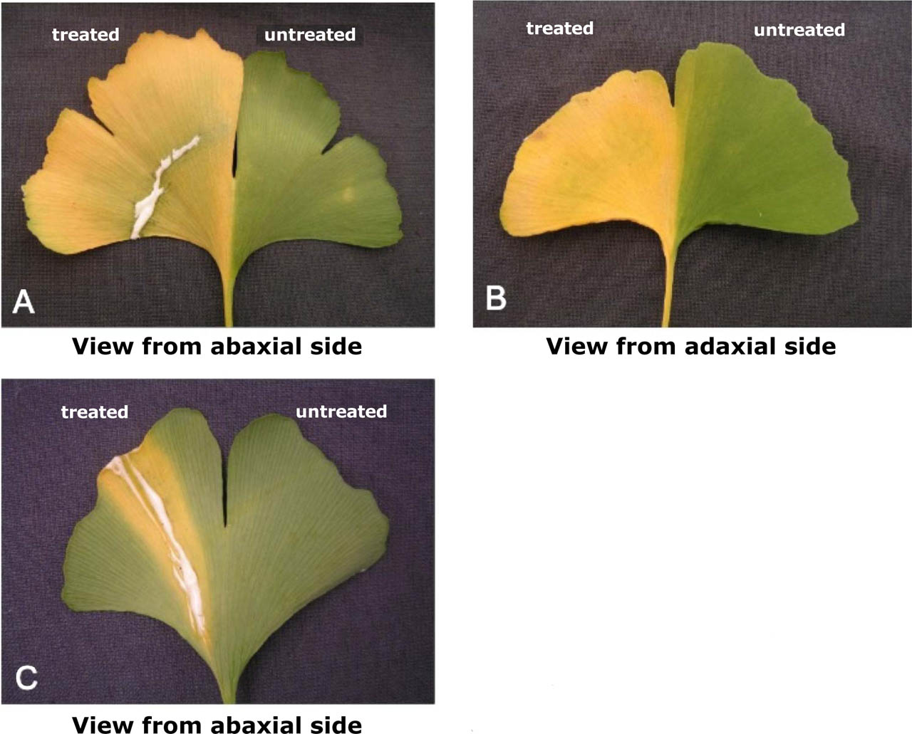
Figure 7.
Methyl jasmonate-induced senescence of Ginkgo biloba leaves.
Methyl jasmonate was applied across (A and B) or along (C) to the vein of leaf blade of abaxial side of a half leaf in 12 years old Ginkgo biloba tree. Treatment with methyl jasmonate was carried out in the abaxial side of half leaf blade (left); opposite half leaf blade was of without treatment as control (right). Photographs were taken 3 weeks after the treatment.
(denoted by Saniewski et al., 2020 with modifications)
3.2. Other chemical compounds
Metabolic inhibitors and antibiotics, such as Actinomycin D (Takegami, 1975), chloramphenicol and thiouracil (Wollgiehn, Parthier, 1964) are also known to accelerate the yellowing of various plant tissues. These substances can eliminate the antisenescence action of cytokinins. A high concentration of L-serine has been reported to possess a similar effect in oat leaf segments, especially in the presence of cytokinin, possibly through enhancement of pro-tease biosynthesis (Martin, Thimann, 1972). In barley leaf segments a high concentration of EDTA has a bleaching effect only in the light (Kotaka, Krueger, 1969). Aliphatic compounds (Satler, Thimann, 1980; Ueda et al., 1984) and other organic compounds (Garg, Kapoor, 1972; Knypl, 1969; Singh, Mishra, 1975) have been reported to affect chlorophyll preservation and/or degradation in plant tissues.
4. BRIEF HISTORY OF SENESCENCE RESEARCH AND PROGRAMMED CELL DEATH IN PLANTS
The history of research in this field is summarized in Table 1. Until the 1950s, researches of plant senescence focused on phenomena such as chlorophyll loss and nutrient mobilization. From the 1950s through the 1970s, studies shifted toward the hormonal regulation of senescence in plants, followed by the 1980s and 1990s, which centered on genetic programming. Since 2000, researches have evolved to investigate the molecular mechanisms underlying the aging process. The first significant research appears to be Molisch's (1929) work on the delay of plant lifespans. Systematic research truly began following a 1967 symposium in the UK titled “Aspects of the Biology of Aging.”
Table 1.
Brief history of plant senescence researches focusing on key chemical compound.
| Year | Author | Achievement |
|---|---|---|
| 1924 | Denny F.E. | Reported the promotion of degreening (yellowing) in lemon fruits by ethylene |
| 1929 | Molisch H. | Published “The Longevity of Plants” (Die Lebensdauer der Pflanze) |
| 1933a, b | Laibach F. | Reported auxin production in Orchid pollen, and it inhibited petiole abscission |
| 1935 | Yemm E.W. | Studied metabolism in starved barley leaves |
| 1937 | Vickery H.B. et al. | Researched chemical changes in tobacco leaves kept in light and darkness |
| 1949 | Hemberg T. | Studied growth-inhibiting substances in the cortical tissue of potato tubers |
| 1953 | Bennet-Clark T.A. and Kefford N.P. | Named a growth inhibitor found in many plants as “inhibitor-β” |
| 1954 | Chibnall A.C. | Hypothesized the existence of a new plant hormone supplied from roots to leaves |
| 1957 | Richmond A.E. and Lang A. | Discovered that kinetin (a cytokinin) inhibits plant senescence, supporting Chibnall's hypothesis. |
| 1958 | Phillips I.D.J. and Wareing P.E. | Studied growth inhibitors in the terminal buds and leaves of Acer pseudoplatanus (Sycamore Maple) |
| 1961 | Carns H.R. et al. | Reported the promotion of abscission in cotton by gibberellin |
| 1963 | Eagles C.E. and Wareing P.E. | Named a dormancy-inducing substance “dormin” (later identified as abscisic acid) |
| 1963 | Ohkuma K. et al. | Isolated abscisin II (later identified as abscisic acid) |
| 1965 | Ohkuma K. et al. | Determined the chemical structure of abscisin II (abscisic acid) |
| 1966 | Fletcher R.A. and Osborne D.J. | Reported that gibberellin inhibits chlorophyll degradation |
| 1967 | Woolhouse H.W. | Organized the symposium “Aspects of the Biology of Aging” |
| 1970 | Shibaoka H. and Thimann K.V. | Conducted systematic research on plant senescence using leaf segments |
| 1980 | Ueda J. and Kato J. | Isolation and identification, and discovered the powerful senescence-promoting effect of methyl jasmonate |
| 1997 | Pennell R.I. and Lamb C. | Published a review on Programmed Cell Death (PCD) in plants |
| 1997 | Yamamoto R. et al. | Reported the regulation of programmed cell death by brassinosteroids |
| 2007 | Kusaba et al. | Identified the NYC1 and NOL genes using rice mutants that remain green (stay-green). |
| 2007 | Sato Y. et al. | Researched the green-cotyledon peas used by Mendel to propose the “Laws of Inheritance” (identified the STAY-GREEN gene) |
| 2016 | Shimoda Y. et al. | Proved that the STAY-GREEN gene encodes magnesium-dechelatase, an enzyme that removes magnesium from the chlorophyll |
4.1. Milestones in plant hormone research
Auxin: Reported to inhibit senescence. In 1933, it was shown that applying auxin to the petioles of Coleus (after removing the leaf blade) prevented the petiole from falling off (Laibach, 1933a, b). However, at high concentrations, synthetic auxins like NAA promote fruit abscission, and 2,4-D has been used as a herbicide (and famously as a component of “Agent Orange” during the Vietnam War).
Gibberellin: In 1961, it was found to promote abscission in cotton (Carns et al., 1961), but by 1966, it was also reported to inhibit the discoloration of excised dandelion (Taraxacum officinale) leaves (Fletcher, Osborne, 1966).
Cytokinin: In 1954, Chibnall hypothesized that hormones from the roots regulate protein metabolism in leaves. This was verified in 1957 by Richmond and Lang using kinetin, proving that root-supplied cytokinins strongly inhibit senescence (Richmond, Lang, 1957).
Ethylene, Abscisic Acid, and Methyl Jasmonate: These were identified as potent promoters of senescence between 1924 and 1980. These substances strongly induce abscission zones in petioles, subsequently leading to leaf abscission. They also inhibit the anti-aging effects of cytokinins.
4.2. Modern molecular perspectives: Programmed cell death
Since 1997, plant senescence has been increasingly understood as Programmed Cell Death – a form of “cellular suicide” where the cell actively uses its own genetic products to die for the benefit of the whole organism (often referred to as apoptosis). In plants, apoptosis is observed during the development of root caps, vascular tissues (xylem and phloem), and reproductive cells. This process is also under hormonal control. For example, research on Zinnia mesophyll cells shows that brassinosteroids induce genes related to cell wall synthesis and the breakdown of proteins and nucleic acids, eventually leading to cell death via vacuole collapse to differentiate the cell into a water-conducting vessel (Yamamoto et al., 1997).
4.3. Mendel and the STAY-GREEN gene
Beyond the mutants mentioned earlier, rice is also known to possess the sgr (stay-green) mutant. Significant research has been conducted on the causative gene, identified as the STAY-GREEN (SGR) gene.
In 1865, Gregor Mendel in Brno proposed the “Laws of Inheritance” by investigating various traits in garden peas (Pisum sativum). One of the primary traits he observed was cotyledon color – specifically, whether the seeds were yellow or green (Figure 8).
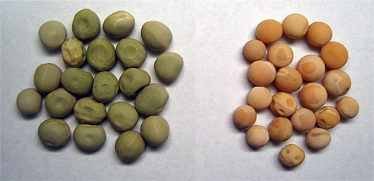
Figure 8.
Dried pea (Pisum sativum) seeds with green (left) and yellow (right) cotyledons. Photograph: J. Ueda.
In 2007, it was discovered that the sgr mutant, in which cotyledons remain green even after drying, results from a mutation in the SGR gene (Kusaba et al., 2007). Subsequent research in 2016 using Arabidopsis thaliana revealed that the SGR gene encodes magnesium-dechelatase. This enzyme is responsible for removing the magnesium (Mg2+) atom from the center of the chlorophyll a molecule. As previously discussed, senescence involves the loss of green pigment as chlorophyll decomposes. In this pathway, magnesium is removed from chlorophyll a to form pheophytin a, which is subsequently metabolized into pheophorbide a. The green peas that led Mendel to his historic discovery were actually plants with a mutated SGR gene. Because these plants cannot produce a functional enzyme, magnesium remains bound to the chlorophyll, thereby preserving the green color even in dormant or dried states (Shimoda, et al., 2016).
5. ABSCISSION OF LEAVES AND FRUITS
5.1. The formation of the abscission zone
As the senescence of leaves and fruits progresses, they are eventually severed from the stems or branches. This phenomenon is known as organ abscission. In biological terms, a group of cells with similar shapes and functions is called a tissue, and a collection of tissues working together is an organ. Usually, the location where an organ detaches is predetermined. As senescence advances, a specialized zone of cells with thin cell walls differentiates and develops at the point of detachment. This is called the abscission zone. Once this zone is fully developed, even a slight force can cause the leaf or fruit to fall. Microscopic observation reveals that in petioles, the abscission zone forms in the cortex (the zone between the epidermis and the central vascular cylinder) but is rarely observed in the vascular bundles that run through the center. Vascular bundles are responsible for transporting water and nutrients, while the petiole is the stalk that connects the leaf blade to the stem. An abscission zone may already be present when the organ is very young (Figure 9), or it may develop gradually as senescence progresses (Addicott, 1982).
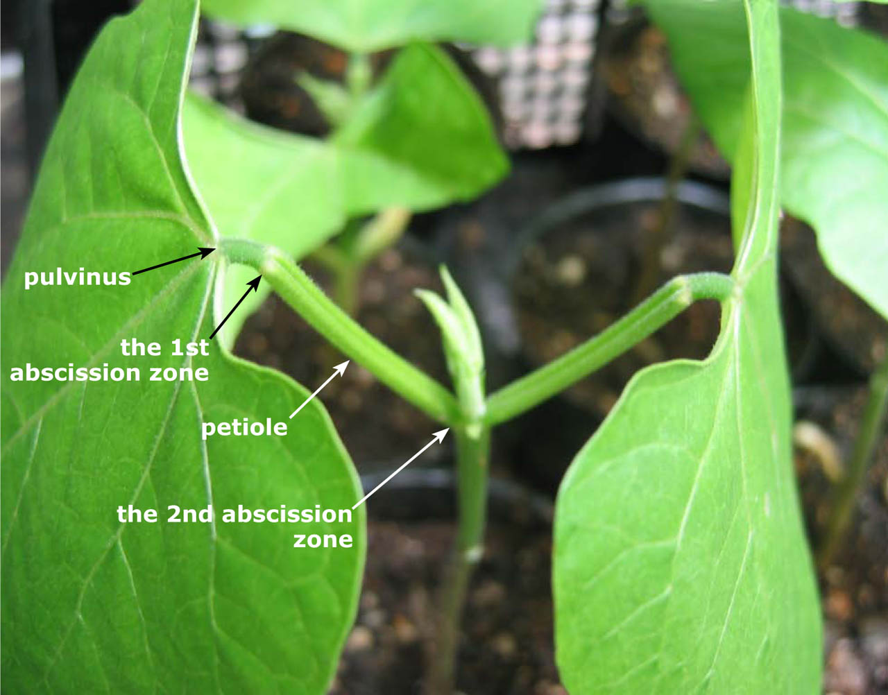
Figure 9.
Two abscission zones observed in the primary leaf of Phaseolus vulgaris seedling. Photograph: J. Ueda.
The primary abscission zone is located at the junction of the pulvinus and the petiole, and the secondary abscission zone is located at the junction of the petiole and the stem. The pulvinus refers to the thickened section at the base of a leaflet or petiole.
The series of physiochemical processes leading to organ detachment is closely governed by the dynamics of key regulatory substances. Abscission requires the de novo synthesis of RNA and proteins within the cells of the abscission zone, leading to a surge in cellulase and pectinase activity. These enzymes degrade the cell walls and the middle lamella, eventually resulting in the mechanical breakdown of vascular tissues until the organ detaches (Kozlowski, 2012).
Promoters such as abscisic acid (ABA), ethylene, and methyl jasmonate enhance this cellulase activity (Ueda et al., 1996). Conversely, while auxin applied to a leaf can sometimes induce ethylene production, it generally functions to suppress cellulase activity and inhibit abscission. Recent studies have also indicated that hydrogen peroxide and fatty acid peroxides act as signaling molecules that promote abscission zone formation (Ueda et al., 1991b).
5.2. Secondary abscission
If artificial treatments are applied – such as the application of specific chemicals – an abscission zone can sometimes form in tissues where it would never occur under natural conditions. It is referred as secondary abscission zone formation (Saniewski et al., 2000). It was found that methyl jasmonate induced the secondary abscission zone in not only explants but also decapitated intact one of Bryophyllum calycinum, the concentration gradient and balance of auxin between tissues on either side of the potential site are critical. When a paste made of methyl jasmonate and lanolin (refined wool wax) is applied to a stem, secondary abscission zones form at two points in the middle of the stem where they do not naturally occur (Figure 10, denoted by Saniewski et al., 2000). Auxin exogenously applied extremely prevented the formation of secondary abscission zones and senescence in the stem tissues induced by methyl jasmonate (Saniewski et al., 2020). Evidence for a close functional relationship between jasmonates signaling pathway and auxin homeostasis has been documented in literature (see Saniewski et al., 2020).
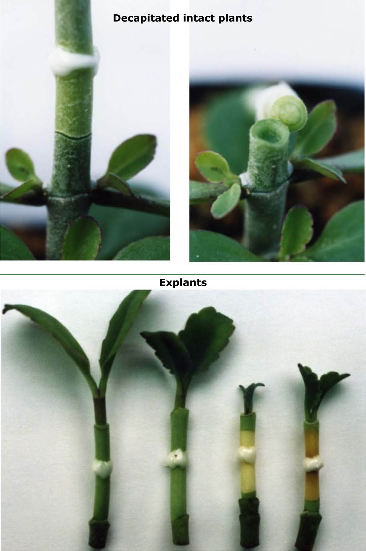
Figure 10.
Secondary abscission in Byophyllum calycinum treated with methyl jasmonate
(Upper): Decapitated intact plants (two pictures above) When methyl jasmonate in lanolin paste was applied, the secondary abscission zone was formed in the middle of the stem (left). The upper part of the stem treated methyl jasmonate was abscissed (right) (Lower): Explants
When methyl jasmonate in lanolin paste was applied to explants with small leaves, two secondary abscission zones formed in the middle of the stem. The sections between these zones turned yellow, indicating advanced senescence (the two on the right). When methyl jasmonate was applied to explants with larger leaves, no secondary abscission occurred, and the stem remained green and healthy (the two on the left); auxin produced in leaves is transported basipetally and counteracts the action of methyl jasmonate, and no two abscission zones.
(denoted by Saniewski et al., 2000)
6. AGRICULTURAL APPLICATIONS OF CHEMICAL COMPOUNDS INDUCING PLANT SENESCENCE
At present, there is a strong demand for labor-saving and increased efficiency in agricultural production and harvest as described below. Regarding the practical applications of abscission zone formation in plants, notable examples include cotton harvesting on large-scale farms in the United States and the use of thinning agents in Japanese Satsuma mandarin (Citrus unshiu) cultivation.
6.1. Post-harvest ripening of banana (Musa spp.)
Bananas are transported from tropical regions to Japan. They are imported as “green bananas” because shipping them in a ripe state would cause them to damage or rot during transit. Upon arrival in Japan, they are placed in specialized warehouses known as “ripening rooms”. There, they are exposed to a low concentration of ethylene gas for a specific period to trigger the ripening process (fruit senescence) (Abraham et al., 2022).
6.2. Cotton crops (Gossypium spp.) harvesting and abscission
In cotton cultivation, harvesting historically required an immense amount of time and labor; in the United States, this was once the primary reason for the exploitation of many enslaved people. Today, modern methods involve using harvest aids (defoliants) to forcibly drop the leaves that hinder harvesting, allowing large-scale machinery to harvest the crop all at once. Interestingly, although the plant hormone abscisic acid was originally discovered through research on abscission zone formation in cotton (Addicott, 1982), it proved to be ineffective as a practical defoliant. Instead, chemically synthesized agents such as thidiazuron and diuron are currently utilized as defoliants for cotton crops (Murthy et al., 1998; Liu et al., 2021).
6.3. Fruit thinning in satsuma mandarin (Citrus unshiu) cultivation
On the other hand, in the cultivation of Satsuma mandarins – which exhibit parthenocarpy (the ability to produce fruit without pollination) – chemical thinning agents are used to ensure that a tree bears an appropriate number of fruits. For this purpose, Figaron containing 20% ethychlozate (ethyl 5-chloro-3(1H)-indazoleacetate) has been used (Hirose et al., 1978).
7. PERSPECTIVES/HYPOTHESES
Plant senescence is regulated by various environmental factors (Figure 5). Although several papers suggest that gravity appears to have an effect on plant senescence (Kordyum et al., 2019; Miyamoto et al., 2001; Mao et al., 2024), definitive findings have yet to be established.
Simulated microgravity conditions significantly alter plant senescence, generally accelerating leaf aging, cellular senescence and apoptosis while reducing cell proliferation (Kordyum et al., 2019; Miyamoto et al., 2001; Mao et al., 2024). Unfortunately, the role of gravity is still being explored. Ueda et al. (1999) and Ueda (2020), in collaboration with NASA and JAXA, have investigated plant growth and hormonal regulation in space environments using the Space Shuttle (STS-95) and the International Space Station (ISS) in 1998 and 2016, respectively. While it is yet to be confirmed if chlorophyll degradation or abscission zone formation are directly influenced by gravity, we can infer that in a microgravity environment, senesced leaves and fruits might not “fall” in the traditional sense. Without external forces like wind or gravitational pull, a tree in space might theoretically remain covered in senescent red leaves and ripe fruit indefinitely – a fascinating biological prospect.
8. CONCLUSIVE REMARKS
While this review focuses specifically on the classical aspects of plant senescence, consolidating these perspectives is significant for future research in the field. It is difficult to reach a definitive understanding of plant senescence solely through the lens of gene expression and regulatory mechanisms. Leaving such recent molecular findings to other reviews (Nguyen, Ha, 2026; Qiu et al., 2025; Sun et al., 2026; Tunc, von Wirén, 2025), this review article aims to reorganize our understanding of the actual phenomena of plant senescence, with a primary focus on visual data.
Plant senescence is thought to be controlled by various environmental factors that influence the dynamics of functional substances, including plant hormones. These substances ultimately regulate gene expression to govern the aging process. For detailed regulatory mechanisms, such as gene expression patterns that modulate plant senescence, please refer to recent reviews described above. For instance, jasmonates are known to strongly promote plant senescence. According to recent findings, these compounds are synthesized from α-linolenic acid derived from chloroplast membrane lipids, which is then metabolized and transported to peroxisomes to become jasmonic acid. It has been demonstrated that jasmonic acid moves from the peroxisomes to the cytosol, where jasmonoyl-L-isoleucine is biosynthesized. This metabolite then translocates into the nucleus to regulate the expression of jasmonate-related genes. Ultimately, it has been clarified that jasmonic acid itself does not directly affect gene expression; rather, its metabolite acts as the regulator (reviewed by Ueda and Saniewski, 2026, in press).
The various manifestations of plant senescence described here are controlled by the dynamics of functional substances like jasmonates. Elucidating which types of senescence are controlled at the molecular level through the dynamics of specific functional substances may eventually reveal the complete picture of the plant senescence process. As delineated above, plant senescence represents a fundamental and critical physiological process within the natural world. Elucidating its intricate underlying mechanisms, not only fosters a more profound understanding of plant biology but also offers the potential for transformative advancements in agricultural productivity and global food security.