The oil palm industry is one of the most important in the Colombian agricultural sector. Currently, Colombia is the fourth producer of palm oil and the first in Latin America, with more than 535,000 hectares planted in 112 towns of 20 states, placing it as one of the main agricultural lines of the country (Fedepalma, 2018; Sistema de información estadística del sector palmero-SISPA, 2019). One of the most important aspects of Colombian palm production is related to phytopathological problems. These diseases are considered as the main threat and have been responsible for crop losses, including the so-called lethal ones such as sudden wilt (Phytomonas sp.), lethal wilt (unknown etiology), and the red ring disease associated with the migratory endoparasite nematode Bursaphelenchus cocophilus (Cobb, 1919; Goodey, 1960; Baujard, 1989) (=Rhadinaphelenchus cocophilus [Cobb]) (Martínez, 2010).
The red ring disease has been reported in Central America (Guatemala, Nicaragua, Belize, Costa Rica, El Salvador, Honduras, Mexico, Panama), Latin America (Brazil, Ecuador, Guyana, French Guyana, Peru, Venezuela, and Colombia), and in the southern Caribbean (Granada, San Vicente, Tobago, Suriname, Trinidad, Dominican Republic) (Kraaijenga and den Ouden, 1966; Kastelein, 1987; Giblin-Davis, 2001; Sánchez and Cerda, 2002; Brammer and Crow, 2002a).
In Colombia, there is a history of the red ring since 1960s. In 1967, more than 10,000 ha of coconut palm disappeared on the Pacific coast. In 1986, red ring disease was detected in oil palm in Palmeras de la Costa plantation (El Copey, state of Cesar) and currently, in the Eastern Plains and the state of North Santander, causing in addition to the red ring, the short leaf syndrome (Varón de Agudelo and Granada, 1986; Chinchilla, 1992; Calvache et al., 1995; Aldana et al., 2015).
The red ring disease has caused losses that reached 8 million dollars between 1990 and 2002 in the North and Eastern production areas (Aldana et al., 2015). It has been registered mainly in crops from North, Central, and Eastern zone of Colombia, where management strategies are mainly preventive through frequent monitoring and trapping of the Rynchophorus palmarum nematode vector (Coleoptera: Curculionidae) (Blair and Darling, 1968; Griffith, 1969; Oehlschlager et al., 2002). Other management practices include early detection and timely eradication of plants, followed by proper waste disposal (Van Hoof and Seinhorst, 1962; Griffith, 1987; Chinchilla, 1992; Chinchilla and Escobar, 2007; Saenz, 2016).
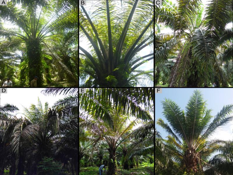
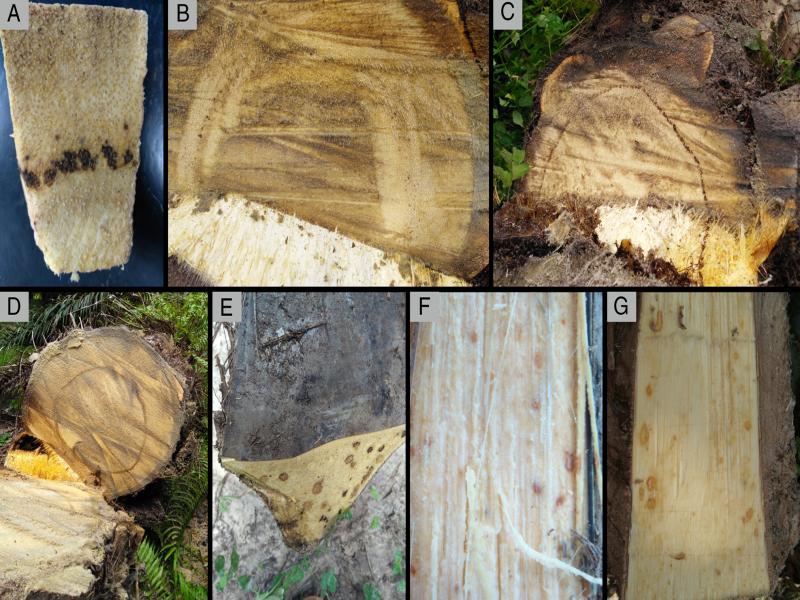
The red ring symptoms in oil palm are varied in Colombia, in the case of the Eastern Plains the main symptom is the shortening of leaves. In the North Zone, there is shortening and copper coloration of the leaflets. In some cases, there is accumulation of spear leaf and when they open are very short and chlorotic (Chinchilla, 1992; Calvache et al., 1995). However, external symptoms are not considered a diagnostic method of the disease because there are other pathogens that induce similar symptoms in the foliage (Dean, 1979). The most frequent symptoms are young leaf chlorosis, drying of lower leaves, breaking of petioles, and leaf folding. Internally in the petioles and spear leaf, brown or reddish brown spots may be observed, and in the stem continuous or discontinuous, brown or reddish brown rings (Brathwaite and Siddiqi, 1975; Chinchilla, 1992; Calvache et al., 1995; Brammer and Crow, 2002b). The final state of the disease is the death of plants (Thorne, 1961; Giblin-Davis, 1990; Sáenz, 2005; Giblin-Davis et al., 2010).
Bursaphelenchus cocophilus can be found infecting the parenchymal tissues of the stem, petioles and sometimes in roots, the intercellular spaces, and in an advanced state of the disease, they act as intracellular parasites (Griffith, 1987; Griffith and Koshy, 1990), causing the interruption of the transport of nutrients, water, and sap, which induces drying of the leaves. In palms with short leaves, the nematodes are found mainly in the leaf buds, which makes the palm generate shorter, deformed, yellow, and dry leaves (Brammer and Crow, 2001; Oehlschlager et al., 2002). It have been reported that in raining season the nematode populations increase and they move toward arrows, central leaves, tissue in the leaf buds, and the spear leaf (Volcy, 1998) and B. cocophilus achieves the central arrow may access to any petiole (Menjivar et al., 1988; Cuthbert, 1993; Ferris, 1999).
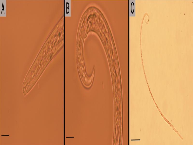
Morphologically, males of B. cocophilus are characterized by having sclerotized cephalic region, paired spicules, absent gubernaculum, a conoid to sharply pointed tail, and strongly ventrally curved, which contains a terminal bursa, presents two pairs of ventrosubmediales papillae very close at the base of the bursa and a pair preanal, anterior to the cloacal aperture (Opperman, 1995). The females are characterized by having a thin cuticle, marked with transverse striations, lateral fields with four incisures, the labial region is high and smooth, covered vulva by a protuding anterior vulvar lip, presence of a long post-uterine sac, a narrow and elongated tail with a clindroid and rounded terminus (Opperman, 1995).
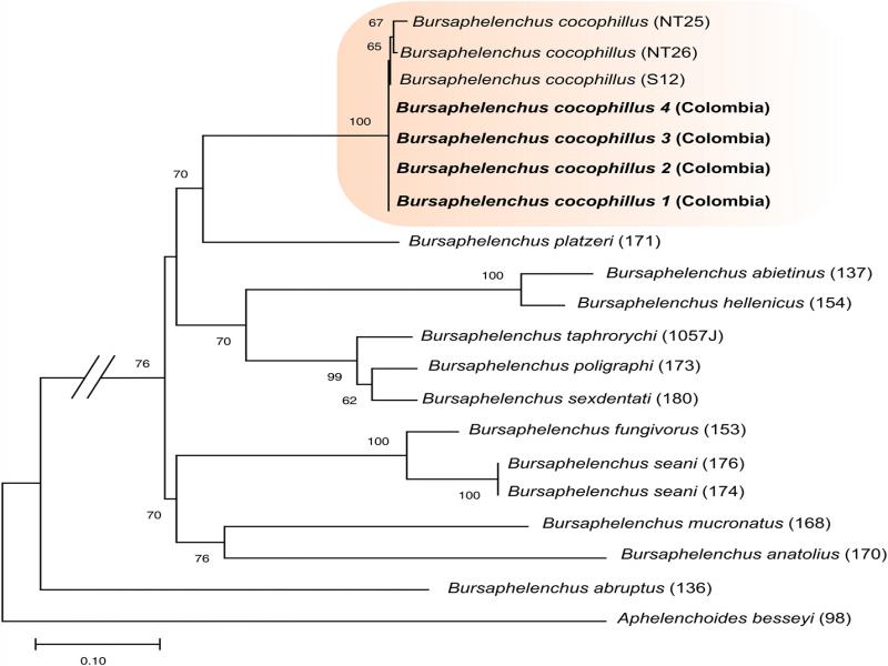
Currently, DNA sequences of segment D2-D3, Internal Transcribed Spacer-(ITS) of ribosomal RNA and Cytochrome oxidase subunit I-COI of mitochondrial DNA have been obtained for B. cocophilus in coconut palm and deposited in public molecular databases, which are used as a reference for the identification of the nematode (Ye et al., 2007; Silva et al., 2016). However, to date in Colombia no molecular characterization or phylogenetic analysis of B. cocophilus from oil palm has been performed.
In Tibu, oil palm plants with symptoms similar to those induced by the disease known as the red ring had been eliminated, to prevent their spread. However, palm growers have observed that some plants remain diseased for a longer time without showing the severity of the disease, so there is doubt about the diagnosis and identity of the causal agent, especially because sometimes the continuous ring is not observed or because the nematode is not found in sampling surveys. Due to the observations made by the palm growers, this research had as main objective the diagnostic of the disease by the verification of the symptoms, the identification of the nematode and confirmation of its identity at the morphometrical and molecular levels using diagnostic characters recommended and the amplification of the large subunit D2-D3 of the 28S.
Materials and methods
Symptoms observation and sampling
In total, 24 lots were visited located in 12 farms of Tibu (Norte de Santander), each of about 10 ha; in each plantation, plants with abnormal aspect were identified, and previously selected by the health censors (expert persons). In each case, description of the external and internal symptoms was recorded when the palms were knocked down with a chainsaw at the base of the stem and then cutting in sections to observe the internal symptoms. Similarly, cuts were made in petioles, bunches, buds, and spear leaf.
Symptomatic tissue samples as petioles, base of spear leaf, inflorescences, peduncle of inflorescences and bunches and stem from eradicated palms, were collected and kept in plastic bags, properly identified and refrigerated until processing. To collect tissues samples in palms that were not eradicated, a manual drill was used to perforate the stem at a height of approximately 1.5 m from the base at a depth close to 30 cm.
Sample processing
Nematodes were extracted using the oxygenation-decantation method (Ravichandra, 2014). Five grams of tissue from each of the sampled organs were cutted into small portions and placed in a decantation sieve, with and without facial paper, rested on a decantation plate with enough water to cover the sample. After 24 h, the nematode suspension contained in the decantation plate was removed and concentrated at 20 mL with the 400-mesh sieve (Varón de Agudelo and Castillo, 2001).
Quantification and nematode identification
To quantify the population of nematodes present in five grams of fresh tissue in each sample, three aliquots of 1 mL were taken and counting in chamber under a light microscope, Olympus PX40 and Olympus DP 73. For the morphological and morphometrical identification, the nematodes were killed with heat at 60°C for 4 min and fixated in 2% formalin. Then, semipermanent preparations were done and morphometric data were registered following Basil (1960), Brathwaite and Siddiqi (1975), Mai and Lyon (1975), Gerber et al. (1989). The morphometric data were taken using a compound microscope ZEISS Axio (China, microscope reference: 3136006171 Axio.A1).
Molecular identification
DNA extraction was performed by the proteinase K method (Riascos-Ortiz et al., 2019). A specimen was divided into three parts with a sterile scalpel, transferring the sections to Eppendorf tubes with 15 µL lysis buffer (50 mM KCl, 10 mM Tris pH 8.0, 15 mM MgCl2, 0.5% Triton x – 100, 4.5% Tween – 20, 0.09% Proteinase K). Subsequently, the tubes were incubated at −80°C (15 min), 65°C (1 h), and 95°C (15 min), centrifuged at 16,000 g (1 min) and stored at –20°C. The PCR amplification of the expansion segment D2-D3 of the large subunit of ribosomal DNA (28S) was performed with the forward D2A primers: (5′-ACAAGTACCGTGAGGGAAAGTTG-3′) and reverse D3B: (5′-TCCTCGGAAGGAACCAGCTACTA-3′) according to De Ley et al. (1999). The PCR conditions were initial denaturation during 2 min at 94°C followed by 40 cycles of 45 sec at 94°C, 45 sec at 55°C and 1 min at 72°C and final extension of 10 min at 72°C. The PCR products were sequenced in both directions by the company Bionner (South Korea).
Phylogenetic analysis
The sequences obtained were edited using the Geneious software (Kearse et al., 2012). Once the sequences edition were carry out, their identity was confirmed using the software BLASTn (http://www.ncbi.nlm.nih.gov/BLAST). Subsequently, the sequences presented under the accession numbers in Table 1 were aligned and analyzed using the MUSCLE algorithm included in the program MEGA6 (Tamura et al., 2013). Based on the matrix obtained, the nucleotide substitution model was determined by taking into account the Bayesian information criterion (BIC) using ModelGenerator v.0.851 software (Keane et al., 2006). The phylogenetic relationship was determinate by the maximum likelihood (ML) method based on the general time reversible (GTR) model, and the Gamma distribution, which was used to model the differences in evolutionary speed between locations. The internal support of the nodes was carry out using the bootstrap method with 1,000 replicates. Aphelenchoides besseyi sequence was used as external group (AY508109).
Table 1.
Information of partial sequences D2-D3 of ribosomal DNA downloaded from GenBank and obtained in the present study for Bursaphelenchus.
| Isolate | Species name | Location | Plant-host | Insect host | GenBank accession number | Reference or source |
|---|---|---|---|---|---|---|
| 1 | B. cocophilus | Tibu, Norte de Santander, Colombia | Elaeis guineensis | R. palmarum | MN612640 | Present study |
| 2 | B. cocophilus | Tibu, Norte de Santander, Colombia | Elaeis guineensis | R. palmarum | MN612641 | Present study |
| 3 | B. cocophilus | Tibu, Norte de Santander, Colombia | Elaeis guineensis | R. palmarum | MN612642 | Present study |
| 4 | B. cocophilus | Tibu, Norte de Santander, Colombia | Elaeis guineensis | R. palmarum | MN612643 | Present study |
| 136 | B. abruptus | USA | None | Anthophora abrupta | AY508073 | Ye et al. (2007) |
| 137 | B. abietinus | Austria | Pityokteines vorontzowi | Abies alba | AY508074 | Ye et al. (2007) |
| 170 | B. anatolius | Turkey | Halictus sp. | None | AY508093 | Ye et al. (2007) |
| S12 | B. cocophilus | Brazil | Cocos nucifera | R. palmarum | KT156772 | Silva et al. (2016) |
| NT25 | B. cocophilus | Colombia | Cocos nucifera | R. palmarum | KT156775 | Silva et al. (2016) |
| NT26 | B. cocophilus | Colombia | Cocos nucifera | R. palmarum | KT156776 | Silva et al. (2016) |
| 153 | B. fungivorus | Germany | Greenhouse soil | Unknown | AY508082 | Ye et al. (2007) |
| 154 | B. hellenicus | Greece | Tomicus piniperda | Pinus brutia | AY508083 | Ye et al. (2007) |
| 168 | B. mucronatus | Germany | Picea abies | Monochamus galloprovincialis | AY508091 | Ye et al. (2007) |
| 173 | B. poligraphi | Germany | Picea abies | Polygraphus poligraphus | AY508096 | Ye et al. (2007) |
| 171 | B. platzeri | USA | None | Carpohilus humeralis | AY508094 | Ye et al. (2007) |
| 174 | B. seani | USA | None | Anthophora bomboides | AY508097 | Ye et al. (2007) |
| 176 | B. seani | USA | None | Anthophora bomboides | AY508099 | Ye et al. (2007) |
| 180 | B. sexdentati | Italy | Pinus pinaster | Unknow | AY508103 | Ye et al. (2007) |
| 1057 J | B. taphrorychi | Poland | Fagus sylvatica | Taphrorychus bicolor | MF422699 | Tomalak et al. (2017) |
| 98 | Aphelenchoides besseyi | USA | Fragaria ananassa | – | AY508109 | Ye et al. (2007) |
| Extraction method (Decantation) | Sampling method | ||||
|---|---|---|---|---|---|
| Tissue class | With facial paper | Without facial paper | Palms knocked down | Palms standing (Drill) | |
| Peduncle | 10 | 8 | X | ||
| Petiolar Base | 16 | 501 | X | ||
| Spear leaf base | 5 | 0 | X | ||
| Inflorescences | 110 | – | X | ||
| Stem | 469 | 51 | X | ||
| Stem | 1 |
|
| ||
| Extraction method (Decantation) | Sampling method | ||||
|---|---|---|---|---|---|
| With facial paper | Without facial paper | Palms knocked down | Palms standing (Drill) | ||
| Maximum Population | 1,295 | 720 | X | ||
| Maximum population | 2 | 5 | X | ||
| Absolute density | 150 | 150 | X | ||
| Absolute density | 0.86 | 4 | X | ||
| Absolute frequency | 58.8 | 71.4 | X | ||
| Absolute frequency | 42.9 | 85.7 | X | ||
| Locality/Publication | Tibu-North Santander, Colombia oil palm (Present study) n=12 | Brazil Coconut Red Ring Disease After (Lordello and Zamith, 1954) n=5 | Trinidad coconut Red Ring Disease (Basil, 1960) n=10 | Venezuela oil palm Red Ring Disease (Gerber et al., 1989) n=10 | Venezuela oil palm Little leaf (Gerber et al., 1989) n=10 |
|---|---|---|---|---|---|
| Body length | 964.67±70.07 (807.09-1062.67) | 820-1420 | 1020 (840-1160) | 1017±77 (841-1111) | 866±71 (789-965) |
| a | 83.89±10.68 (69.53-99.78) | 92-143 | 120 (100-179) | 129.7±13.3 (113.8-150.0) | 99.3±11.8 (78.9-116.1) |
| c | 29.33±1.72 (27.02-32.25) | 22-47 | 28 (24-35) | 26.1±2.2 (22.5-29.2) | 22.4±2.8 (18.8-26.5) |
| c′ | 3.70±0.31 (3.19-4.31) | – | – | 5.6±0.5 (4.8-6.4) | 5.4±0.8 (4.7-6.8) |
| T | 96.58±0.20 (96.30-96.90) | – | – | – | – |
| Max. Body diam. | 11.62±1.34 (9.97-14.22) | – | – | 8±1.0 (7.0-9.0) | 9.0±1.0 (7.0-10.0) |
| Stylet | 10.90±0.88 (9.07-12.12) | 10.7-13.8 | 12-13 | 11±1.0 (11-12) | 12.0±1.0 (11.0-12.0) |
| Lip region width | 4.61±0.52 (3.81-5.22) | – | 5.5 | – | – |
| Lip region height | 3.13±0.43 (2.43-365) | – | 3.0 | – | – |
| Median bulb length | 12.16±0.79 (10.75-13.04) | – | – | – | – |
| Median bulb diam. | 5.99±0.84 (4.66-7.64) | – | – | – | – |
| Tail length | 32.83±2.35 (28.94-37.90) | – | – | 39.0±4.0 (32.0-46.0) | 39.0±4.0 (33.0-46.0) |
| Cloacal or anal body diam. | 8.90±0.71 (7.75-10.16) | – | – | 7.0±1.0 (5.0-8.0) | 7.0±1.0 (6.0-9.0) |
| Spicule length | 15.21±1.41 (13.29-17.32) | – | – | 12.0±1.0 (11.0-13.0) | 11±1.0 (10.0-13.0) |