Rice [Oryza sativa L.] is a staple food for about 3.5 billion people in the world (Singh and Singh, 2017), with an annual production of about 480 metric tons of milled rice consumption (Muthayya et al., 2014). Rice contributes about 50% of dietary caloric supply and forms part of substantial protein intake for an estimate of 520 million people in Asia (Muthayya et al., 2014). In South Korea, rice is a staple grain and a largest starch source (Choi et al., 2016). In 2019, rice production in Korea was estimated at 3,744,450 metric tons from 730,000 ha area under production (USDA Foreign Agricultural Service, 2019). A big area of 729,586 ha was under paddy rice compared to 229 ha only for Upland rice production (USDA Foreign Agricultural Service, 2019). In addition to being a table food, rice is important in the production of beverages such as Soju and Makgeoli/raw rice wine and in baking industries (Choi et al., 2016). Under different areas and environments of production, rice is exposed to various pests and diseases including soil bone plant pathogens such as plant-parasitic nematodes. Plant-parasitic nematodes have been documented as an important constraint to rice production in various parts of the world and can cause an estimated yield loss of 20 to 90% depending on the nematode species present country, season, crop variety, cultivation practices, and soil conditions (Gilces et al., 2016). The most important nematode species in rice production include Meloidogyne graminicola, Hirschmanniella oryzae, Pratylenchus zea, Aphelechoides besseyi, Ditylenchus angustus, Pratylenchus indicus, and some cyst nematodes especially Heterodera oryzicola (Prasad et al., 1987; Jairajpuri, 2019).
Four species of cyst nematodes infect rice viz Heterodera elachista, H. oryzicola, H. sacchari, and H. oryzae (Nobbs et al., 1992). Although cyst nematodes have been reported in rice from other countries neighboring South Korea such as China and Japan; no practical studies have indicated their presence in South Korea though Choi et al. (1989) listed H. elachista and H. oryzae among the rice nematodes in the country.
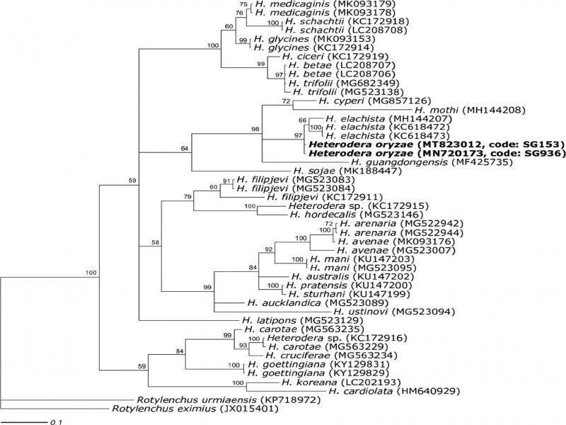
In a survey to identify plant-parasitic nematodes on rice in South Korea, cyst nematodes were found in two rice fields in Dang-jin city and this required a detailed study of the samples in order to get accurate information on the nematode species. Nematode species identification is a requirement before designing and undertaking any management strategy in crop production. Species identification using morphological characters alone is very difficult and does not give correct results. A combination of both morphological and molecular approaches is very crucial for proper and accurate identification (Subbotin et al., 2015). The use of mitochondrial DNA cytochrome c oxidase subunit I (mtCOI) gene, ITS region, and D2-D3 expansion segments of 28S rDNA region sequences are trending today in molecular and phylogenetic studies (Hajibabaei et al., 2007; Subbotin et al., 2007). The objectives of this study were to identify cyst nematode species associated with rice in South Korea, provide morphological and morphometrical information, and characterize the nematodes using DNA barcoding.
Materials and methods
Nematode sampling, extraction, and pure culture establishment
Soil samples were taken from two rice fields in Dang-jin city – South Korea using soil auger from about 15 cm in depth and placed in plastic bags. Samples were labeled as SG936 and SG153 (Table 1). Cysts were extracted from 100 cm3 of a soil sample using the sieving method (20 and 60 mesh). To establish pure cultures, second-stage juveniles (J2s) obtained from a single cyst were used to infect rice plants growing in pots in a plant growth room kept at 25°C. The plants were left to grow for five months before extracting cysts from pot soil. After extraction, cysts were collected with forceps into a watch glass containing tap water using a stereomicroscope (MZ12; Leica, Wetzlar, Germany) and were kept at 4°C until further use.
Morphological and morphometric traits for cysts and J2s
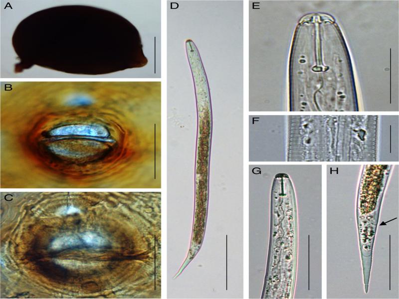
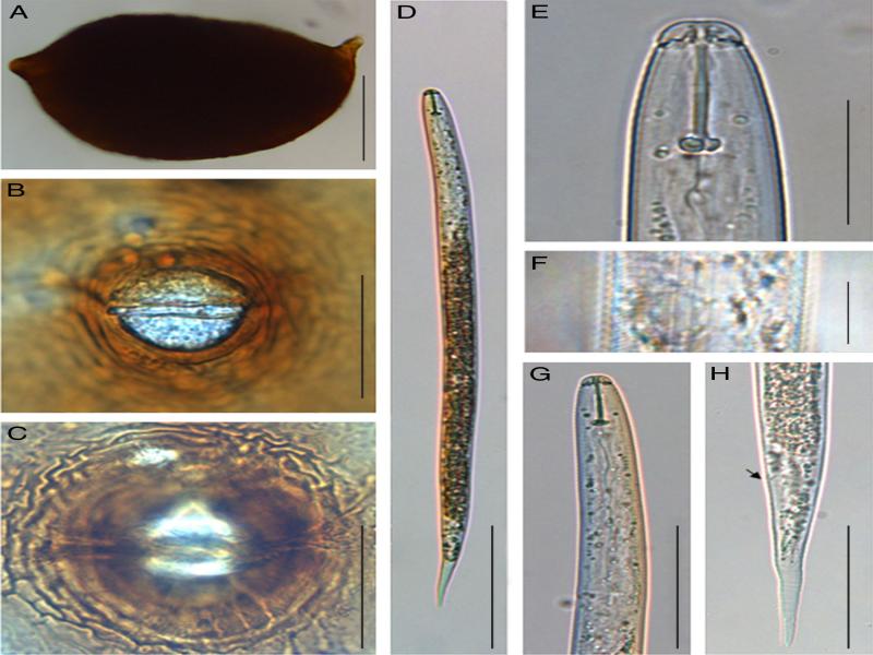
In total, 13 cysts for SG936 and 25 cysts for SG153 were used for full body measurement, vulval cone making, and J2s collection. Full shapes for cysts were captured to measure their length and width.
Vulval cones were cut under a stereomicroscope, transferred to glycerin on slide glasses for microscopic examination. Important characters for distinguishing cyst nematode species such as the underbridge length, presence of bullae, fenestral length, semifenestral width, and vulval slit length were observed under a light microscope, imaged and measured (Sekimoto et al., 2017).
Important taxonomic diagnostic characters for J2s were obtained by temporarily mounting juveniles on slide glasses for examination under a light microscope (DM5000; Leica, Wetzlar, Germany). An automatic camera attached to a LEICA DM5000 compound microscope was used to capture images for measurements. J2s measurements were made for body length, middle body width, stylet length, tail length, hyaline tail length, and excretory pore distance from the anterior part of the body. Measurements were recorded in µm, arithmetic mean, standard deviation, and measurement ranges obtained. Nematodes were identified morphologically based on Heterodera sp. identification key authored by Subbotin et al. (2010).
Molecular characterizations
DNA extraction
To extract DNA, a single cyst was transferred to a glass slide on a small drop of distilled water, opened and its contents crushed using a filter paper chip (2 mm × 2 mm) and forceps. Using forceps, the chip having crushed eggs and J2s was transferred into a PCR tube containing 30 µl lysis buffer (autoclaved triple distilled water, 1 M Tris-HCl, 10% Triton-X 100, 100 µg/ml Proteinase K, 2 M KCl, 1 M MgCl2) for extracting nematode DNA (Iwahori et al., 2000). The tubes were then incubated in a PCR machine (PTC-200; MJ Research, Alameda, CA, USA) at 60°C for 1 hr and 94°C for 10 min. The DNA was stored at −20°C for later use in PCR reactions.
PCR amplification
ITS region
The internal transcribed spacer (ITS) region was amplified using 0.5 µl each of primers TW81 [5′-GTTTCCGTAGGTGAACCTGC-3′] and AB28 [5′-ATTGCTTAAGTTCAGCGGGT-3′] (Joyce et al., 1994). In a PCR tube containing 15 µl of PCR mixture ready to use (Ready-2x-Go with Taqplus; NanohelixTM, Daejeon, Korea) was added 2 µl of nematode DNA extract, 32 µl of triple distilled water, and 0.5 µl of each primer making a total volume of 50 µl. Cycling included pre-denaturation step at 94°C for 5 min followed by 40 cycles of 94°C for 1 min, 57°C for 1 min, and 72°C for 2 min, and finished with one cycle at 72°C for 10 min.
28S large ribosomal subunit D2-D3 expansion segment
Primers D2A [5′-ACAAGTACCGTAGGGAAAGTT-3′] and D3B [5′-TCGGAAGGAACCAGCTACTA-3′] (De Ley et al., 1999) were used to amplify the region. PCR conditions were pre-denaturation at 94°C for 6 min followed by 40 cycles of 94°C for 1 min, 57°C for 1 min, and 72°C for 1 min and finished with one cycle at 72°C for 6 min.
COI gene of the mtDNA
A primer set of JB3 (5′-TTTTTTGGGCATCCTGAGGTTTAT-3′) and JB5 (5′-AGCACCTAAACTTAAAACATAATGAAAATG-3′) (Derycke et al., 2005) was used in the PCR reaction. PCR conditions were pre-denaturation step at 94°C for 4 min followed by 40 cycles of 94°C for 30 sec, 57°C for 30 sec, and 72°C for 45 sec and finished with one cycle at 72°C for 5 min.
PCR products were analyzed by electrophoresis on 1% agarose gel with 1X TAE buffer for 25 min. The gels were visualized using UV transilluminator (UVCI-1100; Major science, New Taipei City, Taiwan).
Phylogenetic analysis
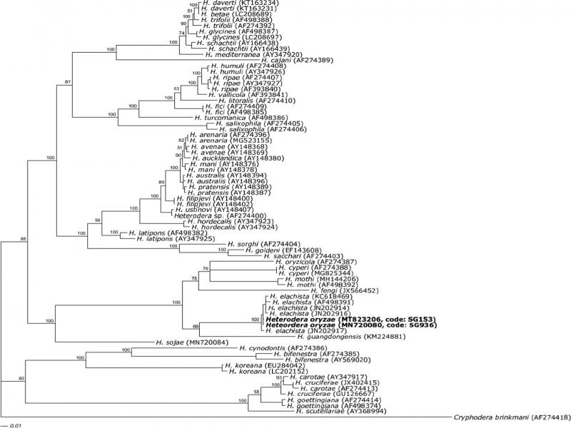
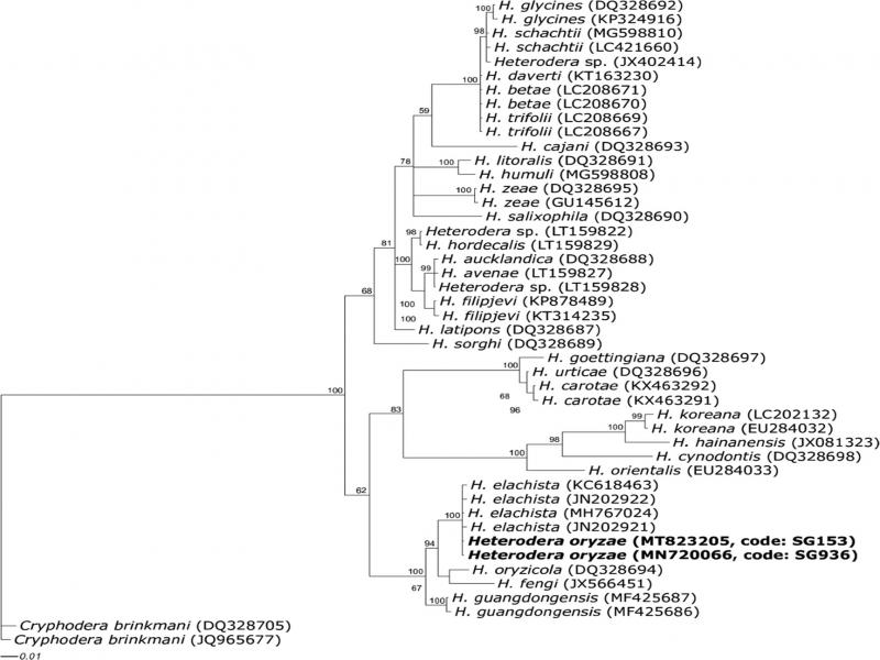
After amplification, PCR products were purified using PCR purification kit following the manufacturer’s manual (NucleoSpin Gel and PCR clean up; Macherey-Nagel, Duren, Germany). Purified DNA was sent for sequence analysis at GenoTech Corporation (Daejeon, Korea). Obtained sequences were edited using EditSeq application to remove low quality bases and assembled in SeqMan computer program. Assembled sequences were posted in National Center for Biotechnology Information (NCBI) to obtain similarity match with other Heterodera species whose sequence nucleotides are already in the Gene Bank. Sequences of ITS rDNA, 28S, and COI genes for Heterodera spp. registered in NCBI were obtained and merged with our sample sequences of ITS rDNA, 28S, and COI, respectively. With the help of previous studies, an outgroup taxon for each data set was obtained. Rotylenchus urmiaensis and Cryphodera brinkmani were used as outgroups for COI, ITS, and 28S data sets, respectively (Madani et al., 2018). The merged sequences were aligned using ClustalX 1.83 program and Bayesian analysis of the sequence datasets performed using MrBayes ver. 3.2.6. The general time reversible substitution model (GTR + I + G) was used for this analysis and ran with four chains for 1 × 10⁶ generations. MCMC (Markov Chain Monte Carlo) method was used to estimate the posterior probability of phylogenetic tree and a consensus tree was generated with a 50% majority rule. The trees were visualized and edited using Dendroscope version 3.5.7 (Daniel and Celine, 2012).
Results
Morphological and morphometrical traits
Morphological characters and morphometric features of J2s (stylet length, body length, tail length, and number of lateral lines) together with cysts and their vulval cone features were examined and measured for species identification. The color of cysts varied from light to dark brown having either oval or lemon shape. Vulval cones were ambifenestrate with bullae and a strong underbridge (Figs. 1, 2). Second-stage juveniles had a strong stylet with an average length of 21 µm. The body length ranged from 414 to 478 µm and 433 to 495 µm for SG936 and SG153, respectively (Table 2). The juveniles for the two samples had three incisors in the lateral field (Figs. 1F, 2F).
Table 1.
Nematode samples used in this study.
| Population code | Collection date | Host plant | Coordinates | ||
|---|---|---|---|---|---|
| SG936 | July 27, 2016 | Rice | 36.902188, 126.661624 | ||
| SG153 | August 16, 2017 | Rice | 36.901005, 126.671008 | ||
| Population | SG936, South Korea, This study | SG153, South Korea, This study | H. oryzae, Côte d’Ivoire, Nobbs et al. (1992) | H. elachista, Japan, Nobbs et al. (1992) | H. elachista, Iran, Tanha Maafi et al. (2004) |
|---|---|---|---|---|---|
| Cysts (n) | 13 | 25 | 50 | 50 | 39 |
| Length (excl. neck) | 498.9 ± 96.4 (307.0-617.2) | 523.3 ± 54.5 (438.9-643.3) | 714.8 ± 71.5 (442-999) | 446 ± 30.3 (328-557) | 431 ± 48 (340-586) |
| Width | 429.3 ± 97.3 (251.2-550.1) | 360.2 ± 47.2 (288.3-447.8) | 426.6 ± 53.8 (229-655) | 322.6 ± 26.0 (229-449) | 311 ± 48 (225-540) |
| Length/width | 1.2 ± 0.1 (1.0-1.4) | 1.5 ± 0.2 (1.3-1.8) | 1.71 ± 0.12 (1.1-2.3) | 1.4 ± 0.1 (1.1-1.9) | 1.4 ± 0.2 (1.1-1.9) |
| Vulval areas (n) | 11 | 16 | 10 | 10 | 10 |
| Fenestral length | 36.5 ± 4.7 (28.2-40.9) | 34.2 ± 4.9 (24.8-43.6) | 38.8 ± 1.85 (32-46) | 29.3 ± 2.0 (23.36) | 40 ± 6.5 (30-50) |
| Semifenestral width | 34.8 ± 4.4 (29.3-44.2) | 35.7 ± 4.4 (25.9-41.0) | 51.6 ± 5.20 (36-69) | 32.0 ± 2.5 (26-39) | 31.2 ± 4.7 (26-42) |
| Vulval slit length | 33.1 ± 5.0 (27.6-45.6) | 34.3 ± 3.8 (27.8-41.3) | 50.7 ± 4.0 (41-65) | 37.5 ± 2.8 (26-46) | 40 ± 3.8 (32-45) |
| Vulval bridge width | 9.0 ± 4.5 (4.4-17.9) | 7.2 ± 3.3 (4.0-15.0) | 12.14 ± 1.9 (6-16) | 5.8 ± 2.0 (2-16) | – |
| Underbridge length | 80.1 ± 7.2 (70.8-96.4) | 83.0 ± 7.9 (63.4-95.3) | 112.5 ± 9.0 (87-153) | 72.0 ± 2.9 (65-78) | 85 |
| Vulva to anus distance | 34.9 ± 3.0 (30.5-41.6) | 36.6 ± 5.9 (30.1-50.3) | – | – | – |
| J2 (n) | 11 | 11 | 20 | 20 | 10 |
| L | 450.6 ± 16.6 (414.7-478.6) | 472.4 ± 19.3 (433.1-495.1) | 554 ± 9.4 (523-589) | 402 ± 10.0 (377-450) | 391 ± 11 (372-410) |
| a | 23.4 ± 0.7 (22.4-24.6) | 23.3 ± 1.6 (20.1-25.9) | 34.0 ± 1.03 (30.1-39.3) | 24.6 ± 0.9 (21.3-28.1) | 22.5 ± 1.0 (20.8-24) |
| b | – | – | |||
| c | 6.8 ± 0.3 (6.3-7.4) | 6.8 ± 0.3 (6.3-7.2) | 9.4 ± 0.3 (8.6-10.7) | 7.0 ± 0.4 (5.4-8.4) | 6.7 ± 0.3 (5.9-7.2) |
| Stylet (S) | 21.1 ± 0.5 (20.4-22.4) | 21.1 ± 0.8 (19.5-22.6) | 20.5 ± 1.15 (16-25) | 19.2 ± 0.8 (16-21) | 20 ± 0.7 (18-21) |
| Labial region height | 4.7 ± 0.5 (3.8-5.4) | 4.7 ± 0.3 (4.3-5.3) | – | – | 3 |
| Labial region diam. | 9.0 ± 0.7 (7.8-10.1) | 9.1 ± 0.3 (8.7-9.6) | – | – | 7.8 ± 0.4 (7-8) |
| DGO | 7.3 ± 0.7 (6.5-8.4) | 6.0 ± 0.8 (4.9-7.7) | – | – | 5.2 ± 0.8 (4-7) |
| Anterior end to | |||||
| Excretory pore | 97.5 ± 4.8 (87.5-103.0) | 99.3 ± 6.6 (84.8-110.8) | – | – | 84 ± 2.8 (79-88) |
| Median bulb valve (MB) | 65.4 ± 2.7 (62.1-72.6) | 64.4 ± 2.2 (60.6-68.0) | – | – | 59 ± 3.6 (52-67) |
| Body diam. at | |||||
| Mid-body | 19.2 ± 0.8 (18.1-20.7) | 20.4 ± 1.9 (18.2-24.3) | 16.3 ± 0.43 (15-18) | – | 17.4 ± 0.8 (16-19) |
| Anus (BDA) | 12.6 ± 0.9 (11.7-14.5) | 13.3 ± 0.8 (12.0-14.6) | 12.3 ± 0.72 (10-15) | 11.4 ± 0.8 (10-13) | 10.2 ± 0.6 (9-11) |
| Hyaline region (H) | 36.9 ± 3.2 (29.3-41.0) | 35.8 ± 3.4 (31.6-42.4) | 32.8 ± 1.3 (26-44) | – | 32 ± 3.0 (25-39) |
| Tail length (Tl) | 66.9 ± 3.8 (60.5-73.4) | 69.6 ± 4.7 (60.6-78.9) | 58.8 ± 2.25 (49-66) | 57.8 ± 2.9 (47-70) | – |
| H/S | 1.7 ± 0.1 (1.4-1.9) | 1.7 ± 0.2 (1.4-2.0) | 1.6 ± 0.11 (1.4-2.2) | – | 1.6 ± 0.1 (1.3-1.9) |
| L/MB | 6.9 ± 0.2 (6.6-7.2) | 7.3 ± 0.3 (6.9-8.1) | – | – | 6.7 ± 0.4 (5.6-7.4) |
| Tl/H | 1.8 ± 0.2 (1.6-2.5) | 2.0 ± 0.1 (1.7-2.2) | – | – | – |