Root-knot nematodes (RKN; Meloidogyne spp.) cause economic damage to a wide range of economically important open field and greenhouse vegetable crops and are considered one of the most damaging agricultural pests worldwide. The genus Meloidogyne has an extremely broad host range of over 2000 plant species, which can cause significant yield losses (Chitwood, 2003; Bleve-Zacheo et al., 2007).
The control of nematodes has become increasingly difficult due to many reasons. Many nematicides and soil fumigants have been removed from the market due to their toxicity and their harmful impact on the environment. An example is methyl bromide, a common fumigant that has been banned since 2005 (Karpouzas et al., 2007). The control against nematodes is further complicated by the lack of inherent resistant mechanism in most vegetable against nematode invasion or the cost-ineffective aspects of preventative approaches such as the long-term crop rotations or the scarcity of biological control agents in the market. As such, there is a great need for new alternatives (Sikora and Fernandez, 2005; Davies and Spiegel, 2011; Giannakou, 2011). In face of the recent European Union (EU) environmental restrictions, it is necessary to develop more ecologically rational alternative systems, like biological control and natural products (Faria et al., 2016).
In the actual context of research and development, a large number of essential oils and their constituents have been studied against a number of pests. In particular, terpenes, which are the primary volatile constituents of essential oils (EO), are among the most promising compounds for biorational pest control (Isman, 2000; Shaaya and Rafaeli, 2007; Sousa et al., 2015). One of these terpenes is eugenol (C10H12O2; 4-allyl-2-methoxyphenol) (Fig. 1), a volatile phenolic constituent and a flavoring agent of great economic importance in the food and cosmetic industries, mainly obtained from Syzygium aromaticum (commonly known as clove) (Chatterjee and Bhattacharjee, 2015).
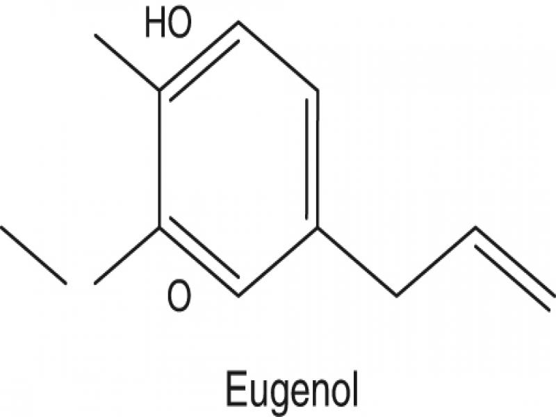
Figure 1:
Molecular structure of eugenol.
Eugenol has shown interesting biological activities, such as antimicrobial (Marchese et al., 2017), anti-inflammatory (Daniel et al., 2009), antioxidant (Ogata et al., 2000), antibacterial (Da Silva et al., 2018), insecticidal (Park et al., 2000), and nematicidal (Tsao and Yu, 2000; Ntalli et al., 2010) among others. Previous lines of research have shown that eugenol at concentration of 660 µg/ml was nematotoxic to Meloidogyne incognita (Leela and Ramana, 2000). Ntalli et al. (2011) reported that mixtures of terpenes have significant synergistic effect against Meloidogyne incognita which cause paralysis. Nematostatic activity was evaluated to show efficacy of nematicides against root-knot nematodes (McGarvey et al., 1984).
The objectives of the present study were to determine (i) the nematicidal and nematostatic activity of eugenol on second-stage juveniles (J2) of M. javanica, (ii) the differentiation of eugenol inhibition on undeveloped eggs, (iii) the hatch inhibition activity of eugenol in egg masses, (iv) the effect of contact and vapor activity of eugenol on M. javanica infectivity, and (v) the effect of sublethal doses of eugenol on M. javanica infectivity.
Materials and methods
Nematode cultures
A population of M. javanica was reared on tomato (Solanum lycopersicum L.) cv. Belladonna in a greenhouse in Agricultural University of Athens, Greece and all seedlings were maintained in a greenhouse (25 ± 2°C, 16 hr photoperiod), in plastic pots (9 cm depth and 8 cm diameter) containing peat-based compost. The seedlings used for inoculations were 6 weeks old, at the four leaf stage. After 40 days, the inoculated plants were uprooted and the galled roots with mature egg masses were gently washed free of soil and cut into 2 cm pieces. Eggs of M. javanica were extracted with 1% sodium hypochlorite solution (Hussey and Barker, 1973). Second-stage juveniles (J2) were obtained by placing eggs on a Baermann funnel at the ambient temperature (27 ± 1°C). Freshly hatched J2 were obtained every 2 days and used in the experiments.
Viability bioassays (nematicidal activity)
Solutions of eugenol were tested for J2 motility at the doses of 62.5, 125, 250, 500 and 1,000 ppm. Eugenol (Merck; Germany) was dissolved in ethanol (Sigma-Aldrich, Italy) and serially diluted in distilled water containing Tween-20 to produce test solutions of the above doses. In all cases, final terpene solutions were prepared containing double the test concentration. Eugenol solution (0.5 ml) was pipetted into each well of a Cellstar® 24-well plate together with J2 suspension (0.5 ml containing approximately 40 J2) at a ratio of 1:1 (v/v), to produce the desired concentration of J2-terpene suspension. Tween-20 is a polysorbate nonionic surfactant and is used as an emulsifying agent for the preparation of stable oil-in-water emulsions. Final concentrations of ethanol and Tween-20 never exceeded 1 and 0.3%, respectively. Second-stage juveniles exposed to these concentrations of ethanol and Tween-20 were not affected, as preliminary tests and previous work indicate (Ntalli et al., 2010). As controls we used either distilled water or water with ethanol and Tween-20 at concentrations identical to those used in the treatment wells. All treated and control plates were covered with a lid to diminish terpene volatilization and incubated at 26 ± 1°C. Juveniles were observed with the aid of an inverted microscope (Zeiss, Germany) at 100 × magnification after 12, 24, 48, and 96 hr and were ranked into two distinct categories: motile or dead. For a window of 10 sec, we checked the juveniles for motility by probing them with a needle. Lack of movement was considered as a strong indication of juveniles’ death. The experiment was conducted twice and all treatments were replicated five times.
Nematode motility bioassays (nematostatic activity)
Eugenol was dissolved in ethanol and diluted serially in distilled water containing Tween-20 resulting in solutions at the doses of 62.5, 125, 250, 500, and 1,000 ppm. In total, 50 ml of each test solution was placed in 250-ml Erlenmeyer flask and then newly hatched J2 were added. In all cases, working solutions were prepared containing double the test concentration and then mixed in flasks at a ratio of 1:1 (v/v) with a 50 ml suspension containing approximately 1,200 J2. A plastic tube connected to an air pump for oxygen supply was inserted into each flask. This was necessary to avoid lack of oxygen since each flask contained a high number of nematodes. Evaporation was avoided by covering the flasks with cotton plug and incubating at 26 ± 1°C temperature settings in dark. Solutions of water with ethanol (1%) and Tween-20 (0.3%) at the same concentrations as the ones in the treatment flasks as well as distilled water were used as controls.
After 12 hr, two solutions of 5 ml each were removed from every flask and used independently. The first one was divided into five aliquots of 1 ml each and placed into wells (containing approximately 40 J2 per ml/well). Second-stage juveniles were observed under an inverted microscope (100 ×) and ranked as motile or dead, as previously described. The second 5-ml solution was placed on a 38 μm sieve and J2 were rinsed with tap water to remove excess of eugenol. Then J2 were placed in a beaker and 5 aliquots of 1 ml each, containing approximately 35 J2, were placed in wells covered with a lid to avoid evaporation. Motile and dead J2 were counted under an inverted microscope (100 ×) after 12 hr to monitor recovery. The same procedure was repeated after 24, 48, and 96 hr. If any J2 regained motility, the effect was considered as nematostatic. Two solutions of 5 ml each was removed from flasks after 24, 48, and 96 hr and the same procedure, as described above, was followed. The experiment was conducted twice and all treatments were replicated five times.
Effect of eugenol on egg development
Following the procedure described by Hussey and Barker (1973), we used sodium hypochlorite solution to extract Meloidogyne javanica eggs, from infested tomato roots (Solanum lycopersicum cv. Belladona). Eggs suspension was sieved through 53 and 38 μm, rinsed thoroughly with tap water and was collected into a 100 ml beaker. We estimated the number of eggs in the suspension using an inverted microscope (100 ×). Inoculum level was adjusted to 100 eggs per ml and used directly in the bioassay experiment.
The effect of eugenol solutions on the development of eggs at the doses of 62.5, 125, 250, 500, and 1,000 ppm was tested. Eugenol was initially dissolved in ethanol and brought to the desired volume using Tween-20 in water, as previously described. In all cases, working solutions were prepared containing double the test concentration and then mixed in Cellstar® 24-well plates at a ratio of 1:1 (v/v) with suspension of eggs added to each well. Eggs suspension (0.5 ml containing approximately 50 eggs) was pipetted into each well and immediately eugenol solution (0.5 ml) was added. Distilled water and water with ethanol plus Tween-20 at concentrations equivalent to those in the treatment wells, served as the controls. All plates were covered with a lid to avoid evaporation and maintained at 26 ± 1°C. In total, 90% of eggs were undifferentiated at the beginning of the experiment. The number of either eggs having a fully developed juvenile or emerged J2 were counted in each well every 7 days (Tzortzakakis and Trudgill, 2005) using an inverted microscope (100 ×). For monitoring the egg development, eggs were observed on day 0 and were categorized either as differentiated (fully developed juvenile) or undifferentiated (eggs containing only cells). Undifferentiated eggs were considered those with cell division (one, two, or more cells).
The experiment was terminated after three weeks. It was conducted twice and each treatment was replicated four times.
Effect of eugenol on egg hatching from egg masses
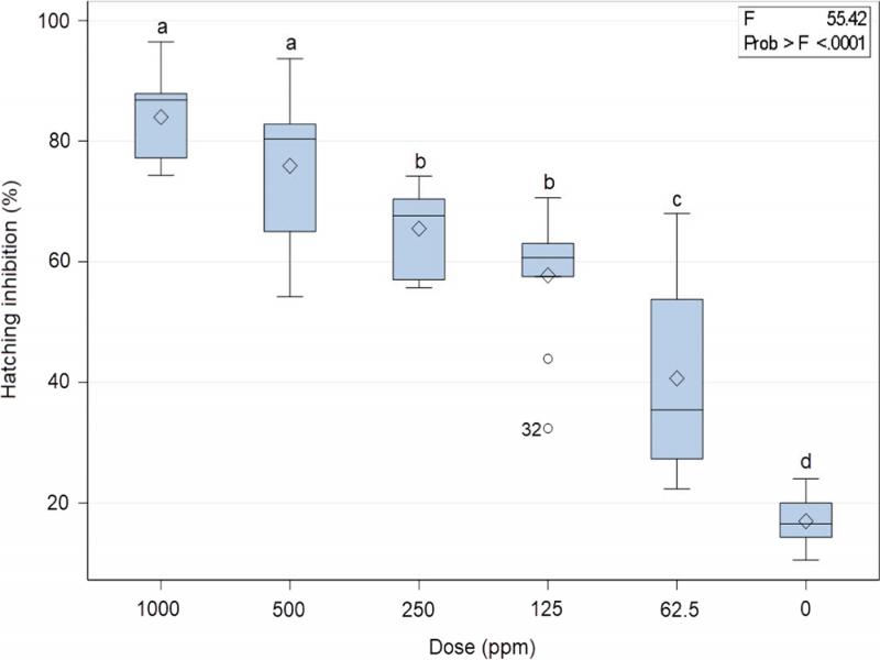
Mature egg masses were handpicked using sterilized forceps, from roots previously rinsed thoroughly, and placed in small plastic extraction trays made by 6 cm Petri dishes (one mature egg mass per extracting tray). Eugenol solutions (62.5, 125, 250, 500, and 1,000 ppm), initially dissolved in ethanol and brought to volume using Tween-20 in water (as described previously), were added to each extracting tray to cover the egg mass (10 ml eugenol solution/extracting tray). Egg masses were maintained for seven days and then test solutions were discarded. Then each egg mass was carefully submerged twice in clean water to remove excess of eugenol and finally was placed in extracting tray filled with clean water. The extracting trays were covered to avoid loss of water and placed in incubator at 26 ± 1°C. Hatched J2 were counted every week, they were discarded and the water was renewed with fresh one. The experiment was terminated after 5 weeks. Then every egg mass was removed from the extracting tray to a drop of water on a glass microscope slide, gently squashed with a coverslip and the number of unhatched eggs per egg mass was counted under an inverted microscope. The experiment was conducted twice and all treatments were replicated five times.
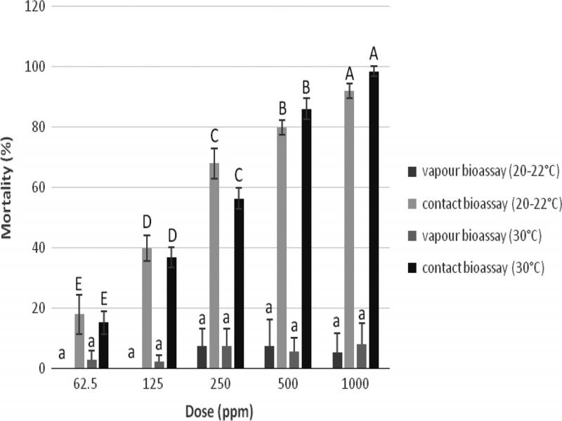
Contact and vapor effect of eugenol against M. javanica
Sandy soil was collected from a field in Gargalianoi village, Messinia, Southern Greece. It was sieved using a 2 mm sieve to separate soil from debris and it was sterilized in an autoclave for 20 min at 100°C. Subsamples were removed to determine the soil moisture (oven drying at 50°C for 24 hr) and maximum water holding capacity (MWHC) (gravimetrical measurement following saturation of the soil with water and allowing to drain for 24 hr) (Pantelelis et al., 2006).
In total, 48 plastic pots (7 cm depth and 5 cm diameter) were filled with 40 g soil each. Every pot was inoculated with a M. javanica suspension containing approximately 500 J2. In half of them, the bottom was replaced with a plastic mesh net (size 1 mm). Subsequently, eugenol solutions were added to the pots with plastic bottom (contact mortality bioassay), while plain water was added to the ones with the mesh net (vapor mortality bioassay). Each pot with a mesh net was placed on top of a pot with a plastic bottom, sealed with parafilm to prevent moisture loss and covered with aluminum foil to prevent light effect (contact-vapor mortality bioassay). The bottom of the netted pot was not in touch with the soil surface of the bottom pot so that nematodes could not migrate from the upper pot to the bottom pot (Fig. 2). The moisture content of the soil never exceeded the 20% of MWHC.
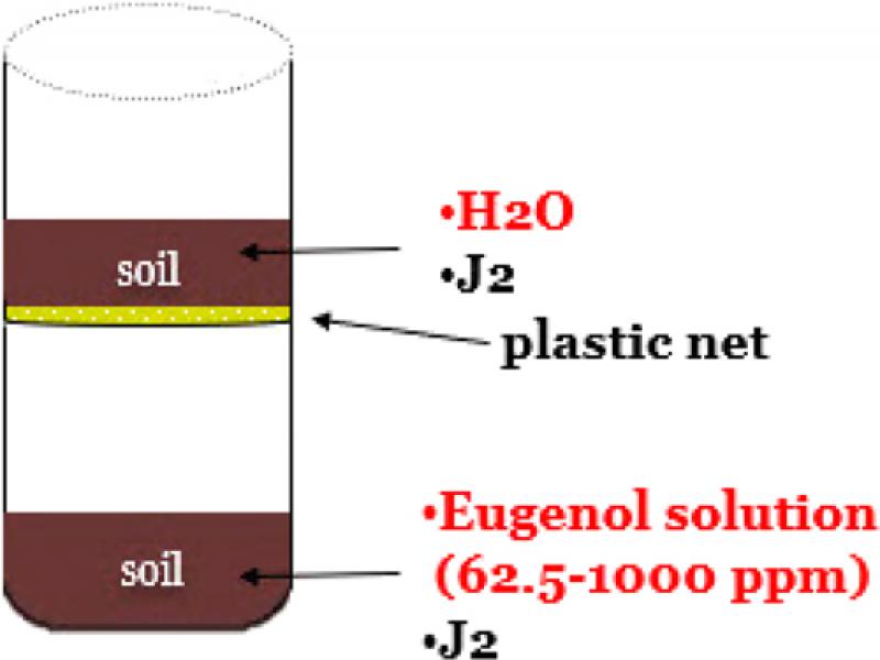
Figure 2:
Pot trial methodology on the contact and vapor effect of eugenol against M. javanica.
The efficacy of eugenol against nematodes was tested at concentrations of 62.5, 125, 250, 500, and 1,000 ppm. Eugenol stock solution was prepared in ethanol and Tween-20 (0.6%) to overcome insolubility, while distilled water with Tween-20 (0.6%) was used for further dilutions according to the above method. Control pots were consisted of soil with J2 only. The above experimental procedure was performed at two levels of temperature (20-22°C and 30°C).
For both temperatures, the plastic pots remained in climate room for three days. For the experiment at the 30°C, the plastic pots were placed on a metal tray with dimensions 60 cm × 40 cm × 8 cm (L × W × H). At the bottom of the metal tray a flexible silicone resistance was placed, connected with an automatic thermostat. Throughout the experiment the tray was filled with wet sand and plastic pots were immersed in the sand. After three days soil from every pot was removed and nematodes were extracted following a modification of Cobb’s decanting and sieving methods (Flegg, 1967) as suggested by Brown and Boag (1988). Juveniles were collected after 2 days and counted with the aid of an inverted microscope at 100 × magnification. The experiment was conducted twice and every treatment was replicated four times. Before statistical analysis, all numbers were calculated according to the Abbott (1925) formula:
Sublethal doses effect of eugenol on juvenile invasion (pot experiment)
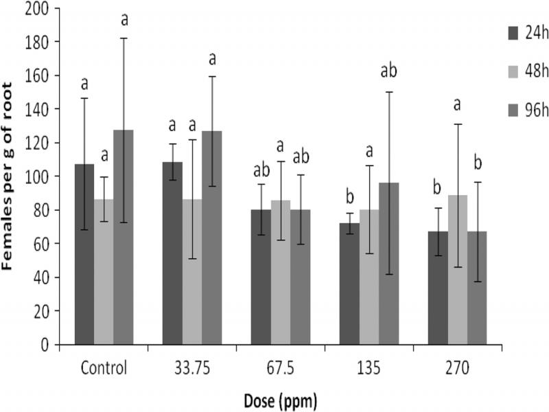
The efficacy of eugenol was evaluated using tomato seedlings cv. Belladona grown in plastic pots (10 cm depth and 6 cm diameter). All seedlings were 6 weeks old and at the four leaf stage. Initially, 15,000 newly hatched J2 were transferred from a graduate cylinder into four 250-ml Erlenmeyer flasks containing a total volume of 100 ml solutions of eugenol with concentration of 33.75, 67.5, 135, and 270 ppm and incubated at 26 ± 1°C. Eugenol was dissolved in ethanol (Sigma-Aldrich, Italy) and serially diluted in distilled water containing Tween-20 to produce test solutions of the above concentrations. Distilled water, as well as a solution of water with ethanol and Tween-20, at concentrations equivalent to those in the treatment flasks, served as control. After 24 hr, 1/3 of the suspension from each flask was placed in a 38 μm sieve and excess of eugenol was removed by washing with tap water. Then, nematodes were transferred to a beaker and four aliquots of 1 ml containing approximately 40 J2 were transferred to a 24-well plate. Second-stage juveniles were scored as motile or dead using an inverted microscope. The remaining J2 were maintained in the flasks for another 48 and 96 hr when the same procedure as previously described was repeated.
In total, 10 ml of a nematode suspension containing approximately 300 motile J2 was used to infect 6-weeks-old tomato plants. All plants were maintained for 30 days in a growth room at 26 ± 2°C. Plants were uprooted and roots were carefully washed free of soil and stained with acid fuchsin solution as described in Byrd et al. (1983). Roots were then washed in water and placed in vials containing equal volumes of glycerol and distilled water. The female nematodes were counted in the whole root system of each plant using a stereoscopic microscope at 12.5 × magnification. The experiment was conducted once in a randomized block design with five replicates per each treatment.
Statistical analysis
All experiments were conducted using the completely randomized design. Data were subjected to one-way analysis of variance (ANOVA) using the General Linear Model (GLM). Treatments means were compared using the LSD test. Statistical analysis in all cases was conducted using SAS statistical package (SAS University Edition). All experiments (except the pot experiment) were conducted twice and they were combined and analyzed together since no variation was revealed between data.
Results
Viability bioassays (nematicidal activity)
The effect of eugenol on J2 motility of M. javanica is presented in Table 1. The percentage of dead J2 increased by increasing the exposure time. At the dose of 1,000 ppm, eugenol showed significant nematicidal activity; more than 99% of J2 were killed after exposure for 12 hr. Furthermore, eugenol gave more than 90% mortality at the 500 ppm dose after 24 hr, while at the same dose 99.8% of J2 were dead after 48 hr exposure. Additionally, approximately 80% of J2 were dead after 96 hr exposure at the dose of 250 ppm, while at doses of 125 and 62.5 ppm no significant effect on juvenile mortality was observed (Table 1). Dead juveniles had a specific shape defined as either straight (I-shape), bent (banana-shape), and L-shape which can separate between dead or motile nematodes.
Table 1.
Effect of eugenol on the motility of Meloidogyne javanica J2 after immersion in test solutions at the doses of 1,000, 500, 250, 125, 62.5, and 0 ppm for 12, 24, 48, and 96 hr.
| Exposure time (hr) | ||||
|---|---|---|---|---|
| 12 | 24 | 48 | 96 | |
| Dose (ppm) | Dead J2 (%) | Dead J2 (%) | Dead J2 (%) | Dead J2 (%) |
| 0 | 0 d | 0.8 d | 1.3 d | 4.7 e |
| 62.5 | 0.4 d | 1.6 d | 2.4 d | 10.7 d |
| 125 | 0.7 d | 2 d | 6.4 c | 19.8 c |
| 250 | 6.5 c | 6.9 c | 24.5 b | 80.8 b |
| 500 | 74.1 b | 92.2 b | 99.8 a | 100 a |
| 1000 | 99.5 a | 100 a | 100 a | 100 a |
| Exposure time (21 days) | |
|---|---|
| Dose (ppm) | Eggs differentiation (%) |
| 0 | 92.1 a |
| 62.5 | 90.6 a |
| 125 | 90.5 a |
| 250 | 82.1 a |
| 500 | 66.2 b |
| 1000 | 21.4 c |