The cysts forming nematodes of the genus Heterodera Schmidt, 1871 (Nematoda: Heteroderidae) are an economically important plant-parasitic nematode (PPN) group with a worldwide distribution and a broad host range causing prominent damages to the host plants ranging from stunted and reduced growth to wilting, chlorosis, and reduced root system (Perry et al., 2018; Sikora et al., 2018). The vermiform second-stage juveniles (J2) of this PPN migrate in the root system of a host plant to feed on the vascular cylinder where they become obese sedentary females; subsequently, following fertilization and egg production, these females turn into protective cysts of more or less lemon shape, housing numerous embryonated eggs. These eggs can remain viable for years inside the cysts, until favorable environmental conditions initiate hatching of the cysts to continue further life cycle (Subbotin et al., 2010; Perry et al., 2018).
Within this genus, 85 nominal species, eight species inquirendae, and a nomen nudum have been listed in a recent update by Handoo and Subbotin (2018). Using morphological and molecular characteristics, the species of this genus have been divided into nine groups, i.e., Afenestrata, Avenae, Bifenestra, Cardiolata, Cyperi, Goettingiana, Humuli, Sacchari, and Schachtii. Morphological characterization of Heterodera species is mainly done based on vulva-slit length, vulval cone fenestration, presence or absence of bullae and underbridge in female cysts, and stylet length, lateral field differentiation, tail length, and hyaline tail length in J2 (Subbotin et al., 2010). Since the last two decades, employing molecular data such as ITS and 28S of ribosomal DNA and COI gene of mitochondrial DNA to characterize Heterodera species has been a common practice, including DNA barcoding, phylogeny, and even phylogeography (Ferris et al., 1999; Toumi et al., 2013a; Subbotin et al., 2017, 2018).
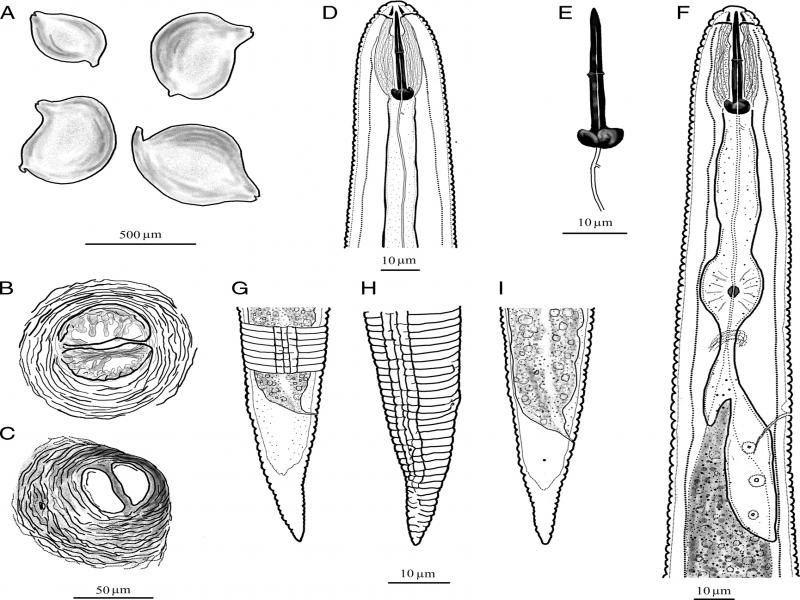
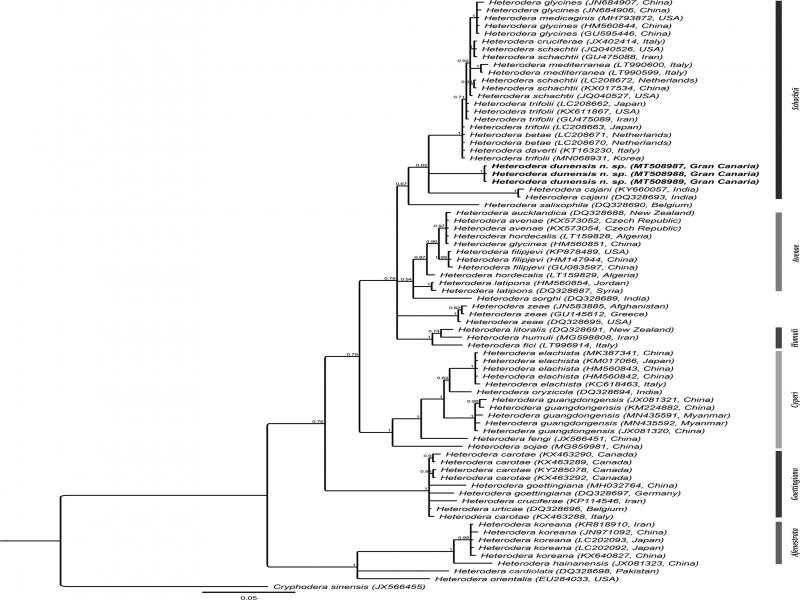
Herein, we characterize Heterodera dunensis n. sp. discovered in a recent exploratory survey of PPN from Canarian coastal dunes of Gran Canaria in May 2019. The species characterization is done based on light microscopy (LM), scanning electron microscopy (SEM), and molecular information of ITS, 18S, and 28S of ribosomal DNA and COI of mitochondrial DNA.
Materials and methods
Nematode extraction and morphological analysis
A sandy soil sample was collected from around the root system of Tetraena fontanesii (Webb & Berthel.) Beier & Thulin, commonly known as Sea Grape or Canarian Bean-Caper. This halophilic succulent plant was growing on a dune (GPS coordinates: 27°44′19.11″ N; 15°35′0.3″ W), about 200 m away from the Maspalomas beach of Gran Canaria. Vermiform J2 was extracted from the sand using the modified Bearmann method (Whitehead and Hemming, 1965) and stored at 4°C during the course of analysis.
For collecting female cysts, sand dried at room temperature was mixed thoroughly in water using a spoon, and after letting the sand settle, floating female cysts were picked out using a fine brush.
Morphological study of J2 was done using both heat-relaxed and fixed specimens. Individual live nematodes were heat-relaxed in a drop of water on a glass slide and examined, photographed, and measured using an Olympus BX51 DIC Microscope (Olympus Optical, Tokyo, Japan), equipped with an Olympus C5060Wz camera and a drawing tube as described in the study of Singh et al. (2018). After recording morphological information, the specimens were recovered from the slide and their genomic DNA was extracted as described in the next section. The remaining J2 juveniles were concentrated in a drop of water in a glass embryo dish, followed by adding a few drops of freshly prepared Trump’s fixative [2% paraformaldehyde, 2.5% glutaraldehyde in 0.1 M Sorenson buffer (sodium phosphate buffer at pH = 7.3)]. The nematodes were then immediately heated in a microwave (700 watts) for about 4 sec, left to rest for 1 hr at room temperature and at 4°C for 24 hr, followed by gradually transferring to anhydrous glycerin to be mounted on glass slides as described in the study of Singh et al. (2018). Vulva cones of female cysts were cut in a drop of water under a stereomicroscope using a blade, and the cones were mounted in glycerol-gelatin (1:1) mix on glass slides. The mounted J2 juveniles and the female vulva cones of the cysts were studied and drawn using the above-mentioned camera- and drawing tube-equipped microscope. Illustrations were improved using Adobe Photoshop CS6 Version 13.0 x 64. For SEM, specimens fixed in Trump’s fixative were washed in 0.1 M phosphate buffer (pH = 7.3), and cysts were additionally sonicated for 8 min to remove any attached dirt, and dehydrated in a graded series of ethanol solutions, critical-point-dried with liquid CO2, mounted on stubs with carbon tabs (double conductive tapes), coated with gold of 25 nm, and photographed with a JSM-840 EM (JEOL) at 12 kV (Singh et al., 2018).
Molecular analysis
Heat-relaxed nematodes after morphological analysis were recovered from temporary slides. Each individual nematode was cut into pieces in distilled water using a blade, and the pieces were transferred to a PCR tube with 20 µl of worm lysis buffer [50 mM KCl, 10 mM Tris at pH = 8.3, 2.5 mM MgCl2, 0.45% NP 40 (Tergitol Sigma), and 0.45% Tween 20]. DNA extraction from cysts containing embryonated eggs was also done for which an individual cyst was crushed and transferred in the PCR tube containing worm lysis buffer as mentioned. The PCR tubes were then incubated at −20°C (10 min) followed by adding 1 µl of proteinase K (1.2 mg/ml), incubation at 65°C (1 hr) and 95°C (10 min), and ending by centrifuging the mixture at 14,000 rpm for 1 min (Singh et al., 2018). PCR amplifications of partial ITS and 18S regions of ribosomal DNA were done using the primer pairs, Vrain2F: 5′-CTTTGTACACACCGCCCGTCGCT-3′/Vrain2R: 5′-TTTCACTCGCCGTTACTAAGGGAATC-3′ (Vrain et al., 1992) and SSU18A: 5′-AAAGATTAAGCCATGCATG-3′/SSU26R: 5′-CATTCTTGGCAAATGCTTTCG-3′ (Mayer et al., 2007) and with thermal profiles described in the study of Singh et al. (2018, 2019). For amplification of D2-D3 expansion segment of 28S of ribosomal DNA, two primer sets were used, the primer pair, 391: 5′-AGCGGAGGAAAAGAAACTAA-3′/501: 5′-TCGGAAGGAACCAGCTACTA-3′ was used as described in the study of Nadler et al. (2006) and D2A: 5′-ACAAGTACCGTGAGGGAAAGTTG-3′/D3B: 5′-TCCTCGGAAGGAACCAGCTACTA-3′ (Nunn, 1992) with the thermal profile from Singh et al. (2019). For the amplification of the COI region of mitochondrial DNA, the primer pair, JB3: 5′-TTTTTTGGGCATCCTGAGGTTTAT-3′/JB4.5: 5′-TAAAGAAAGAACATAATGAAAATG-3′ was used according to Bowles et al. (1992). The PCR products were enzymatically cleaned with alkaline phosphatase (1 U/ml) and exonuclease I (20 U/ml) for 15 min at 37°C followed by 15 min at 80°C and sent for sequencing at Macrogen (https://dna.macrogen.com), and contigs were made from the newly produced forward and backward sequences using Geneious Prime 2020.0.5 (https://www.geneious.com) and deposited in GenBank.
Phylogenetic analysis
The phylogenetic relationships of the new species with other related species were analyzed based on the D2-D3, ITS, 18S, and COI sequences. Phylogenetic programs implemented in Geneious Prime 2020.0.5 were used. The obtained consensus contigs were subjected to BLAST search to check for closely related species on GenBank, and all the collected sequences for each gene fragment were aligned using MUSCLE alignment of Geneious Prime 2020.0.5 using default parameters, followed by manually trimming off of the poorly aligned ends. The best nucleotide substitution model of each gene alignment (see Figures) was determined by jModelTest 2.1.10. Bayesian phylogenetic analysis (MrBayes 3.2.6) was carried out using the selected models, analyses were run under 1 × 10⁶ generations (4 runs), and Markov chains were sampled every 100 generations, and 20% of the converged runs were regarded as burn-in (Huelsenbeck and Ronquist, 2001).
Results
Systematics
Heterodera dunensis n. sp.
Table 1.
Morphometric data of Heterodera dunensis n. sp. from fixed specimens mounted in glycerin, except for cysts that were not fixed.
| Cysts | |||||||||||
| n | 21 | ||||||||||
| Length without neck (L) | 482 ± 75 (316-610) | ||||||||||
| Maximum cyst width (W) | 360 ± 72.1 (241-478) | ||||||||||
| Neck length | 110 ± 31.9 (55-180) | ||||||||||
| L/W | 1.4 ± 0.2 (1.2-1.9) | ||||||||||
| Vulval area | |||||||||||
| n | 13 | ||||||||||
| Fenestral length | 42.1 ± 4.3 (37-51) | ||||||||||
| Mean semifenestral width | 41.8 ± 5.2 (34-53) | ||||||||||
| Vulva bridge width | 8.1 ± 1.6 (5.2-11) | ||||||||||
| Vulva slit length | 52.1 ± 7.3 (35-63) | ||||||||||
| Underbridge length | 63.4 ± 9.8 (44-83) | ||||||||||
| Underbridge width | 18.2 ± 6.2 (8.8-29) | ||||||||||
| Vulva-anus distance | 58.4 ± 5.7 (50-68) | ||||||||||
| Juveniles | |||||||||||
| n | 21 | ||||||||||
| Body length (L) | 471 ± 25.3 (426-520) | ||||||||||
| a = L/MBD | 20.1 ± 1.4 (19-23) | ||||||||||
| b = L/Anterior end to pharynx-intestine junction | 3.6 ± 0.4 (3.1-4.8) | ||||||||||
| b′ = L/Anterior end to the end of pharyngeal gland | 2.7 ± 0.2 (2.4-3.1) | ||||||||||
| c = L/Tail length | 11.6 ± 0.6 (11-12) | ||||||||||
| c′ = Tail length/ABD | 33.6 ± 2.6 (27-39) | ||||||||||
| Stylet length | 29.1 ± 1.1 (27-31) | ||||||||||
| Stylet knob width | 6.4 ± 0.4 (5.9-7) | ||||||||||
| Lip region height | 4.8 ± 0.3 (4.3-5.3) | ||||||||||
| Lip region width | 9.1 ± 0.4 (8.6-10) | ||||||||||
| Dorsal gland opening from stylet base | 3.7 ± 0.4 (3.2-4.4) | ||||||||||
| Anterior end to median valve | 74.2 ± 2.4 (70-79) | ||||||||||
| Anterior end to secretory-excretory pore | 109 ± 3.9 (101-117) | ||||||||||
| Anterior end to pharynx-intestine junction | 131 ± 11.1 (103-155) | ||||||||||
| Anterior end to pharyngeal gland end | 177 ± 9.2 (156-193) | ||||||||||
| Maximum body diameter (MBD) | 23.5 ± 1.4 (21-26) | ||||||||||
| Anal body diameter (ABD) | 14.1 ± 0.8 (13-16) | ||||||||||
| Tail length | 40.7 ± 2.4 (35-45) | ||||||||||
| Hyaline tail length | 20.6 ± 1.9 (16-23) | ||||||||||
| Character | Cyst length (L) | Cyst width (W) | L/W ratio | Fenestral length | Fenestral width | Vulval slit length | J2 body length | J2 stylet length | J2 tail length | J2 tail hyaline length | Hyaline% of the tail |
|---|---|---|---|---|---|---|---|---|---|---|---|
| H. agrostis | 429-800 | 320-541 | 1.2-2.6 | 34-54 | 16-24 | 39-48 | 384-472 | 25-26 | 46-61 | 27-40 | 59-66 |
| H. betae | 830-878 | 455-518 | 1.6-1.9 | 44-55 | 38-43 | 48-57 | 547-607 | 29-31 | 70-74 | 38-42 | 54-57 |
| H. cajani | 448-670 | 209-422 | 1.4-2.1 | 27-65 | 25-40 | 43-55 | 420-519 | 22-27 | 42-52 | 23-31 | 55-60 |
| H. ciceri | 570-930 | 350-550 | 1.6-2.4 | 32-52 | 20-37 | 43-60 | 440-585 | 27-30 | 53-72 | 31-42 | 58-58 |
| H. daverti | 650-749 | 380-491 | 1.4-1.5 | 42-54 | 31-40 | 47-52 | 457-476 | 25-26 | 54-57 | 30-33 | 56-58 |
| H. dunensis n. sp. | 316-610 | 241-478 | 1.2-1.9 | 37-51 | 34-53 | 35-63 | 426-520 | 27-31 | 35-45 | 16-23 | 46-51 |
| H. galeopsidis | 576-797 | 408-556 | 1.4-1.5 | 41-50 | 31-38 | 39-50 | 485-553 | 26-28 | 61-75 | 35-40 | 57-53 |
| H. glycines | 474-709 | 327-535 | 1.3-1.7 | 34-58 | 16-41 | 38-50 | 386-471 | 21-23 | 39-51 | 22-30 | 56-59 |
| H. lespedezae | 678-719 | 371-522 | 1.4-1.8 | 43-59 | 35-41 | 45-47 | 457-481 | 24-25 | 54-56 | 26-30 | 48-54 |
| H. medicaginis | 568-728 | 364-570 | 1.4-1.5 | 39-55 | 30-40 | 39-55 | 417-512 | 24-26 | 41-60 | 22-33 | 54-55 |
| H. mediterranea | 430-690 | 240-570 | 1.2-1.6 | 38-45 | 37-42 | 42-48 | 360-430 | 25-27 | 38-45 | 19-26 | 50-58 |
| H. rosii | 537-1173 | 403-634 | 1.0-1.7 | 48-65 | 40-45 | 45-59 | 430-662 | 27-34 | 58-77 | 37-45 | 64-58 |
| H. schachtii | 768-815 | 512-529 | 1.5-1.6 | 35-38 | 25-31 | 41-44 | 436-489 | 25-26 | 45-49 | 24-27 | 53-55 |
| H. sonchophila | 732-1032 | 381-616 | 1.6 | 37-58 | 29-50 | 42-56 | 437-492 | 24-27 | 47-56 | 26-30 | 55-54 |
| H. spiraeae | 467-861 | 283-566 | 1.1-1.7 | 33-60 | 20-45 | 39-45 | 371-446 | 22-23 | 39-49 | 21-27 | 54-55 |
| H. swarupi | 520-700 | 320-475 | 1.6-2.1 | 45 | 35 | 41 | 400-440 | 21-23 | 39-54 | 20-29 | 51-54 |
| H. trifolii | 608-841 | 341-536 | 1.3-1.8 | 43-53 | 33-44 | 40-53 | 492-613 | 25-28 | 60-72 | 32-37 | 53-51 |