The grass root-knot nematode, Meloidogyne graminis and the Maryland root-knot nematode (M. marylandi), have long been recognized as pathogens on turfgrasses (Sledge and Golden, 1964; Grisham et al., 1974; Jepson and Golden, 1987). Recently, these nematodes have increased in importance as pathogens on ultradwarf bermudagrass (Cynodon dactylon × C. transvaalensis) used on golf greens (Crow, 2018). Non-systematic surveying of root-knot nematodes on turf and forage bermudagrass in Florida (W. T. Crow, unpublished data) have found M. graminis to be the most common species, although M. marylandi has also been reported (Sekora et al., 2012). These same two species are reported as the predominate Meloidogyne spp. associated with golf course bermudagrass in North and South Carolina (Zeng et al., 2012; Ye et al., 2015), Texas (Faske and Starr, 2009), and California and Hawaii (McClure et al., 2012).
A typical turfgrass nematode diagnosis conducted by the University of Florida Nematode Assay Laboratory (NAL) and most other nematode diagnostic laboratories is based on nematodes recovered per volume of soil using centrifugal flotation (Jenkins, 1964) or a similar passive-extraction method. Ideally, turf samples consist of multiple cores collected from the sample area that are mixed to form a composite sample and nematodes are extracted from a standard soil volume of soil. Typically the upper portions of the cores, containing the live turf, thatch and high-organic fraction, are discarded as they add so much debris to the samples that accurate nematode quantification becomes impractical.
During multiple field visits to golf courses with ultradwarf bermudagrass putting greens over the past two decades, chlorotic blotches, often round to oval in shape, that were not associated with known fungal pathogens, were observed by the Sr. author (Fig. 1). These blotches were very similar to those reported on creeping bentgrass greens in the United Kingdom infested with M. minor (Karssen et al., 2004). While Meloidogyne spp. were suspected as the causal agent of this chlorotic symptom, soil nematode assays often recovered very few Meloidogyne J2 although visual inspection of the root systems revealed galls containing many Meloidogyne spp. of sedentary life stages. Further, it was noted that infected roots tend to rot off below the galls causing the root systems to be shallow and sometimes roots growing primarily in the thatch layer with very few penetrating into the soil below (Fig. 2).
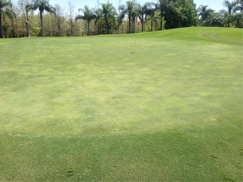
Figure 1:
Symptoms of Meloidogyne graminis on an ultradwarf bermudagrass golf green. Only low numbers of M. graminis vermiform stages were recovered from soil, while staining revealed the roots were infested with large numbers of sedentary stages.
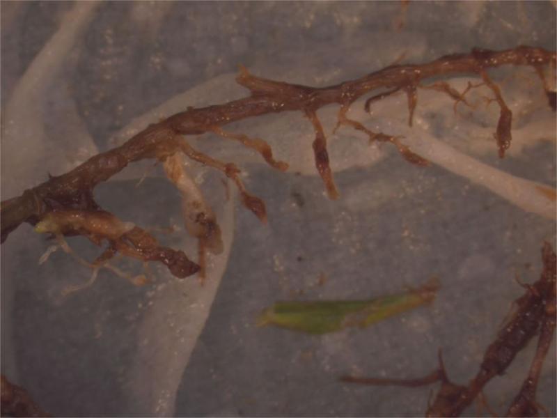
Figure 2:
Golf course bermudagrass roots infested by Meloidogyne graminis and M. marylandi typically rot off just below small galls near the soil-thatch interface, creating an extremely abbreviated root system.
From 2008 through 2015, dozens of turfgrass nematicides trials were conducted at the University of Florida evaluating abamectin for management of sting nematode (Belonolaimus longicaudatus). Abamectin greatly increased turf health in many of these trials, while having no measurable effect on sting nematode (Gu and Crow, 2018). In these trials, close observation revealed the turf roots were severely infested by M. graminis despite finding few, if any, M. graminis J2 in the soil, and those found often were unthrifty in appearance, being burdened by Pasteuria spp. attached to their cuticle or by fungal infection.
Laughlin and Williams (1971) reported that throughout the bermudagrass growing season the majority of M. graminis J2 recovered from soil extractions were found in the upper 5-cm of soil profile. We hypothesized that the majority of M. graminis and M. marylandi activity is confined to the extreme upper soil profile and thatch layer where live roots are found, and those J2 found deeper in the soil are often those that are sick and non-infective. This being the case, soil extraction might not provide the most accurate method of diagnosing this nematode or evaluating nematicide efficacy. In the nematicide trials mentioned above, it was hypothesized that the turf visual improvement observed was likely from impacts on M. graminis inhabiting the upper profile where abamectin accumulates (Gannon et al., 2017) and not from impacts on sting nematode that predominated deeper in the soil profile.
The NAL set out to develop an effective, efficient, and practical method for extracting Meloidogyne spp. vermiform stages for diagnosis on golf course turf, and for evaluation of treatment efficacy in field research. After evaluating several methods, it was determined that a modified Seinhorst (1950) mist method met these criteria. The objective of this research is to compare extraction of Meloidogyne spp. vermiform stages using mist extraction from turf plugs with extraction from soil using centrifugal flotation (Jenkins, 1964).
Materials and methods
Mist chamber
The NAL mist chamber (Fig. 3) was built from 0.63-cm-thick polycarbonite sheets that form a chamber 202-cm-wide, 102-cm-deep, and 89-cm-high. The chamber has hinged doors for entry, and a center shelf 29-cm from the bottom of the chamber to hold funnels and samples. The chamber floor is a galvanized metal tray on a wood frame with a drain in the bottom. In total, 40 10-cm-diam. holes were cut into the center shelf at 19-cm centers to hold funnels, and there are four rows with 10 funnel holes each. A 0.63-cm-high ring of 10-cm-diam. PVC pipe was attached to the surface of each hole and sealed with waterproof caulking to prevent water from the shelf from running down the outside of the funnels (Fig. 4). Drain holes (0.63-cm-diam.) were drilled into the shelf to prevent water accumulation on the shelf. Misting rails are 1.27-cm-diam. PVC pipes attached to the ceiling of the chamber running length ways and placed immediately above the spaces between every second funnel row so that drips do not fall into funnels (Fig. 5). Superfine, no drip, low flow mist emitters (DripWorks, Willits CA) were fitted directly into the PVC rails immediately above the gap between each second and third funnel, and at both funnel row ends.
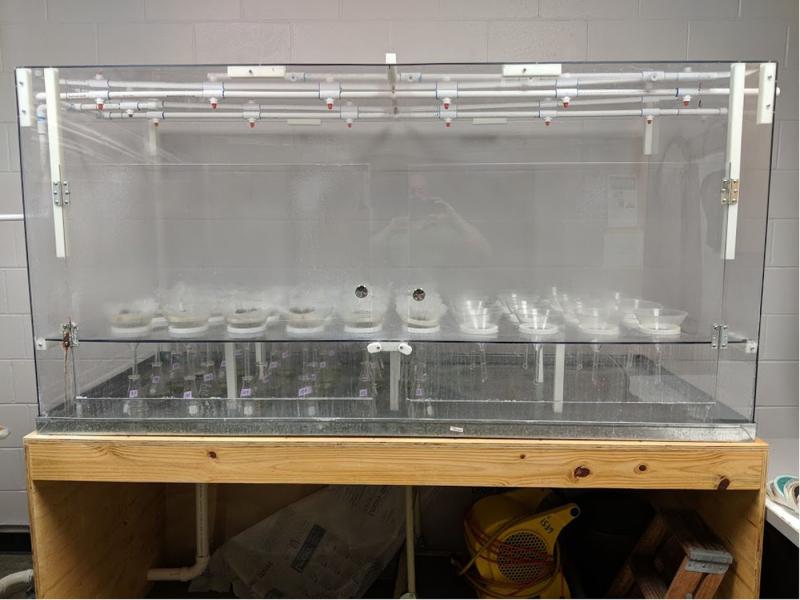
Figure 3:
The University of Florida Nematode Assay Lab mist chamber.
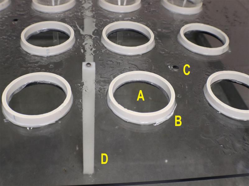
Figure 4:
(A) Funnel hole cut in center shelf. (B) Water flow prevention ring. (C) Drain hole. (D) Shelf support.
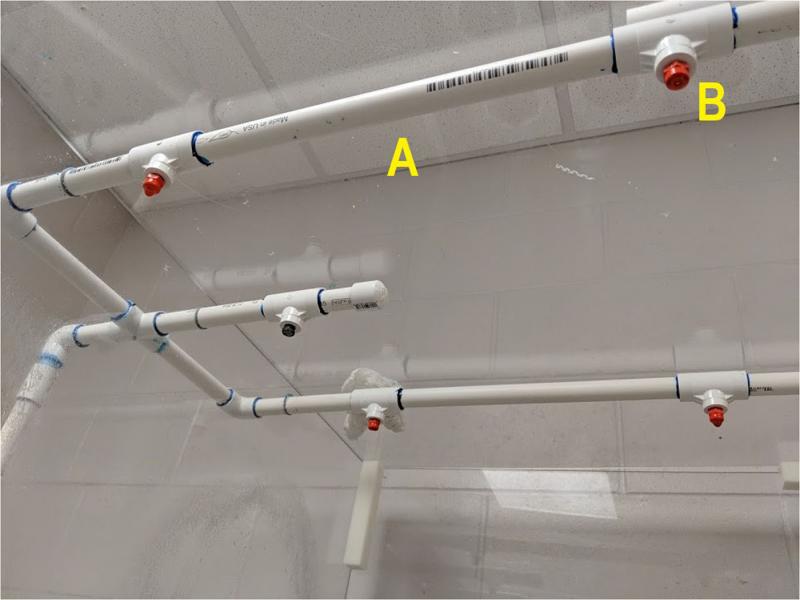
Figure 5:
(A) Misting rail. (B) Superfine mist emitter.
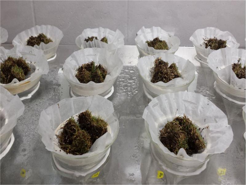
Glass funnels (15-cm-diam. at top) with 15-cm stems were placed into the funnel holes and 250 ml flasks were inserted under the funnel spouts to collect the emerging nematodes. The stem of the funnels inserted slightly into the flask openings. The water supply was circulated through a sediment filter to remove particulate debris and then into an Autopilot 24-hr recycling timer (Hydrofarm, Petaluma, CA), that regulated the mist length and interval. Sample support rings made of windowscreen glued to 0.63-cm-high and 10-cm-diam. ring, were placed into funnels (Fig. 6), and then a coffee filter was placed onto each screen to hold the sample. Samples were placed directly onto the coffee filter (Fig. 7). Vermiform nematodes emerging from plant material migrated through the filter and then moved via water flow into the flask below. The mist timer was set to apply mist for 10 sec each hour.
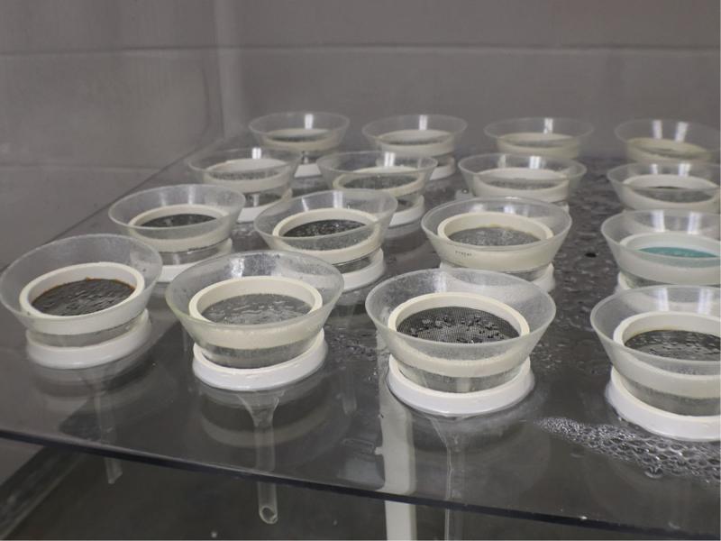
Figure 6:
Funnels with sample support rings.
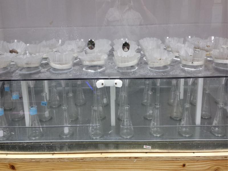
Figure 7:
Samples are placed onto coffee filters on the sample support rings. Funnel necks insert slightly into the 250-ml-flaskes.
Field plots
Nematode samples for both soil and mist extraction were collected from multiple golf course nematicide trials over multiple years in multiple locations. In all the trial locations, the root-knot nematodes were speciated using mitochondrial DNA haplotyping (Baidoo et al., 2016). The predominant Meloidogyne sp. infesting the sites was M. graminis, except for the ‘Sunday’ bermudagrass site (Table 1) where it was M. marylandi. In this paper only data from the initial, pre-nematicide application, sampling were used. All the trials were conducted on bermudagrass putting greens of varying cultivars and number of plots. Plots were 1.5 m2 with 0.61-m borders between adjacent plots. Four cores 6.3-cm-deep were collected from each plot using a 3.8-cm-diam. coring device (Duich Ball Mark Plugger; Turf-Tec International, Tallahassee, FL). Soil was removed from the turf plugs and used for soil extraction, and the plugs, composed of live turf, thatch and roots, were gently washed and used for mist extraction (Fig. 8).
Table 1.
Comparison of the number of Meloidogyne spp. J2 and males recovered from either 100 cm3 of soil using centrifugal flotation or from four 3.8-cm-diam. turf plugs using mist extraction.
| J2 | Males | |||||||||
|---|---|---|---|---|---|---|---|---|---|---|
| Year | Cultivar | n | Soil | Mist | P < | r 2 | Soil | Mist | P < | R 2 |
| 2017 | Jones Dwarf | 77a | 6b | 436 | 0.0001c | 0.014d | 1 | 40 | 0.0001 | 0.059 |
| 2017 | Tifdwarf | 49 | 225 | 945 | 0.0001 | 0.003 | 100 | 184 | 0.0001 | 0.085 |
| 2017 | Tifdwarf | 45 | 92 | 387 | 0.0001 | 0.002 | 21 | 92 | 0.0001 | 0.150 |
| 2017 | TifEagle | 59 | 69 | 368 | 0.0001 | 0.048 | 2 | 25 | 0.0001 | 0.059 |
| 2018 | Tifdwarf | 40 | 130 | 466 | 0.0001 | 0.005 | 28 | 129 | 0.0001 | 0.000 |
| 2019 | TifEagle | 71 | 70 | 723 | 0.0001 | 0.332 | 22 | 108 | 0.0001 | 0.122 |
| 2019 | TifEagle | 100 | 24 | 128 | 0.0001 | 0.604 | 2 | 13 | 0.0001 | 0.374 |
| 2019 | Sunday | 120 | 14 | 81 | 0.0001 | 0.202 | <1 | <1 | 0.0001 | 0.003 |
| 2020 | Jones Dwarf | 35 | 22 | 286 | 0.0001 | 0.560 | 5 | 26 | 0.0001 | 0.244 |