Nematodes of the genus Pelodera (Schneider, 1866) are widespread, mainly occurring as free-living saprophytes, which are usually found in the biological cycle in organic substances (Sudhaus and Schulte, 1988; Georgi and Georgi, 1991; Saari and Nikander, 2006). Some are facultative, opportunistic parasites, while others show a period of obligate parasitism in their life cycles. Several Pelodera species are also associated with termites (Carta et al., 2010) and rodents (Sudhaus et al., 1987; Casanova et al., 1996), but often are also reported as parasites of domestic animals and birds (Saari and Nikander, 2006). In Sudhaus’s (2011) revision of Rhabditidae, Pelodera is considered a genus comprising three major monophyletic groups the ‘strongyloides’, the ‘coarctata’and the‘teres’ groups. The ‘strongyloides’ group consists of six morphologically similar species associated with vertebrate and invertebrate hosts: the parasites P. nidicolis (Sudhaus and Schulte, 1986), P. orbitalis (Sudhaus and Schulte, 1986), P. cutanea (Sudhaus et al., 1987), P. strongyloides (Schneider, 1860; Sudhaus and Schulte, 1988), P. termitis (Carta et al., 2010), and P. scrofulata (Tahseen et al., 2014). Of particular interest is P. strongyloides, a small saprophytic nematode that usually reproduces on decaying organic matter, but there is a strain named P. strongyloides dermatitica (Talvik et al., 2005), the third-stage larvae of which are able to invade the skin of several animals causing dermatitis in dogs, sheeps, and humans (Sudhaus and Schulte, 1988; Georgi and Georgi, 1991; Tanaka et al., 2004; Saari and Nikander, 2006).
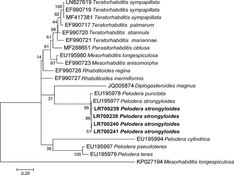
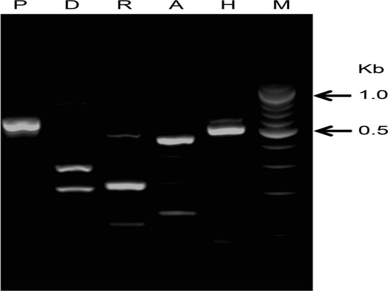
During a nematode survey in Georgia, a nematode species was recovered from the body of a dead individual of Lucanus ibericus (Coleoptera: Lucanidae) and identified morphologically as P. strongyloides. Considering the potential impact of P. strongyloides on human and animal health, accurate identification of this nematode species is necessary to provide proper management strategies. In the present study, a Georgian population of P. strongyloides recovered for the first time from L. ibericus and was fully characterized by morphology and morphometrics. Furthermore, the PCR amplification and sequencing of the D2 to D3 expansion domains of the 28S rRNA gene, the ITS, and the mitochondrial COI were carried out. The phylogenetic relationships of P. strongyloides from Georgia to other Pelodera species and Rhabditidae were also reconstructed.
Materials and methods
Nematode detection and isolation
Pelodera strongyloides was isolated in 2016 in the municipality of Borjomi, in the territory of the Borjomi-Kharagauli reserve, from the body of a single dead beetle belonging to L. ibericus (Coleoptera: Lucanidae). The beetle species was identified using morphological and morphometric criteria by entomologist of the Institute of Zoology of Ilia State University, Prof. Georgy Chaladze. Nematodes of different stages (L1 through adult), including females and males, were localized in the lipid tissue of the beetle organism. Insect cadaver with nematodes was placed on White trap (Kaya and Stock, 1997) and the emerging juveniles (J) were harvested and stored in 50 ml plastic tubes filled with wet polyurethane sponge at 4 to 6°C.
To obtain an isogenic population of this nematode species, one mature female and male were placed on water traps in the feeding area containing crushed Galleria mellonella L. (Lepidoptera: Pyralidae), Tenebrio molitor L. (Coleoptera: Tenebrionidae), and in agar peptonic plates. After 3 to 4 days, nematodes multiplied in the nutrient medium and nematodes at different stages, including females and males, migrated into water solution. As beetle-derived nutrition appeared to become scarce, the dauer larvae emerged and migrated in the water suspension. Extracted nematodes were morphologically and morphometrically studied in Tbilisi, in the laboratory of entomopathogens of the Institute of Zoology at Ilia State University (Georgia), whereas molecular analysis was carried out at the Institute for Sustainable Plant Protection in Bari, Italy.
Nematode pathogenicity
Nematode pathogenicity of P. strongyloides was evaluated in comparison to Steinernema carpocapsae (Weiser, 1955) Wouts, Mráček, Gerdin, and Bedding, 1982, using last instar larvae of G. mellonella. Four Petri plates with 15 G. mellonella (last instar lavae) each were used for the tests. A distilled water suspension of 3 ml containing about 100 specimens/1 insect larva was inoculated into each Petri plate. Only distilled water was used as a positive control. Petri plates were incubated at 24°C in darkness and larval mortality checked at 3, 5, and 7 days after inoculation. Only 3.3% of the tested waxworm larvae were killed using P. strongyloides after 7 days of inoculation, whereas on day 5, 100% mortality of G. mellonella was observed with S. carpocapsae.
Light microscopy and morphometrics
Measurements were conducted using a light biological research microscope (Motic®-DMB1). Nematodes were measured with an ocular micrometer and illustrated using a drawing (RA-4) tube.
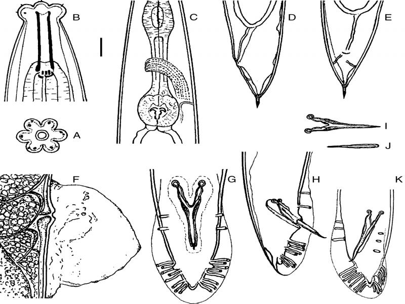
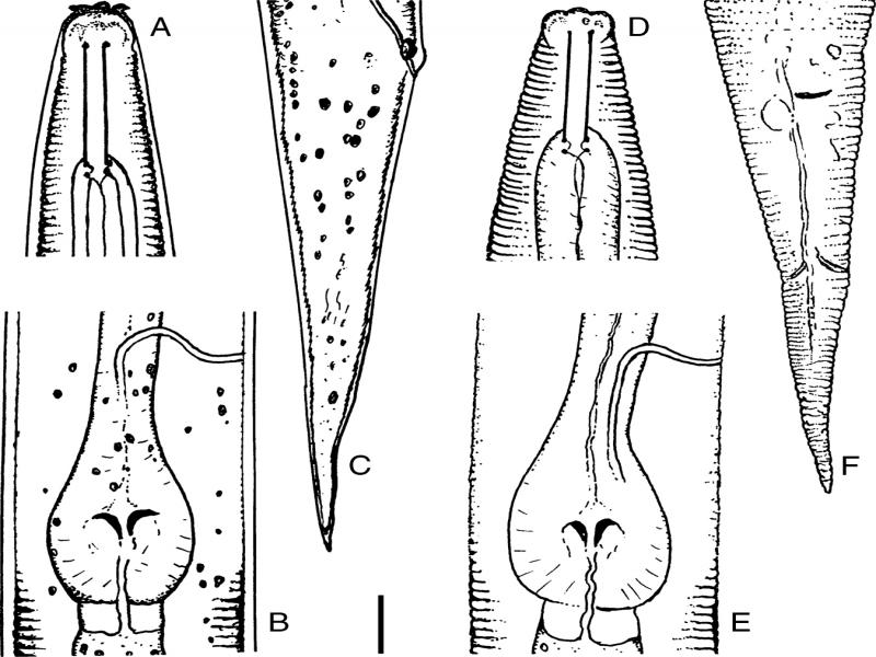
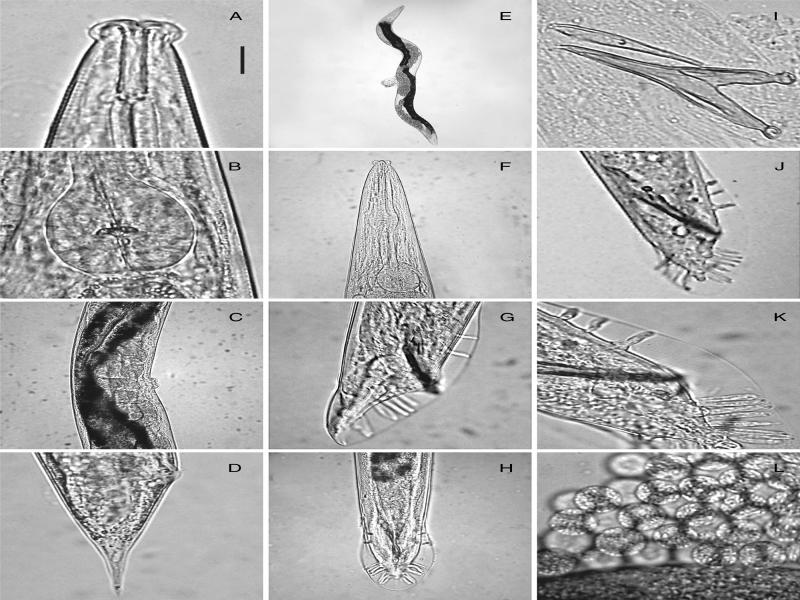
Drawings and photomicrographs of nematodes fixed on glass slides were taken using a digital video camera Genius (G-Shot) DV 1110 (Figs. 1-3). In total, 20 specimens from each stage (adults and 22 specimens of dauer juveniles) were randomly collected from T. molitor larvae. Nematodes were examined live or heat relaxed in Ringer’s solution at 60°C (Filipjev, 1934).
Nematodes were fixed in triethanolamine formalin (TAF) (Courtney et al., 1955) and processed to anhydrous glycerol for mounting (Seinhorst, 1959). Specimens were mounted on glass slides, and the coverslip was supported by glass rods to avoid flattening. Morphometric characters were selected according to Hominick et al. (1997) and reported in Table 1.
Table 1.
Ratios and measurements (in µm) of heat relaxed specimens of Pelodera strongyloides collected from Lucanus ibericus in Georgia.
| Character | Females | Males | 3rd juvenile stage | P. strongyloides (Schneider, 1866) | |||||||||||||||||
|---|---|---|---|---|---|---|---|---|---|---|---|---|---|---|---|---|---|---|---|---|---|
| n | 20 | 20 | 22 | 11 | |||||||||||||||||
| Body length | 1566 ± 120 (1353-1867) | 1280 ± 100 (1080-1494) | 663 ± 51 (566-772) | 1629 (1142-2149) | |||||||||||||||||
| Body width | 96 ± 10 (75-112 | 62 ± 4.8 (55-70) | 30 ± 2.4 (25-35) | 107 (80-132) | |||||||||||||||||
| Lip region diam | 19 ± 1.6 (16-22 | 17 ± 1.5 (15-20) | – | – | |||||||||||||||||
| Stoma length | 31 ± 1 (30-32) | 29 ± 2.2 (25-32) | 24 ± 1.7 (22-30) | 33 (30-38) | |||||||||||||||||
| Stoma diam | 5.1 ± 0.4 (5.0-6.2) | 4.7 ± 0.4 (3.7-5.0) | 2.6 ± 0.4 (2.5-3.7) | – | |||||||||||||||||
| Anterior end to nerve ring | 178 ± 6 (172-197) | 155 ± 10 (137-175) | 102 ± 6 (92-120) | – | |||||||||||||||||
| Stoma length as % pharynx length | 12.2 ± 0.4 (11-13) | 13 ± 1.2 (11-15) | 16 ± 0.8 (14-17) | – | |||||||||||||||||
| Pharynx length | 252 ± 5 (237-257) | 219 ± 17 (192-250) | 152 ± 6 (142-177) | – | |||||||||||||||||
| Corpus length | 144 ± 7 (135-162) | 122 ± 10 (107-137) | 87 ± 2 (80-92) | – | |||||||||||||||||
| Corpus as % pharynx length | 56 ± 2.3 (53-63) | 55 ± 2 (50-59) | 57 ± 2.5 (52-61) | – | |||||||||||||||||
| Median bulb (MB) diam | 27 ± 2.1 (22-30) | 30 ± 4.9 (20-37) | 11 ± 0.8 (10-12) | – | |||||||||||||||||
| Terminal bulb (TB) diam | 39 ± 4.1 (32-47) | 34 ± 2.7 (32-40) | 15 ± 0.6 (12-17) | 36-50 | |||||||||||||||||
| MB diam. as % TB diam | 77 ± 9.5 (54-88) | 79 ± 6.8 (62-93) | – | – | |||||||||||||||||
| Anterior end to excretory pore | 236 ± 14 (212-270) | 205 ± 13 (164-227) | 130 ± 10 (110-150) | 235 (183-295) | |||||||||||||||||
| Ex. pore posit. as % phar. length | 93 ± 5 (83-105) | 93 ± 8 (75-109) | 85 ± 6.7 (75-98) | – | |||||||||||||||||
| Tail length | 69 ± 11 (52-95) | 53 ± 5 (45-62) | 72 ± 4 (65-82) | – | |||||||||||||||||
| Anal (cloacal) body width (ABW) | 40 ± 3 (32-47) | 37 ± 3 (32-42) | 19 ± 1.8 (16-22) | – | |||||||||||||||||
| Gonad lengtha | 1285 ± 198 (995-1658) | 956 ± 87 (747-1268) | – | 890 (653-1157) | |||||||||||||||||
| Gonad length as % body length | 82 ± 15 (61-107) | 72 ± 5 (66-82) | – | 46-63 | |||||||||||||||||
| Gonad length. as % intestine leng.b | 108 ± 23 (72-143) | 93 ± 11 (69-110 ) | – | – | |||||||||||||||||
| Anterior gonad branch length | 800 ± 104 (555-928) | – | – | – | |||||||||||||||||
| Posterior branch length | 546 ± 128 (290-794) | – | – | – | |||||||||||||||||
| Testis flexure length | – | 201 ± 49 (123-260) | – | – | |||||||||||||||||
| Ant. flexure as % of ant. branch | 33 ± 3 (26-42) | 17 ± 4 (13-24) | – | – | |||||||||||||||||
| Post. flex. as % of post. branch | 27 ± 6 (13-34) | – | – | – | |||||||||||||||||
| Sperm diameter | n.d. | 5 ± 0.6 3. (7-7.5) | – | – | |||||||||||||||||
| Egg length | 60 ± 3.4 (55-67) | – | – | – | |||||||||||||||||
| Egg diam | 41 ± 4 (35-47) | – | – | – | |||||||||||||||||
| Rectum length | 38 ± 3.2 (30-42) | n.d. | 24 ± 1.5 (22-25) | – | |||||||||||||||||
| Rectum length/ABW | 0.9 ± 0.1 (0.8-1.3) | n.d. | 0.9 ± 0.1 (7-1.2) | – | |||||||||||||||||
| Anus to phasmid distance | 23 ± 1.8 (20-27) | n.d. | – | – | |||||||||||||||||
| Anus to phasmid distance/ABW | 0.6 ± 0.05 (0.4-0.7) | n.d. | – | – | |||||||||||||||||
| Posit. phasmid as % tail length | 35 ± 6 (27-48) | – | – | 32-61 | |||||||||||||||||
| Spicule length | – | 66 ± 3 (60-72) | – | 55-82 | |||||||||||||||||
| Gubernaculum length | – | 44 ± 2.2 (40-50) | – | – | |||||||||||||||||
| Gubern. leng. as % spic. length | – | 66 ± 4.5( 60-78) | – | – | |||||||||||||||||
| a | 16.3 ± 2 (13.5-20.3) | 20.7 ± 1 (19-23.1) | 22.3 ± 1.5 (19.3-25.8) | 15.1 (12-18.7) | |||||||||||||||||
| b | 6.1 ± 0.4 (5.4-7) | 5.8 ± 0.5 (5.1-7) | 4.2 ± 0.2 (3.7-4.7) | 6.2 (4.9-7.6) | |||||||||||||||||
| c | 23 ± 3.4 (17.6-32.2) | 24.3 ± 2.8 (20.5-30.4) | 9.1 ± 0.5 (8-10) | 33 (26.3-37.9) | |||||||||||||||||
| c′ (= tail length/ABW) | 1.6 ± 0.6 (1.3-2.2) | 1.3 ± 0.1 (1.2-1.7) | 3.7 ± 0.3 (3.2-4.5) | – | |||||||||||||||||
| V (vulva posit. in % body length) | 57 ± 1.9 (51.8-59.1) | – | – | 58 (55-64) |
| Characters | 1 | 2 | 3 | 4 | 5 | 6 | 7 | 8 | 9 | 10 | 11 | 12 | 13 | 14 | 15 | 16 | 17 | 18 | 19 | 20 | 21 |
|---|---|---|---|---|---|---|---|---|---|---|---|---|---|---|---|---|---|---|---|---|---|
| P. cutanea | 1 | 0 | 0 | 1 | 1 | 0 | 0 | 0 | 0 | 2 | 0 | 0 | 2 | 0 | 0 | 0 | 1 | 1 | 1 | 1 | 1 |
| P. nidicolis | 0 | 0 | 1 | 1 | 1 | 0 | 0 | 0 | 0 | 2 | 1 | 0 | 2 | 1 | 0 | 0 | 0 | 0 | 0 | 0 | 0 |
| P. orbitalis | 1 | 1 | 0 | 0 | 0 | 1 | 0 | 0 | 0 | 2 | 0 | 0 | 2 | 0 | 0 | 0 | 1 | 0 | 0 | 0 | 1 |
| P. strongyloides | 0 | 2 | 0 | 0 | 1 | 1 | 1 | 0 | 0 | 2 | 1 | 0 | 2 | 0 | 1 | 1 | 1 | 0 | 0 | 0 | 0 |
| P. strongyloides ex Lucanus ibericus | 0 | 2 | 0 | 0 | 1 | 1 | 1 | 0 | 0 | 1 | 1 | 0 | 0 | 0 | 0 | 1 | 1 | 0 | 0 | 0 | 0 |
| P. termitis | 0 | 0 | 0 | 0 | 1 | 1 | ? | 0 | 1 | 1 | 0 | 1 | 1 | 0 | 1 | 1 | 2 | 0 | 0 | 0 | 0 |
| P. scrofulata | 1 | 1 | 1 | 1 | 1 | 0 | 0 | 1 | 1 | 0 | 0 | 1 | 0 | 0 | 0 | 0 | 1 | 0 | 1 | 1 | 1 |