The genus Deladenus was erected by Thorne (1941) with D. durus (Cobb, 1922) Thorne (1941) as its type species. Compared to other genera in the family Neotylenchidae (Thorne, 1941), the genus is characterized by its low cephalic region, small stylet, pharyngo-intestinal junction anterior to nerve ring, excretory pore at the level with, or immediately behind the nerve ring, pharynx lacking a median bulb, long overlap of the glands, no post-vulval uterine sac (PUS), and males with bursa. The genus has two entomoparasitic and free-living mycetophagous generations (Siddiqi, 2000). The list of the valid species under the genus is given by several authors (Siddiqi, 2000; Miraeiz et al., 2017; Yu et al., 2017; Morris et al., 2018). Almost all old species under the genus have been established based upon traditional approaches. On the contrary, several recently described species include molecular data (e.g., Yu et al., 2009, 2011, 2013, 2014; Morris et al., 2013; Tomar et al., 2015; Kanzaki et al., 2016). The potential biocontrol ability of Deladenus siricidicola (Bedding, 1968) against Sirex noctilio Fabricius (Bedding and Akhurst, 1978) has increased the interests on the genus and studying of several aspects of its biology (e.g., Morris et al., 2013). Deladenus proximus (Bedding, 1974) appears to be an efficient sterilizer against Sirex nigricornis (Zieman et al., 2015). D. canii (Bedding, 1974) has been found parasitizing North American Sirex cyaneus F. and Deladenus nevexii (Bedding, 1974) parasitizes Xeris spectrum L. All species of Deladenus were assumed to be mycetophagous, until Bedding (1968) found the infective form for some species, showing that two phases occur in their life. Around 13 species of the genus are only known by their mycetophagous phase.
Currently, D. durus and D. persicus (Miraeiz et al., 2017) are reported from Iran (Jahanshahi Afshar et al., 2014; Miraeiz et al., 2017).
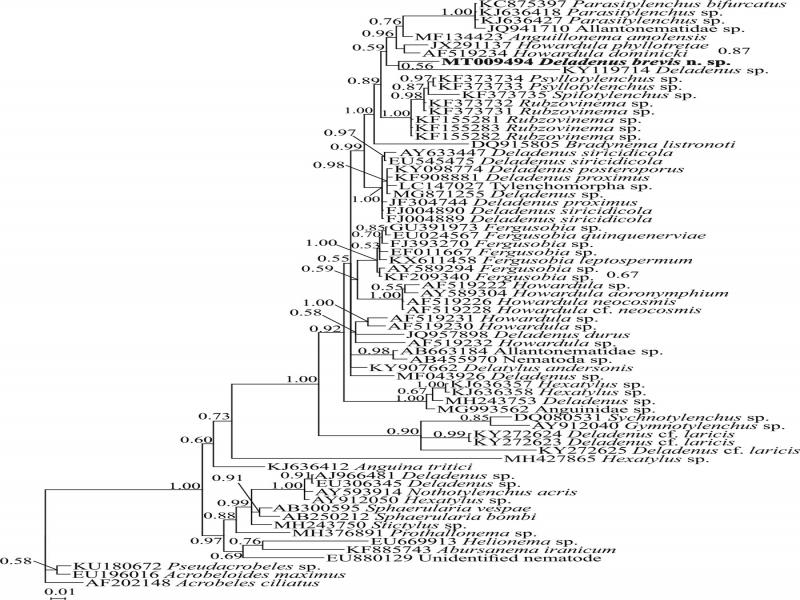
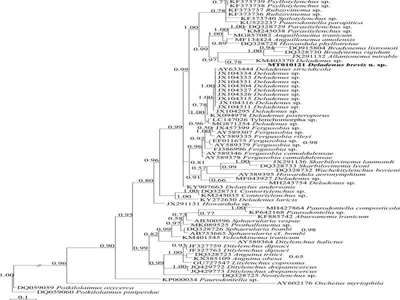
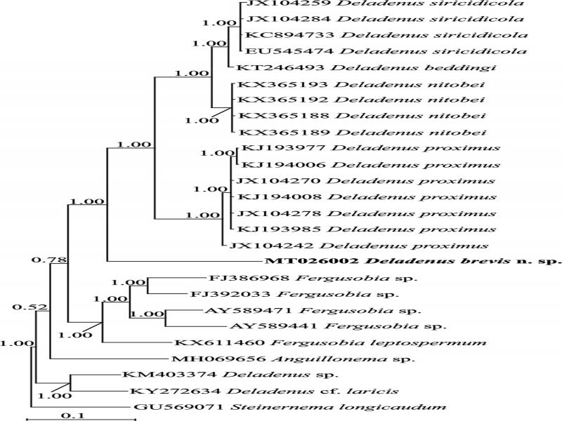
During a survey to recover insect-related nematodes in Iran, a population of an undescribed species of Deladenus was recovered from a deadwood sample of a dead forest tree collected from the forests of Golestan province, northern Iran. Thus, the present paper aims to describe the newly recovered species and resolve its phylogenetic relationships with other relevant species and genera using three SSU, LSU rDNA, and COI mtDNA markers.
Materials and methods
Sampling, nematode extraction, mounting, and drawing
Specimens of Deladenus brevis n. sp. were obtained from the bark and rotten wood samples of a dead forest tree collected in Golestan province, northern Iran using the tray method (Whitehead and Hemming, 1965). Live specimens of interested nematodes were handpicked under a Nikon SMZ1000 stereomicroscope, heat killed by adding boiled 4% formalin solution, transferred to anhydrous glycerin, mounted on permanent slides according to De Grisse (1969), and examined using a Nikon Eclipse E600 light microscope. Photomicrographs were taken using an Olympus DP72 digital camera attached to an Olympus BX51 microscope equipped with differential interference contrast. Drawings were made using a drawing tube attached to a microscope and were redrawn using the CorelDRAW® software version 17.
Scanning electron microscopy (SEM)
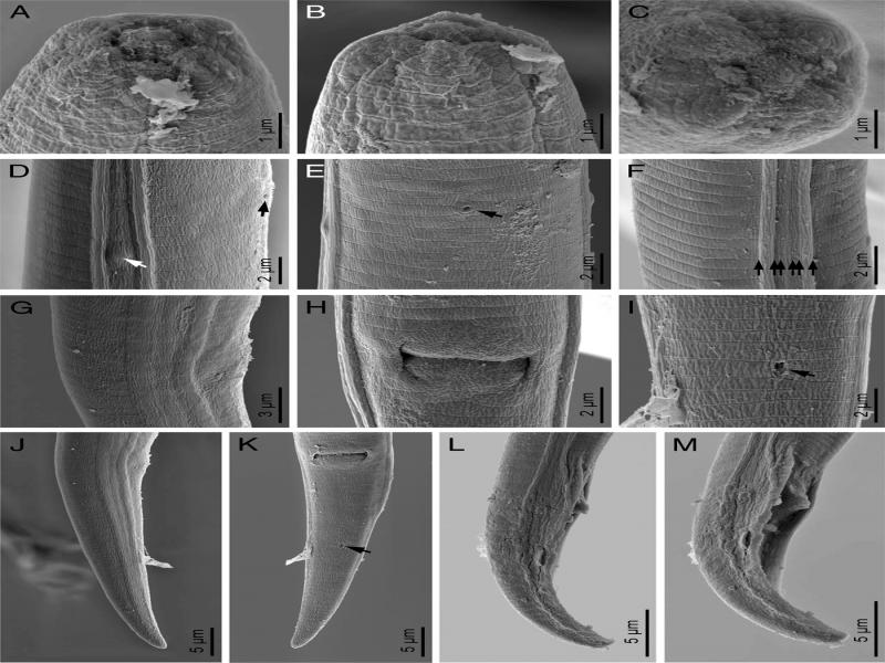
Specimens preserved in glycerine were selected for observation under SEM according to Abolafia (2015). They were hydrated in distilled water, dehydrated in a graded ethanol-acetone series, critical point dried, coated with gold, and observed with a Zeiss Merlin microscope (5 kV) (Zeiss, Oberkochen, Germany).
DNA extraction, PCR, and sequencing
A single live nematode specimen of D. brevis n. sp. was picked out and transferred to a small drop of TE buffer (10 mM Tris-Cl, 0.5 mM EDTA; pH 9.0; Qiagen) on a clean slide and squashed using a clean cover slip. The suspension was collected by adding 15 μl TE buffer. The DNA sample was stored at −20°C until used as PCR template. Three DNA samples were prepared in this manner. Primers for LSU rDNA D2-D3 expansion segments amplification were forward D2A (5′-ACAAGTACCGTGAGGGAAAGTTG-3′) and reverse D3B (5′-TCGGAAGGAACCAGCTACTA-3) (Nunn, 1992), forward KKLSU-1 (5′-AAGGATTCCCTTAGTAACGGCGAGTG-3′) (Kiontke et al., 2004), and reverse 1006R (5′-GTTCGATTAGTCTTTCGCCCCT-3′) (Holterman et al., 2008) primers. Primers for partial amplification of SSU rDNA were forward 1813F (5′-CTGCGTGAGAGGTGAAAT-3′) and reverse 1912R (5′-TTTACGGTCAGAACTAGGG-3′) primers (Holterman et al., 2006); forward primer F22 (5′-TCCAAGGAAGGCAGCAGGC-3′) (Dorris et al., 2002) and reverse primer 18 S 1573 R (5′-TACAAAGGGCAGGGACGTAAT-3′) (Mullin et al., 2005). The COI mtDNA was amplified using HCO (5′-TAAACTTCAGGGTGACCAAAAAATCA-3′) and LCO (5′-GGTCAACAAATCATAAAGATATTGG-3′) primer pairs (Folmer et al., 1994). The PCR condition for the amplification of genomic fragments and COI mtDNA was according to Aliramaji et al. (2020), except that the annealing temperature for COI mtDNA amplification was set to 46°C. The PCR products were purified and sequenced directly for both strands using the same primers used in PCR with an ABI 3730 XL sequencer (Bioneer, South Korae). Sequences were deposited in GenBank database under the accession numbers presented in corresponding phylogenetic trees.
Phylogenetic analyses
The molecular sequences of D. brevis n. sp. were compared with those of other nematode species available in GenBank. The previously used sequences for phylogenetic analyses (Tomar et al., 2015; Kanzaki et al., 2016; Yu et al., 2017; Morris et al., 2018) were retrieved from the database, updated by adding extra sequences and the newly generated sequences of the new species. Three independent SSU, LSU, and COI mtDNA data sets were prepared. The sequences were aligned using the Q-INS-i algorithm of the online version of MAFFT version 7 (http://mafft.cbrc.jp/alignment/server/) (Katoh and Standley, 2013). The poorly aligned positions and divergent regions of SSU and LSU data sets were eliminated using the online version of Gblocks 0.91b (Castresana, 2000) using all three less stringent options. The COI alignment was edited manually using MEGA6 (Tamura et al., 2013). The model of the base substitution was selected using MrModeltest 2 (Nylander, 2004). The Akaike-supported model, a general time-reversible model including among-site rate heterogeneity and estimate of invariant sites (GTR + G + I), was selected for all phylogenetic analyses. Bayesian analysis was performed using MrBayes v3.1.2 (Ronquist and Huelsenbeck, 2003) with a starting random tree and running the chains for four million generations for all. The burn-in phase was set at 25% of the converged runs. The Markov chain Monte Carlo (MCMC) method within a Bayesian framework was used to estimate the posterior probabilities of the phylogenetic tree (Larget and Simon, 1999) using the 50% majority rule. To visualize the results of each run in order to check the effective sample size of each parameter, Tracer v1.5 (Rambaut and Drummond, 2009) was used. The classic rhabditid taxa were selected as outgroups (for species names and accession numbers see trees). The output file of MrBayes was visualized using Dendroscope v3.2.8 (Huson and Scornavacca, 2012) and redrawn in CorelDRAW version 17 (Jahanshahi Afshar et al., 2019a, 2019b).
Results
Systematics
Deladenus brevis n. sp. (Figs. 1 – 3).
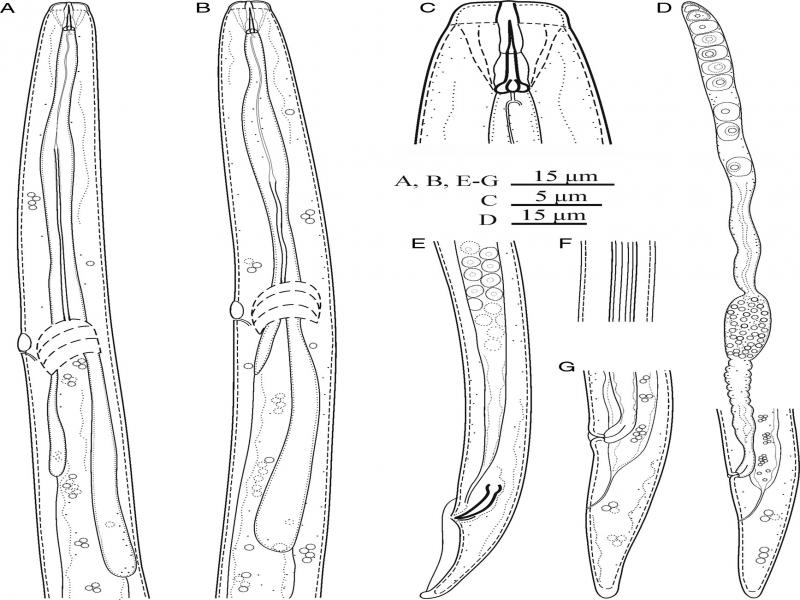
Figure 1:
Line drawings of Deladenus brevis n. sp. (A, B) Pharynx of male and female; (C) Anterior end of female; (D) Female genital tract and posterior body region; (E) Male posterior body region; (F) Lateral lines; (G) Female tail.
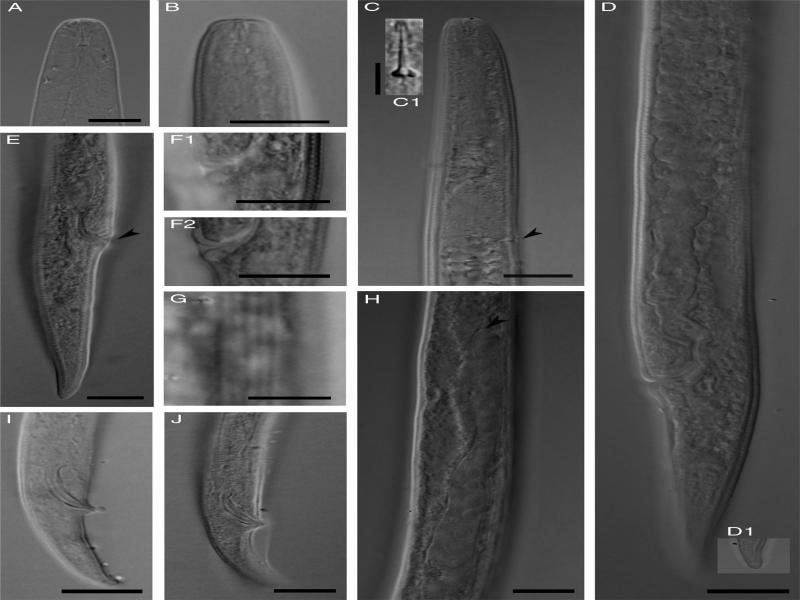
Figure 2:
Photomicrographs of Deladenus brevis n. sp. (A, B) Anterior body region of female (fresh sample in water and permanently preserved specimen in glycerin respectively); (C) Part of pharynx (arrowhead showing excretory pore and C1 showing details of stylet); (D) Part of female reproductive system; (E) Female posterior body region; (F1 and F2) Lateral vulval flaps in ventrolateral and lateral view in two specimens; (G) Lateral lines; (H) Tip of ovary (arrowhead); (I, J) Male posterior body (Scale bars: A-F2, H-J =10 µm, F1, C1, G =5 µm).
Measurements
Measurements of the new species are given in Table 1.
Table 1.
Morphometrics of Deladenus brevis n. sp.
| Female | Male | ||
|---|---|---|---|
| Character |
|
|
|
| n | − | 8 |
|
| L | 435 |
|
|
| 26.0 | 28 ± 3 (22.3-32.3) |
|
| 9.5 | 8.9 ± 0.7 (8.0-10.3) |
|
| 15.0 |
|
|
| 3.2 | 2.8 ± 0.3 (2.4-3.2) |
|
| T or V | 88.9 | 89.0 ± 1.0 (87.2-90.3) |
|
| 39.5 |
|
|
| 1.4 | 1.9 ± 0.2 (1.5-2.1) |
|
| 5.7 | 5.9 ± 0.4 (5.0-6.5) |
|
| 17 | 15 ± 2 (13-18) |
|
| 6.6 | 6.4 ± 0.3 (6-7) |
|
| 2.6 | 2.5 ± 0.2 (2-3) |
|
| 78 | 79.0 ± 5.3 (67-86) |
|
| 71 |
|
|
| 76 | 77.5 ± 6.0 (66-86) |
|
| 13 | 12.5 ± 2. (10-18) |
|
| 9 | 9.0 ± 1.2 (7-10) |
|
| 29 | 25.0 ± 3.4 (20-30) |
|
| − | − |
|
| − | − |
|