The nematode species Bursaphelenchus crenati Rühm, 1956 belonging to Sexdentati group was originally described from the galleries of Hylesinus crenati Fabricius in bark of ash Fraxinus excelsior L. in Bavaria, Germany and later was re-described by Gu et al. (2017) using the populations from Poland. This species was recently detected in several localities of Russia and Belarus (Ryss et al., unpublished). Transmissive dauer juveniles of B. crenati were found in senior larvae, pupae, and female imagoes of the beetle species. From these dauer juveniles, adult nematodes were grown in cultures of the spore-less strain of the fungus Botrytis cinerea on the 2% potato dextrose agar (PDA). All records revealed associations of B. crenati with wilt symptoms of the ash Fraxinus excelsior. In the same beetle vector, the fungi belonging to the family Ophiostomataceae were extracted and cultured in 2% PDA and 2% MEA (malt agar). Nutrition relations between the nematodes and fungi were established, it has been shown that the nematode population grown in slow rate (5 wk) comparing to that culture with Botrytis cinerea (7 d at room temperature). To study a functional role of different juvenile stages of B. crenati in the adaptations to vector and plant and fungus host, as well as in survival of winter seasonal stress, the identification of the life cycle stages would be necessary.
Special attention was given to observe the genital primordium, because according to previous studies this organ is most useful in identification of stage and sex of juveniles (Hirschmann, 1962, 1971; Hirschmann and Triantaphyllou, 1968; Ryss, 1981, 1988; Ryss and Chernetskaya, 2009, 2010; Ryss and Polyanina, 2017). The purpose of this research is to characterize the juvenile stages of the species.
Materials and methods
Nematode samples
The bark and wood samples with the beetle bark tunnels of Hylesinus crenatus Fabricius, in the inner bark layer and sapwood in wilted ash tree, Fraxinus excelsior L. were collected by A.V. Petrov in the forestry plantation “Tellermanovskoye Opytnoye Lesnichestvo ILAN RAS,” Tellermanovsky settlement, Gribanovsky District, Voronezh Oblast, Russia, June 2017, in a location with the GPS. 51°21′46.8″N 42°02′40.3″E. Samples were sent to Zoological Institute RAS. In the laboratory, the nematode Bursaphelenchus crenati dauers were collected from beetle imagoes, where they formed semi-dry spot-like assemblages (“nematangia”) on the inner surface of elytra.
Nematode culturing
Nematodes were multiplied from dauers in the sterile laboratory cultures on the lawn of spore-less strain of the fungus Botrytis cinerea, inoculated in the 2% potato dextrose agar media in petri dish (Ryss, 2015). Over 2 wk after 20 nematode specimen inoculation, nematodes occupied all the petri dish space (diam. 6 cm), devastating the fungus lawn, showing active oviposition and population growth. To maintain the nematode isolate, approximately 100 nematode specimens were transferred to the fresh culture of B. cinerea, multiplying population occupied all the petri dish space in 5 to 7 d at 20 to 22°C.
Morphological study
To study the morphology, nematodes were washed out from petri dish with 1 ml of distilled water and fixed in hot TAF as described by Ryss (2017b). Over 2 d after fixation, nematodes were stained with acetic orcein or methylene blue. A modification of the staining technique (Ryss, 1988; Ryss and Chernetskaya, 2009; Ryss and Polyanina, 2017) was used to stain nematodes with acetic orcein. A 1.5 ml Eppendorf tube with nematodes in TAF was placed in upright position for 10 min to settle nematodes down, and the upper layer of fixative was removed with a syringe up to the two fold level of the nematode suspension height (usually 100 µL). Then 200 µL of 70% acetic acid was added into the tube. After shaking the tube, two drops of saturated acetic orcein solution in 65% acetic acid were added, which is usually used for chromosome staining. The mixture was kept overnight for staining procedure and then a drop of staining suspension was placed into the glycerin in a cavity slide. The stained nematodes picked out by needle and transferred to 5 µL drop of glycerin.
To stain with methylene blue, the nematodes were fixed in TAF and processed to glycerin according to Ryss (2017a). Then, nematodes were picked out and transferred from glycerin suspension into a small 1 µL drop of glycerin on a glass slide, and a 5 µL drop of saturated methylene blue water solution was added from above. The drop was placed for 12 hr in open air at room temperature for water evaporation and staining. Then nematodes were transferred in another small glycerin drop on glass slide, heated softly and quickly for stain contrasting and differentiation.
Stained nematodes were picked out with needle under a stereomicroscope and placed in a minute drop of glycerin on a microscopic slide. The drop was covered with 20 × 20 mm coverslip. The drop was spread under coverslip to press nematodes to obtain flattened worm bodies for detailed study of nematode genital primordia structures. After drop spreading, coverslip was sealed with nail polish along borders. All stained nematodes in slides were studied, measured, and photographed under the automated microscope system Leica DM5000 B with differential interference contrast (DIC) and with the Leica DFC320 (R2) digital camera with Leica DFC Twain Software for PC and Leica IM50 Image Manager for PC. All measurements were made using the software ImageJ 1.48 v (National Institute of Health, USA, http://imagej.nih.gov/ij).
Measurements and values used in morphometry of the individual development stages
Measures: L: body length; BW: body width; pharynx length till the pharyngo-intestinal valve; pharynx length till the end of the pharyngeal gland lobe; tail length; tail width at anus (ABW); GP: genital primordium length; GW: genital primordium width; spicule length along arc; hyaline part of tail in male juveniles (future bursal flap of male). Ratios: a: body length divided to body width; b: body length divided to pharynx length till the pharyngo-intestinal valve; b’: body length divided to pharynx length till the end of pharyngeal glands; c: body length divided to tail length; c’: tail length divided to tail width; V or (V): distance from anterior body end to vulva, or to the center of the genital primordium, divided to the body length, in %; GP/GW: genital primordium length divided to its width; GP/L: genital primordium length divided to body length, in %.
Because the bodies were flattened, the re-calculation of the body and tail diameter was used: the flattened width was considered as a half of the circle (πR or pi R), thus the diameter (2 R) was calculated as the flattened body width (BW) × (2/pi) = 0.64 × BW.
Results
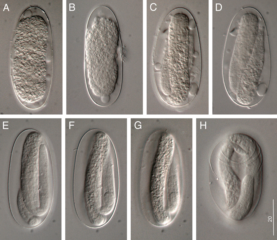
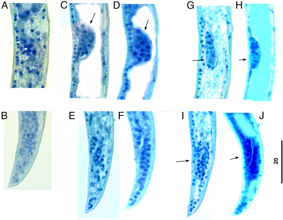
The stained nematodes and eggs were studied. The first molt took place inside the egg shell; thus during hatching the second stage juvenile (J2) broke the egg-shell and came out (Fig. 1E,F,G).
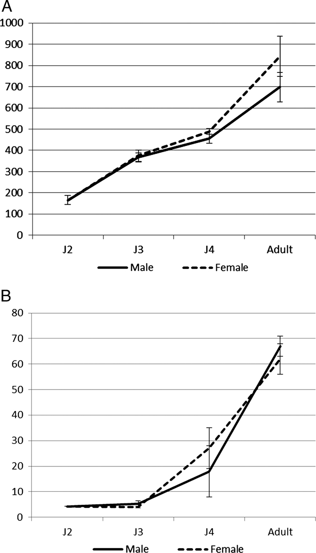
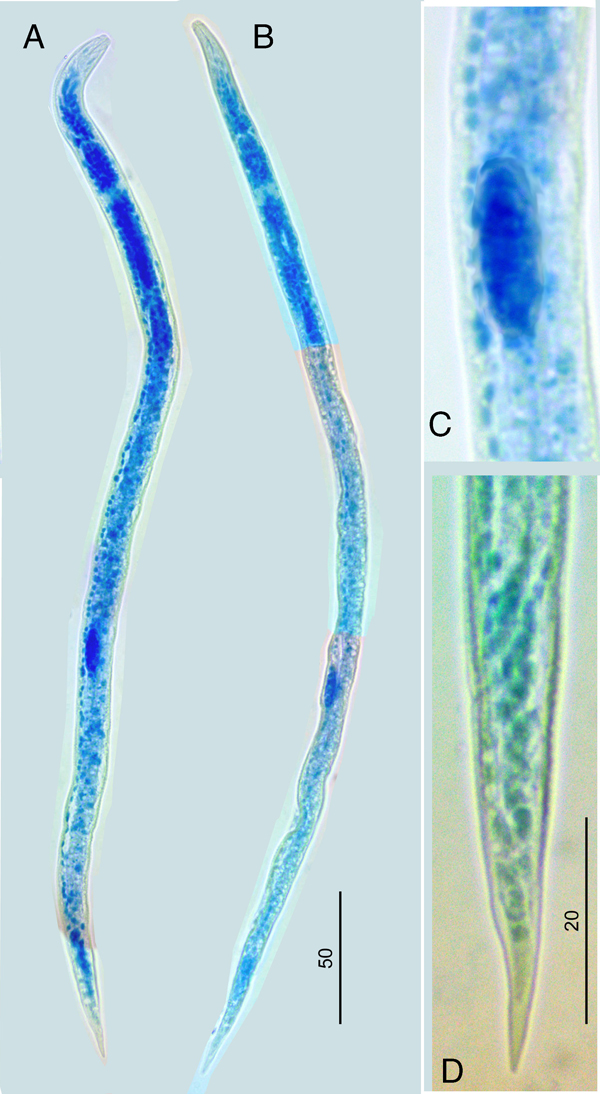
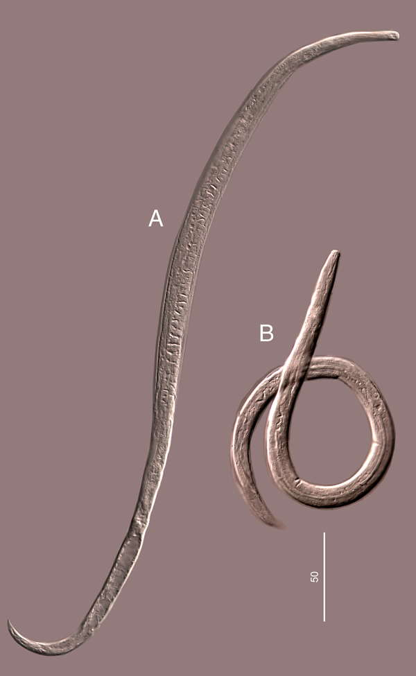
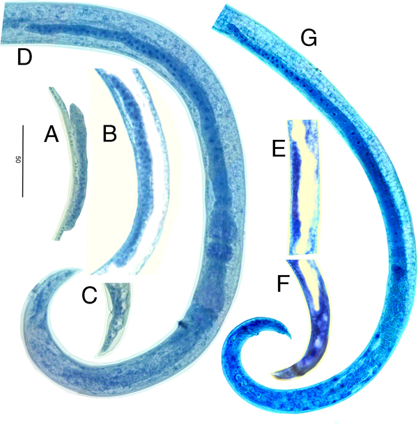
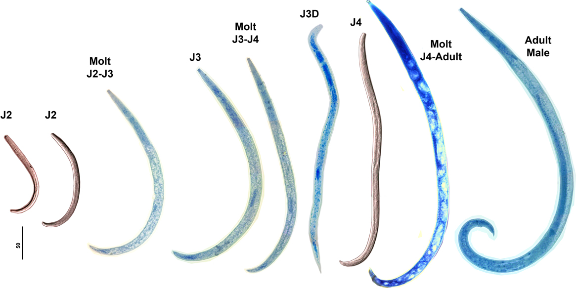
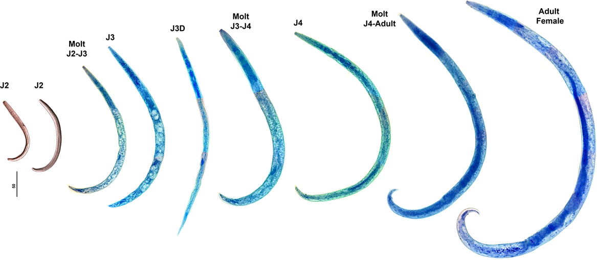
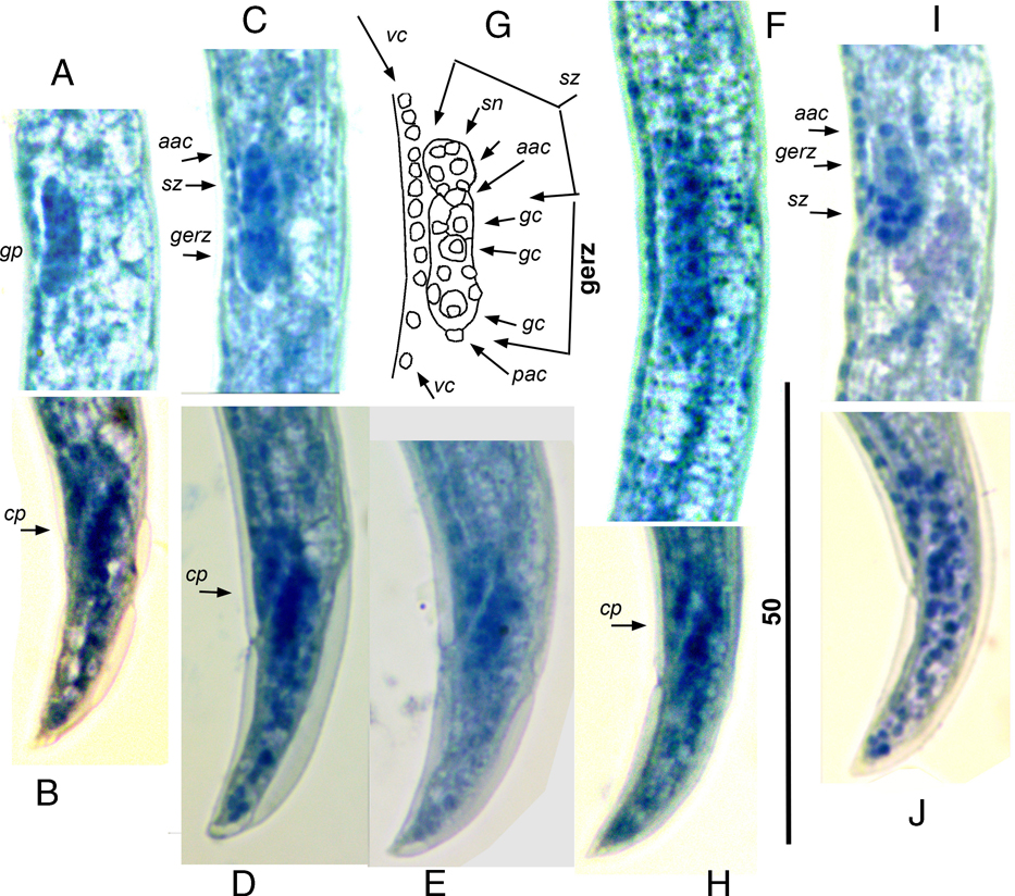
Other stages may be classified as J2, J3, J4, and adult males and females, separated by three molts. Molts were detected by cuticle separation from the body at body extremities, the head and tail tips. The sex of J3 and J4 may be recognized, whereas it cannot be determined in J2. The stage and sex of the juvenile may be determined in the genital primordium structure and its position in the body. The body size and the ratio between genital primordium length and the body are useful to identify the stage. Genital primordia in every stage and sex are described and illustrated below.
Second stage juveniles (J2) (Figs. 2A,B, 4,5; Table 1)
Table 1
Measurements (in µm) and ratios of the development stages of Bursaphelenchus crenati.
| Stage/Character | J2 | J3 male | J3 female | J4 male | J4 female | Male | Female |
|---|---|---|---|---|---|---|---|
| n | 20 | 20 | 20 | 15 | 18 | 15 | 15 |
| L | 166 ± 22 | 367 ± 21 | 375 ± 26 | 455 ± 21 | 489 ± 14 | 698 ±69 | 843 ± 94 |
| (150–181) | (338–400) | (339–402) | (377–532) | (479–508) | (607–804) | (750–1082) | |
| Body width | 7 ± 1 | 10 ± 2 | 11 ± 2 | 14 ± 4 | 12 ± 0.2 | 17 ± 3 | 16 ± 3 |
| (6–8) | (6–12) | (8–13) | (12–17) | (10–15) | (14–216) | (13–20) | |
| Pharynx | 68 ± 4 | 111 ± 5 | 115 ± 6 | 106 ± 0.7 | 125 ± 12 | 122 ± 12 | 136 ± 22 |
| (65–70) | (104–118) | (109–125) | (105–106) | (111–140) | (105–144) | (108–178) | |
| Pharyngeal gland | 30 ± 5 | 50 ± 5 | 52 ± 5 | 49 ± 6 | 50 ± 6 | 50 ± 6 | 59 ± 14 |
| (27–34) | (44–58) | (45–60) | (45–53) | (42–55) | (41–64) | (40–85) | |
| Stylet | 7 ± 1 | 8 ± 1 | 8 ± 0.8 | 10 ± 1 | 10 ± 1 | 10 ± 0.1 | 11 ± 1 |
| (6–8) | (6–9) | (7–9) | (9–11) | (9–11) | (8–12) | (10–13) | |
| Lip region width | 4.0 ± 0.3 | 5 ± 0.2 | 5 ± 0.4 | 5 ± 0.2 | 5.8 ± 0.2 | 7.0 ± 0.6 | 7.0 ± 0.5 |
| (3.5–4.5) | (4–5) | (4–5) | (5–5.5) | (5.5–6.0) | (7–8) | (6.0–7.5) | |
| Lip region height | 2.3 ± 0.5 | 3 ± 0.3 | 2 ± 0.2 | 3 ± 0.2 | 2.5 ± 0.1 | 4 ± 0.5 | 3.6 ± 0.3 |
| (2.0–2.5) | (2–3) | (2–3) | (3–3.5) | (2.4–2.7)– | (3–5) | (3–4) | |
| Median bulb from | 30 ± 2 | 48 ± 2 | 48 ± 1.4 | 40 ± 3 | 56 ± 6 | 58 ± 5 | 60 ± 9 |
| anterior | (29–31) | (45–52) | (46–50) | (38–43) | (51–64) | (51–71) | (51–73) |
| Median bulb length | 9.4 ± 1 | 12 ± 1 | 12 ± 1.3 | 12 ± 0.3 | 12.7 ± 0.7 | 15 ± 2 | 18 ± 4 |
| (9–10) | (10–14) | (10–14) | (11.5–12) | (11.5–13.0) | (13–18) | (14–23) | |
| Median bulb width | 5.7 ± 0.3 | 9 ± 2 | 9 ± 1.2 | 9 ± 2 | 10.3 ± 1.3 | 12 ± 1 | 12 ± 1 |
| (5.5–6.0) | (6–11) | (7–10) | (7–10) | (10–12) | (11–14) | (10–13) | |
| Excretory pore | 35 ± 3 | 53 ± 2.1 | 54.0 ± 3.9 | 46 ± 4 | 61.3 ± 0.9 | 63 ± 2 | 70 ± 11 |
| from anterior | (33–37) | (50–57) | (49–61) | (44–49) | (60–62) | (60–77) | (59–90) |
| Tail | 9 ± 3 | 22 ± 3 | 25 ± 2 | 23 ± 3 | 27.5 ± 1.4 | 29 ± 3 | 31 ± 5 |
| (7–11) | (19–28) | (21–28) | (21–25) | (26–29) | (26–35) | (20–37) | |
| Anal body width | 4 ± 1 | 9 ± 1.1 | 9 ± 2 | 8 ± 1 | 10 ± 2 | 17 ± 2 | 11 ± 2 |
| (4–5) | (7–10) | (6–11) | (7–9) | (9–13) | (12–19) | (8–14) | |
| Genital primordium | 7 ± 1 | 20 ± 5 | 15 ± 2 | 78 ± 25 | 132 ± 40 | 472 ± 68 | 526 ± 90 |
| length (GPL) | (6–8) | (14–28) | (12–17) | (60–95) | (77–160) | (355–581) | (413–630) |
| Genital primordium | 2.7 ± 0.1 | 5 ± 0.4 | 6 ± 1 | 5 ± 1 | 8 ± 2 | 14 ± 3 | 16 ± 3 |
| Width (GPW) | (2.5–3.0) | (4–5) | (4–7) | (4–6) | (6–10) | (11–14) | (12–21) |
| Spicule length | – | – | – | – | – | 17 ± 1 | – |
| along arc | (16–19) | ||||||
| a | 23 ± 2 | 40 ± 8 | 36 ± 4 | 31.6 ±1.2 | 40 ± 7 | 40 ± 5 | 53 ± 8 |
| (22–24) | (32–58) | (32–41) | (31–33) | (33–49) | (34–50) | (41–60) | |
| b | 4.5 ± 0.4 | 6.1 ± 0.3 | 6.0 ± 0.3 | 8.0–1.2 | 6.6 ± 0.6 | 9.7 ± 1.2 | 11.1–2.0 |
| (4.2–4.8) | (5.6–6.5) | (5.6–6.3) | (7.1–8.9) | (5.7–7.0) | (7.7–11.3) | (8.8–14.1) | |
| b’ | 2.5 ± 0.5 | 3.3 ± 0.2 | 3.3 ± 0.2 | 4.3 ± 1.1 | 3.9 ± 0.4 | 5.8 ±0.6 | 6.3 ± 1.0 |
| (2.1–2.8) | (3.1–3.6) | (3.0–3.6) | (3.6–5.1) | 3.4–4.3) | (4.9–6.6) | (4.6–7.6) | |
| c | 19.0 ± 4.3 | 16.6 ± 2.1 | 15.2 ± 0.5 | 20.0 ± 2.4 | 17.9 ±1.5 | 24.3 ± 2.9 | 27.7 ± 5.5 |
| (15.9–22.1) | (12.4–20.5) | (14.6–16.2) | (18.4–21.7) | (16.8–19.9) | (20.4–30.5) | (22–37) | |
| c’ | 2.1 ± 0.4 | 2.7 ± 0.4 | 2.8 ± 0.4 | 2.8 ± 0.1 | 2.8 ± 0.4 | 1.8 ±0.2 | 2.9 ± 0.3 |
| (1.8–2.4) | (2.3–3.4) | (2.4–3.4) | (2.7–2.9) | (2.3–3.1) | (1.4–2.2) | (2.4–3.4) | |
| V % or (V): genital primordium center from anterior, to body length, in % | 65 ± 2 | 61 ± 2 | 66 ± 2 | 58 ± 12 | 75 ± 2 | – | 75 ± 2 |
| (63–66) | (56–64) | (63–68) | (49–66) | (72–77) | (73–77) | ||
| GPL/GPW | 2.6 ± 0.4 | 4.3 ± 1.0 | 2.4 ± 0.2 | 17 ± 10 | 16.8 ± 3.0 | 34 ± 7 | 34 ± 8 |
| (2.4–2.9) | (3.0–5.7) | (2.2–2.8) | (10–24) | (13.6–20.6) | (26–49) | (24–46) | |
| GPL/L, % | 4.3 ± 0.04 | 5.4 ± 1.1 | 4 ± 0.3 | 18 ± 10 | 27 ± 8 | 67 ± 4 | 62 ± 6 |
| (4.26–4.32) | (4.2–7.1) | (3–4) | (11–25) | (16–33) | (58–72) | (55–71) | |
| Lip region length to its | 1.8 ± 0.3 | 1.9 ± 0.2 | 1.9 ± 0.1 | 1.7 ± 0.1 | 2.3 ± 0.1 | 2.0 ± 0.3 | 1.9 ± 0.2 |
| Width | (1.6–2.0) | (1.6–2.3) | (1.8–2.1) | (1.7–1.8) | (2.2–2.5) | (1.6–2.5) | (1.6–2.2) |
| Median bulb length to | 1.6 ± 0.1 | 1.4 ± 0.3 | 1.4 ± 0.1 | 1.4 ± 0.3 | 1.3 ± 0.2 | 1.3 ± 0.1 | 1.4 ± 0.2 |
| Its width | (1.6–1.7) | (1.0–1.9) | (1.2–1.6) | (1.2–1.6) | (1.0–1.5) | (1.1 ± 1.5) | (1.2–1.8) |
| Stage/Character | J2→J3 male | J2→J3 female | J3→J4 male | J3→J4 female | J4→male | J4→female | Dauer J3D |
|---|---|---|---|---|---|---|---|
| n | 16 | 5 | 10 | 20 | 5 | 17 | 20 |
| L | 347 ± 7 | 323 ± 86 | 370 ± 3 | 377 ± 10 | 596 ± 91 | 544 ± 69 | 413 ± 20 |
| (338–354) | (181–410) | (366–374) | (364–398) | (532–660) | (433–614) | (392–463) | |
| Body width | 9 ± 1 | 10 ± 2 | 11 ± 2 | 12 ± 1 | 16 ± 0.3 | 13 ± 3 | 8 ± 1 |
| (8–10) | (8–14) | (9–13) | (11–13) | (14–17) | (9–17) | (6–9) | |
| Pharynx | 107 ± 4 | 103 ± 21 | 110 ± 5 | 110 ± 5 | 112 ± 9 | 117 ± 6 | 119 ± 9 |
| (104–112) | (65–116) | (105–115) | (102–117) | (105–118) | (109–123) | (101–128) | |
| Pharyngeal gland | 49 ± 5 | 51 ± 1 | 49 ± 4 | 48 ± 4 | 46 ± 1 | 48 ± 6 | 43 ± 7 |
| (44–56) | (49–53) | (44–55) | (41–54) | (45–47) | (40–55) | (35–55) | |
| Stylet | 7 ± 1.2 | 8 ± 1 | 7 ± 1 | 9 ± 0.7 | 9 ± 0.4 | 9 ± 1.4 | 9.4 ± 0.4 |
| (6–9) | (6–9) | (6–8) | (8–10) | (9–10) | (8–11) | (9–10) | |
| Lip region width | 5.0 ± 0.3 | 5 ± 0.6 | 5.6 ± 0.5 | 5 ± 0.6 | 5 ± 0.4 | 5.4 ± 0.4 | 4.8 ± 1.0 |
| (4–5) | (4–6) | (4.8–6.0) | (4–6) | (5–6) | (5–6) | (4–7) | |
| Lip region height | 3 ± 0.3 | 2.4 ± 0.2 | 2.4 ± 0.1 | 3 ± 0.3 | 3 ± 1 | 2.9 ± 0.4 | 2.9 ± 0.3 |
| (2–3) | (2–3) | (2.3–2.5) | (2–3) | (2–3)– | (2–3) | (2.5–4) | |
| Median bulb from | 46 ± 1 | 45 ± 8 | 47 ± 0.1 | 47 ± 3 | 46 ± 12 | 53 ± 3 | 49 ± 2 |
| Anterior | (45–47) | (31–50) | (46–48) | (40–50) | (38–54) | (51–58) | (45–51) |
| Median bulb length | 11 ± 1.2 | 11 ± 0.9 | 12 ± 1 | 12 ± 1 | 13 ± 1 | 13 ± 0.2 | 11 ± 2 |
| (10–13) | (10–12) | (11–13) | (10–13) | (12–14) | (12.5–13.5) | (9–15) | |
| Median bulb width | 9 ± 1.1 | 8 ± 1.5 | 10 ± 1 | 10 ± 1 | 9.9 ± 0.2 | 10 ± 1 | 8 ± 2 |
| (8–10) | (6–10) | (9–11) | (7–10) | (9.5–10.0) | (9–11) | (6–10) | |
| Excretory pore | 51 ± 1 | 50 ± 8 | 53 ± 2 | 54 ± 4 | 52 ± 12 | 58 ± 3 | 40 ± 3 |
| from anterior | (50–52) | (37–56) | (51–54) | (45–59) | (44–61) | (54–62) | (37–44) |
| Tail | 22 ± 4 | 21 ± 6 | 22 ± 1 | 26 ± 1.5 | 28 ± 5 | 28 ± 3 | 37 ± 3 |
| (20–28) | (11–27) | (21–24) | (24–28) | (25–32) | (23–31) | (30–41) | |
| Anal body width | 8 ± 0.4 | 8 ± 3 | 11 ± 2 | 11 ± 1.1 | 11 ±3 | 10.1 ±0.9 | 8 ± 1 |
| (7–8) | (5–12) | (8–13) | (10–13) | (9–13) | (9–11) | (5–9) | |
| Genital primordium | 18 ± 4 | 10 ± 3 | 20 ± 2 | 18 ± 5 | 158 ± 138 | 197 ± 105 | 14.8 ± 2.1 |
| Length (GPL) | (14–23) | (6–14) | (17–22) | (13–28) | (60–255) | (122–369) | (11–18) |
| Genital primordium | 5 ± 1 | 5 ± 1 | 4.8 ± 05 | 5 ± 1 | 10 ±5 | 9 ± 3 | 3.9 ± 0.4 |
| Width (GPW) | (4–5) | (3–7) | (4.0–5.3) | (3–6) | (6–13) | (7–14) | (3–5) |
| Spicule length | – | – | – | – | – | – | – |
| Along arc | |||||||
| a | 40 ± 5 | 34 ± 8 | 34.8 ± 5.2 | 32 ± 2 | 39 ± 12 | 41.5 ± 4.6 | 54 ± 8.3 |
| (34–46) | (22–41) | (28.4–41.8) | (29–35) | (31–48) | (36.8–48.9) | (44.6–69.6) | |
| b | 6.0 ± 0.3 | 5.6 ± 0.6 | 6.1 ± 0.2 | 6.1–0.2 | 9.1 ± 0.3 | 7.8 ± 0.6 | 5.5 ± 0.4 |
| (5.7–6.4) | (4.8–6.4) | (5.9–6.3) | (5.8–6.5) | (8.9–9.2) | (6.7–8.3) | (5.1–6.4) | |
| b’ | 3.2 ± 0.1 | 3.1 ± 0.3 | 3.4 ± 0.2 | 3.4 ± 0.2 | 5.3 ± 0.4 | 4.6 ±0.5 | 3.5 ±0.3 |
| (3.1–3.3) | (2.8–3.6) | (3.2–3.6) | (3.1–3.7) | (5.1–5.6) | (4.0–5.2) | (3.0–4.1) | |
| c | 16.0 ± 2.5 | 15.6 ± 0.6 | 16.6 ± 0.9 | 14.8 ± 0.8 | 21.1 ± 0.9 | 19.7 ± 1.7 | 11.2 ± 0.9 |
| (12.4–17.9) | (14.7–16.2) | (15.1–17.3) | (13.8–1.58) | (20.5–21.7) | (18.0–22.3) | (9.9–13.2) | |
| c’ | 2.8 ± 0.4 | 2.8 ± 0.5 | 2.1 ± 0.3 | 2.3 ± 0.2 | 2.6 ±0.1 | 2.8 ± 0.3 | 4.7 ± 0.5 |
| (2.4–3.4) | (2.2–3.4) | (1.7–2.5) | (2.0–2.8) | (2.6–2.7) | (2.3–3.2) | (4.3–5.5) | |
| V % or (V): genital primordium center from anterior, to body length, in % | 61 ± 2 | 64 ± 3 | 62 ± 1 | 66 ± 2 | 52 ± 3 | 73.2 ± 1.6 | 62 ± 5 |
| (59–64) | (59–68) | (60–64) | (63–68) | (49–54) | (70.5–74.3) | (56–72) | |
| GPL/GPW | 4.0 ± 1.1 | 2.3 ± 0.6 | 4.2 ± 0.2 | 3.9 ±1 | 15 ± 6 | 20 ± 4 | 3.8 ± 0.5 |
| (3.0–5.4) | (1.5–2.9) | (3.9–4.5) | (2.7–6.1) | (10–19) | (16–27) | (3–4.5) | |
| GPL/L, % | 5.2 ± 1.2 | 3.3 ± 1.0 | 5.5 ± 0.6 | 5 ± 1 | 25 ± 19 | 35.3 ±15.2 | 4 ± 1 |
| (4.2–6.6) | (1.8–4.3) | (4.5–5.9) | (4–7) | (11–39) | (24.3–60.0) | (3–5) | |
| Lip region length to its | 1.8 ± 0.3 | 2.0 ± 0.3 | 2.3 ± 0.2 | 1.9 ± 0.2 | 2.1 ± 0.6 | 1.9 ± 0.1 | 1.6 ± 0.3 |
| Width | (1.6–2.3) | (1.6–2.4) | (2.0–2.6) | (1.6–2.4) | (1.7–2.5) | (1.8–2.0) | (1.4–2.4) |
| Median bulb length to | 1.4 ± 0.2 | 1.4 ± 0.2 | 1.2 ± 0.1 | 1.2 ± 0.1 | 1.3 ± 0.2 | 1.3 ± 0.1 | 1.4 ± 0.2 |
| Its width | (1.1–1.6) | (1.3–1.7) | (1.2–1.3) | (1.0–1.4) | (1.2–1.4) | (1.2 ± 1.4) | (1.1–1.7) |
| C1 | C2 | C3 | C4 | C5 | C6 | |
|---|---|---|---|---|---|---|
| Stage/Character | Copulative structures | Average body size, in µm | Ratio: genital primordium to body length, in % | Genital primordium cellular structure | Cloaca primordium or cloaca around rectum | Genital primordium somatic part: a division to sections |
| J2 | Absent | 170 (150–180) | 4 | Small with two large germinal cells in center | Absent | Absent |
| J3 male | Absent | 370 (340–400) | 5 (4–7) | 2–5 large germinal cells in the posterior part of primordium, 12 small somatic nuclei in anterior part | Present | Absent |
| J3 female | Absent | 380 (340–400) | 4 (3–4) | 2–5 large germinal cells in the anterior part of primordium, 12 small somatic nuclei in posterior part | Absent | Absent |
| J4 male | Absent | 460 (380–530) | 18 (11–25) | 20–60 large germinal cells in anterior part of primordium and 30–60 somatic cells in posterior part | Present | Absent |
| J4 female | Absent | 490 (480–510) | 27 (16–33) | 20–60 large germinal cells in anterior part of primordium and 30–60 somatic cells in posterior part | Absent | Absent |
| Male, adult | Paired spicules in cloaca, at tail end | 700 (610–800) | 67 (58–72) | More than 60 germinal cells in anterior part | Present | Absent |
| Female, adult | Vulva at 75% body length from anterior end | 840 (750–1100) | 62 (55–71) | More than 60 germinal cells in anterior part | Absent | Present |