
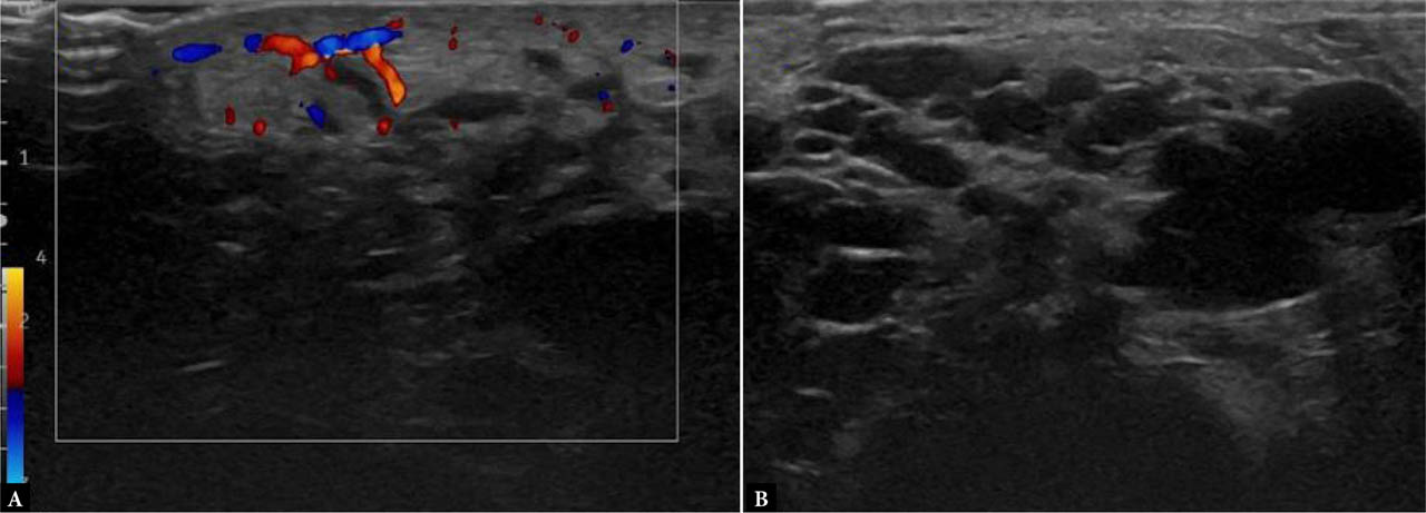
Fig. 1.
Ultrasound of the right gluteal region in a patient with a congenital macrocystic lymphatic malformation complicated by superinfection. A. Color Doppler image showing increased vascular signal within the edematous and thickened skin and subcutaneous tissue (maximum thickness approximately 8 mm), consistent with acute inflammation. B. Corresponding grayscale image without Doppler. No clear sonographic evidence of intra- or perilesional abscess is identified. The underlying macrocystic lymphatic malformation is confirmed

Fig. 2.
Follow-up ultrasound of the right gluteal region at 72 hours. A. Color and power Doppler images showing a hypoechoic lesion with peripheral vascular signal (maximum dimensions 9 × 9 mm), suggestive of abscess formation. B. Grayscale image showing hypoechoic transformation of one of the macrocysts within the lymphatic malformation, containing internal debris. Persistent edematous thickening of the skin and subcutaneous tissue (current thickness approximately 9 mm) with increased vascularity is also noted, consistent with ongoing inflammation
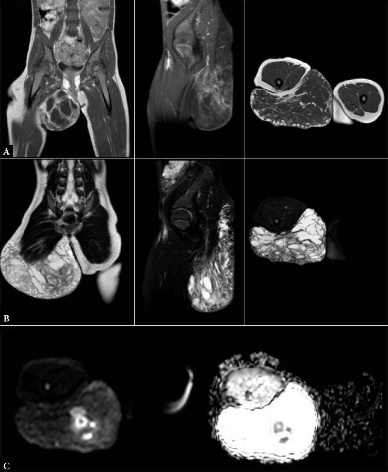
Fig. 3.
MRI performed for definitive lesion characterization in a patient with a right gluteal macrocystic lymphatic malformation complicated by cellulitis, recent trauma, and ongoing dual antibiotic therapy. Sequences included TSE T1-weighted (A), T2-weighted SPAIR (B), and diffusion-weighted imaging with the corresponding apparent diffusion coefficient (DWI/ADC) maps (C), acquired in the coronal, sagittal, and axial planes. Imaging demonstrated: (1) extensive edema involving both the superficial and deep subcutaneous tissues surrounding the lesion (B); (2) marked diffusion restriction within the cystic cavity, compatible with purulent content (C); and (3) no abnormal signal involving the contralateral gluteal region or adjacent osseous structures (A–C). These findings confirmed abscess formation superimposed on a macrocystic lymphatic malformation, consistent with secondary infection following recent trauma

Fig. 4.
MRI images, post-contrast T1-weighted sequence in axial (A) and coronal (B) planes, showing marked peripheral enhancement of the cyst walls
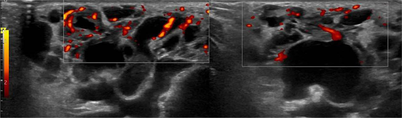
Fig. 5.
Ultrasound of the right gluteal region in a patient with a known macrocystic lymphatic malformation and recent clinical hyperemia in the ipsilateral inguinal area. The cystic components of the malformation demonstrate homogeneously anechoic content. At the fold between the right gluteal and inguinal regions, superficial to the macrocystic lymphatic malformation, mild subcutaneous tissue thickening is present, without significant signs of intra- or perilesional inflammation, including in the posterior gluteal area. In the right inguinal region, several oval-shaped lymph nodes are identified, with a maximum short-axis diameter of approximately 7 mm, consistent with a reactive appearance