
Introduction
Macrocystic lymphatic malformations (MLMs) are congenital low-flow vascular anomalies composed of dilated lymphatic channels. They most commonly involve the cervicofacial and axillary regions but may occasionally present in atypical sites such as the gluteal region(1). Although benign, MLMs can develop secondary complications, including hemorrhage and infection, particularly after trauma(2). The clinical presentation of infection is often non-specific, mimicking cellulitis or abscess, posing diagnostic challenges(3). Multimodal imaging with ultrasound (US) and magnetic resonance imaging (MRI) plays a pivotal role in timely diagnosis, differentiation from other soft-tissue infections, and guiding treatment decisions. We report a case of a 2-year-old girl with a gluteal macrocystic lymphatic malformation complicated by abscess formation, highlighting the critical role of imaging. According to the International Society for the Study of Vascular Anomalies (ISSVA) classification, lymphatic malformations are low-flow vascular anomalies subdivided into macrocystic, microcystic, or mixed forms(4).
Case presentation
A 2-year-old girl with a known congenital macrocystic lymphatic malformation of the right gluteal region presented to the emergency department after sustaining blunt trauma to the same site. Clinical examination revealed severe localized pain, inconsolable crying, fever, and erythema of the overlying skin. Vital signs at presentation included fever and local signs of inflammation without hemo-dynamic instability. Laboratory investigations revealed leukocytosis and elevated inflammatory markers.
Clinical timeline
The clinical timeline of presentation, imaging, and management was as follows:
Day 0: emergency department admission following blunt trauma to the right gluteal region; fever, localized pain, and erythema observed. Baseline ultrasound performed;
Day 3: clinical worsening with persistent fever and local inflammatory signs; follow-up ultrasound showing evolution toward superinfection;
Day 4: contrast-enhanced MRI confirming abscess formation within the macrocystic lymphatic malformation; initiation of intravenous antibiotic therapy and ultrasound-guided percutaneous drainage;
Day 7: clinical and laboratory improvement; resolution of fever and local signs;
Follow-up: complete resolution of inflammatory changes without early recurrence.
Initial ultrasound
An urgent soft-tissue ultrasound demonstrated a voluminous anechoic macrocystic lesion consistent with the known macrocystic lymphatic malformation. Ultrasound was performed using a high-frequency linear transducer, 7–12 MHz, with color and power Doppler evaluation. The known macrocystic lymphatic malformation was confirmed, and the term “lymphangioma” was replaced in accordance with ISSVA nomenclature. No definite signs of abscess collections were detected initially. Surrounding the lesion, an 8-mm thickened hyperechoic subcutaneous layer with increased vascularity on color Doppler suggested acute inflammation without abscess formation (Fig. 1).
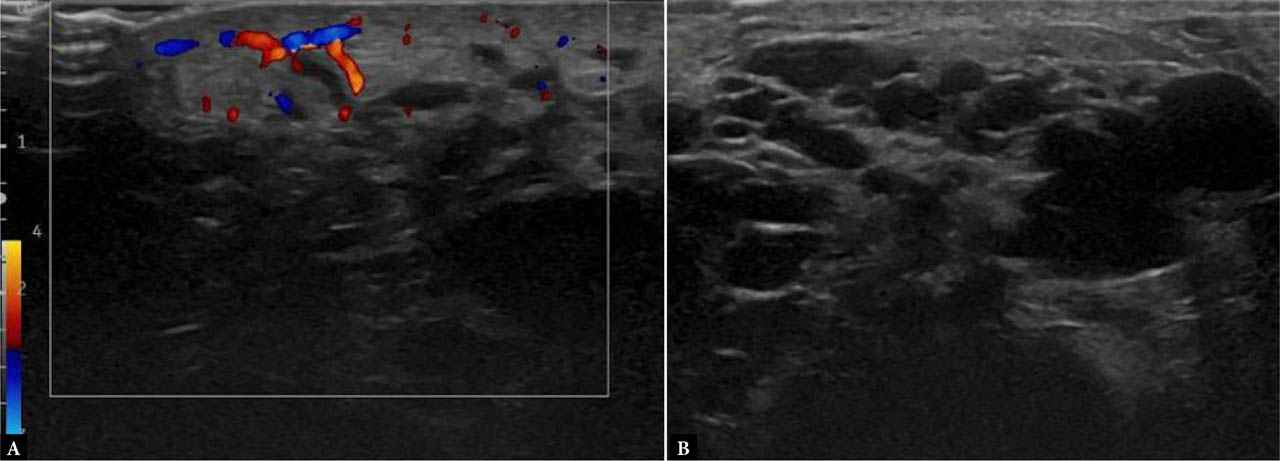
Fig. 1.
Ultrasound of the right gluteal region in a patient with a congenital macrocystic lymphatic malformation complicated by superinfection. A. Color Doppler image showing increased vascular signal within the edematous and thickened skin and subcutaneous tissue (maximum thickness approximately 8 mm), consistent with acute inflammation. B. Corresponding grayscale image without Doppler. No clear sonographic evidence of intra- or perilesional abscess is identified. The underlying macrocystic lymphatic malformation is confirmed
Follow-up ultrasound at 72 hours
At 72 hours, repeat ultrasound revealed progression, demonstrating cyst wall thickening, new vascular signals along the cyst walls on power Doppler, and hypoechoic cyst contents with internal debris suspicious for early abscess formation. Power Doppler confirmed peripheral hyperemia of the cyst walls, supporting the diagnosis of superinfection. Inflammatory changes had progressed, with a 9 × 9 mm hypoechoic lesion showing peripheral Doppler activity, indicating probable abscess formation. Persistent cutaneous and subcutaneous edema with hyperemia was also noted (Fig. 2).
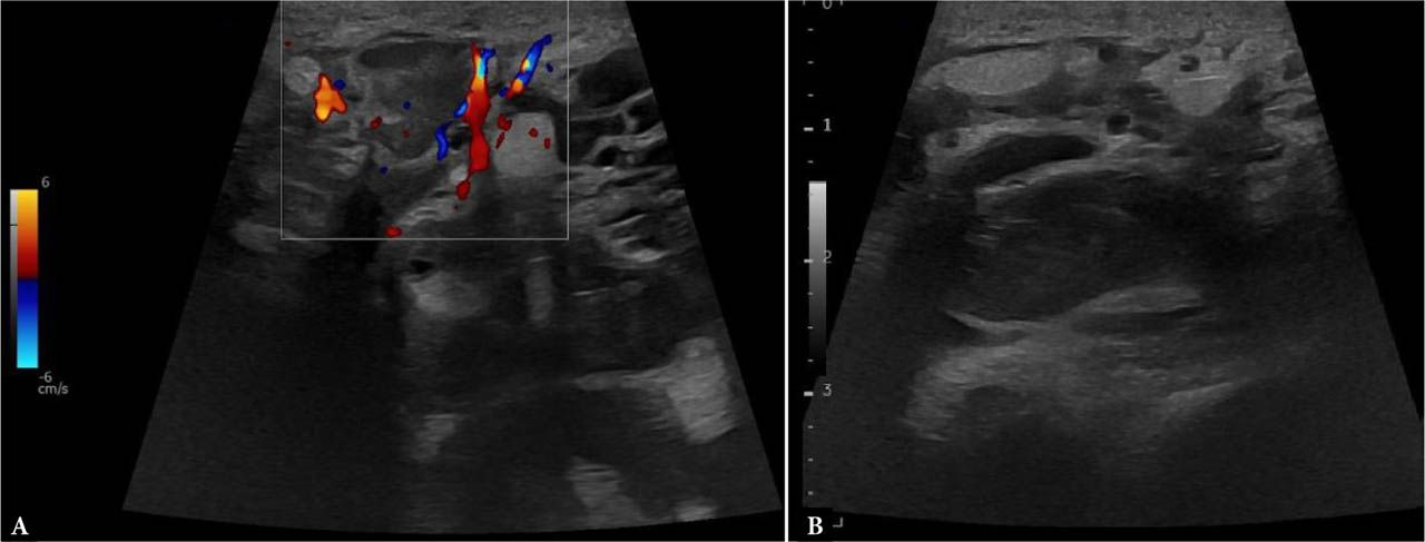
Fig. 2.
Follow-up ultrasound of the right gluteal region at 72 hours. A. Color and power Doppler images showing a hypoechoic lesion with peripheral vascular signal (maximum dimensions 9 × 9 mm), suggestive of abscess formation. B. Grayscale image showing hypoechoic transformation of one of the macrocysts within the lymphatic malformation, containing internal debris. Persistent edematous thickening of the skin and subcutaneous tissue (current thickness approximately 9 mm) with increased vascularity is also noted, consistent with ongoing inflammation
Magnetic resonance imaging
MRI was performed for definitive characterization. Sequences included TSE T1- and T2-weighted imaging, SPAIR T2, and diffusion-weighted imaging (DWI), both pre- and post-contrast administration. MRI revealed:
extensive edema of the superficial and deep subcutaneous fat surrounding the lesion;
marked peripheral enhancement of the cyst walls;
internal cystic contents with restricted diffusion, consistent with purulent material;
no involvement of the contralateral gluteal region or underlying bones.
MRI was performed on a 1.5-T system and included axial and coronal T1-weighted, T2-weighted, fat-suppressed (SPAIR/STIR), DWI, and post-contrast sequences. Restricted diffusion with low ADC values was observed within the lesion, consistent with purulent content. Contrast-enhanced ultrasound (CEUS) was considered but not performed. The imaging findings confirmed a diagnosis of abscess formation complicating the MLM in the context of trauma and infection (Fig. 3, Fig. 4).
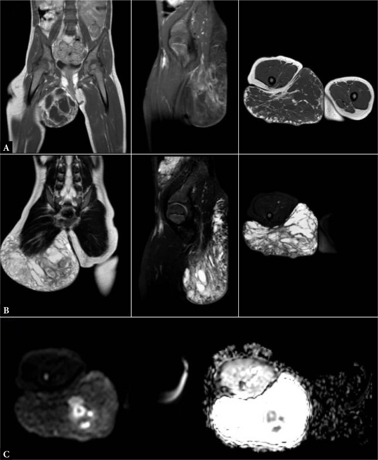
Fig. 3.
MRI performed for definitive lesion characterization in a patient with a right gluteal macrocystic lymphatic malformation complicated by cellulitis, recent trauma, and ongoing dual antibiotic therapy. Sequences included TSE T1-weighted (A), T2-weighted SPAIR (B), and diffusion-weighted imaging with the corresponding apparent diffusion coefficient (DWI/ADC) maps (C), acquired in the coronal, sagittal, and axial planes. Imaging demonstrated: (1) extensive edema involving both the superficial and deep subcutaneous tissues surrounding the lesion (B); (2) marked diffusion restriction within the cystic cavity, compatible with purulent content (C); and (3) no abnormal signal involving the contralateral gluteal region or adjacent osseous structures (A–C). These findings confirmed abscess formation superimposed on a macrocystic lymphatic malformation, consistent with secondary infection following recent trauma
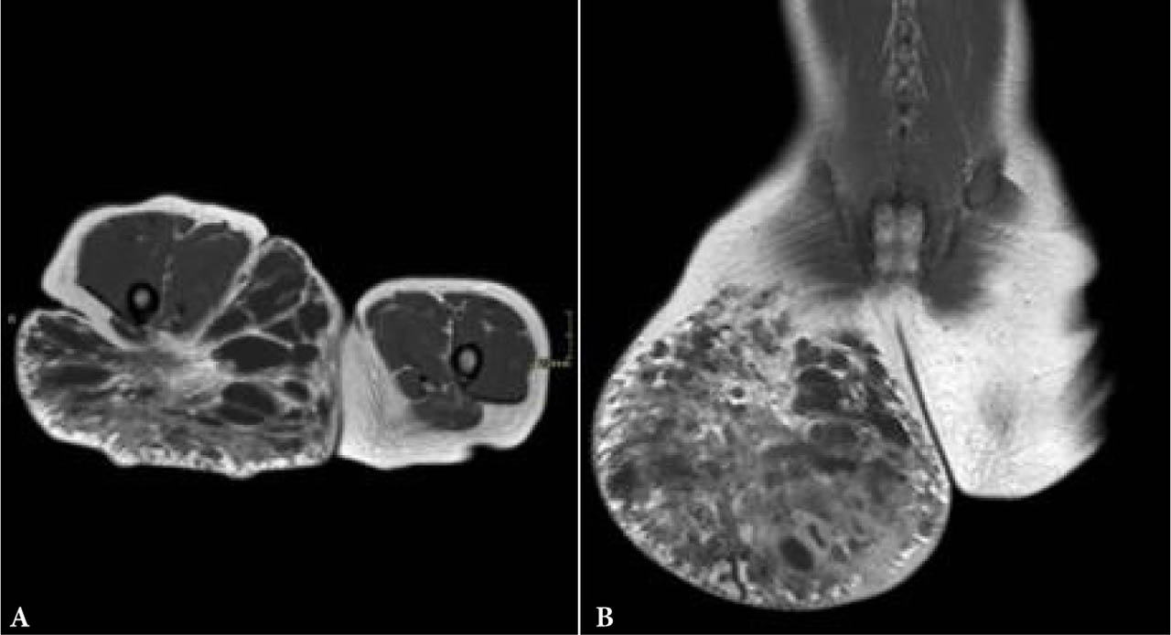
Fig. 4.
MRI images, post-contrast T1-weighted sequence in axial (A) and coronal (B) planes, showing marked peripheral enhancement of the cyst walls
An additional ultrasound examination was performed due to new-onset erythema in the ipsilateral inguinal region, despite the previously performed MRI (Fig. 5).
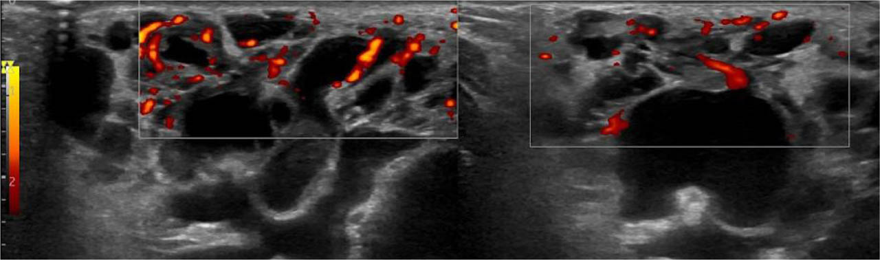
Fig. 5.
Ultrasound of the right gluteal region in a patient with a known macrocystic lymphatic malformation and recent clinical hyperemia in the ipsilateral inguinal area. The cystic components of the malformation demonstrate homogeneously anechoic content. At the fold between the right gluteal and inguinal regions, superficial to the macrocystic lymphatic malformation, mild subcutaneous tissue thickening is present, without significant signs of intra- or perilesional inflammation, including in the posterior gluteal area. In the right inguinal region, several oval-shaped lymph nodes are identified, with a maximum short-axis diameter of approximately 7 mm, consistent with a reactive appearance
Clinical course and management
The patient was treated conservatively with targeted intravenous antibiotics and ultrasound-guided percutaneous drainage of the abscess collections. Despite the infection, the surgical team opted to delay excision of the lymphangioma to minimize cosmetic sequelae, as successful healing without urgent surgery prevented complications such as superinfection or scarring. Ultrasound-guided percutaneous drainage was performed using a needle aspiration technique, yielding purulent material.
Microbiological culture of the aspirated material was performed. The patient received empirical intravenous antibiotic therapy with amoxicillin–clavulanate (100 mg/kg/day) for 7 days, followed by oral therapy for an additional 7 days, with subsequent clinical and laboratory improvement. Microbiological culture of the aspirated purulent material did not isolate pathogenic bacteria, likely due to prior antibiotic administration. Ultrasound-guided percutaneous drainage was performed using a needle aspiration technique, yielding approximately 6 mL of purulent fluid. No procedure-related complications were observed. Follow-up confirmed resolution of inflammatory signs without early recurrence.
Discussion
Macrocystic lymphatic malformations are congenital vascular anomalies characterized by cystic dilation of lymphatic channels. The term “lymphatic malformation” is used consistently throughout this manuscript in accordance with ISSVA recommendations(4), replacing the outdated term “lymphangioma”. Although typically benign and stable, these lesions are prone to secondary infection or abscess formation, particularly in pediatric patients who are more susceptible to minor trauma or superficial infections(1,2). The clinical presentation of infection may mimic other inflammatory or neo-plastic conditions, complicating initial diagnosis. Symptoms such as localized pain, erythema, swelling, and fever are nonspecific and overlap with cellulitis, infected cysts, or soft-tissue tumors(3).
Differential diagnosis
The differential diagnosis included infected epidermoid or pilonidal disease, pyomyositis, post-traumatic hematoma, and venous malformation with thrombosis. Malignant soft-tissue tumors were considered unlikely due to the cystic morphology, absence of solid enhancing components, and imaging features typical of infection rather than neoplasia.
Role of imaging and its evolution
Ultrasound (US) is the cornerstone of initial assessment of MLMs due to its accessibility, lack of ionizing radiation, and ability to provide real-time morphological and hemodynamic information. In macrocystic lesions, US typically demonstrates anechoic or hypoechoic cystic spaces with thin walls. However, once infected, changes such as wall thickening, internal echoes from debris, septations, and increased peripheral vascularity on Doppler imaging become apparent(4). The dynamic nature of US allows clinicians to monitor progression from uncomplicated cystic lesions to abscess formation, providing crucial information for treatment planning(1). Doppler US is particularly valuable in distinguishing sterile fluid collections from infected abscesses by demonstrating hyperemia associated with active inflammation(5).
MRI serves as an adjunct when US findings are equivocal or when deeper tissue involvement is suspected. MRI offers superior soft-tissue contrast, allowing clear delineation of the extent of infection, involvement of adjacent structures, and identification of complications such as fistula or bone involvement(6). Contrast-enhanced MRI sequences demonstrate peripheral rim enhancement typical of abscesses, while DWI shows restricted diffusion within purulent collections, enhancing diagnostic confidence and helping exclude hemorrhage or neo-plastic transformation(7). The role of MRI in treatment planning is also paramount, guiding percutaneous drainage and surgical approaches.
Management implications
Accurate and early diagnosis of abscess formation within MLMs facilitates timely initiation of antibiotic therapy and image-guided percutaneous drainage, avoiding emergent surgical excision with its associated risks and morbidity(1,7). Conservative management allows stabilization of the infectious process and optimization of surgical timing, which can be deferred to achieve better cosmetic and functional outcomes. This tailored approach aligns with current consensus guidelines that emphasize minimally invasive strategies when feasible(8).
Scientific relevance and future directions
This case highlights the importance of maintaining a high index of suspicion for secondary infection in pediatric patients with known MLMs, particularly following trauma. Multimodal imaging, combining US, Doppler, and MRI, enables comprehensive assessment, guiding therapeutic decisions that balance efficacy and morbidity. Advances in CEUS also show promise for real-time evaluation of lesion perfusion and infection status without radiation exposure, although further pediatric studies are warranted(9,10,11,12,13). Ongoing research into minimally invasive techniques and novel imaging biomarkers will likely refine management pathways for these complex cases.
In conclusion, a structured imaging approach integrated with clinical findings is essential for managing macrocystic lymphatic malformations complicated by abscess, ensuring prompt, effective, and patient-tailored care in pediatric emergency settings.
Conclusion
In pediatric patients with macrocystic lymphatic malformations, minor trauma or local inflammation can precipitate secondary infection and abscess formation, representing a potentially serious complication requiring timely diagnosis. A sequential, multimodal imaging strategy, beginning with ultrasound, including Doppler evaluation, and followed by contrast-enhanced MRI when indicated, is essential for early detection, precise lesion characterization, and assessment of the extent of infection. This comprehensive imaging approach informs clinical decision-making by distinguishing simple cystic lesions from infected collections and excluding other mimics such as hemorrhage or neoplasm.
Early and accurate diagnosis facilitates targeted conservative management, including antibiotics and image-guided percutaneous drainage, which can significantly reduce the need for invasive surgical procedures and associated morbidity. Furthermore, an imaging-guided tailored treatment strategy allows optimal timing of elective surgical excision, improving cosmetic and functional outcomes in this vulnerable pediatric population.
Overall, integration of clinical findings with multimodal imaging is paramount for effective management, promoting better patient care and resource utilization. Continued advancements in imaging techniques, including contrast-enhanced ultrasound, hold promise for enhancing noninvasive assessment and follow-up of complicated lymphatic malformations, ultimately improving prognosis and quality of life for affected children.
Notes
[1] Ethical statements
No ethical approval was required. This study was conducted using data obtained for clinical purposes. All patient data were anonymized. Written informed consent for publication of clinical data and images was obtained from the patient's legal guardians.