
Figure 1
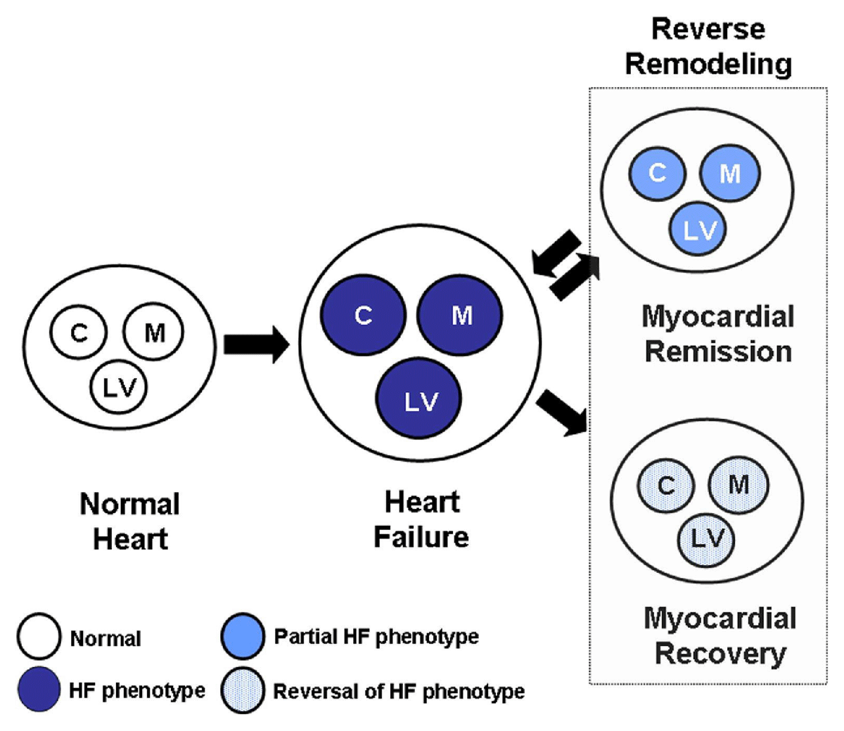
Reverse remodeling and myocardial recovery. Cardiac remodeling occurs secondary to abnormalities that arise in the biology of the cardiac myocyte (C), the myocardium (cardiocytes and extracellular matrix [M]), as well as LV geometry (LV), which have collectively been referred to as the HF phenotype. C: cardiac myocyte; M: myocardium; LV: left ventricular; HF: heart failure. Reproduced with permission from Mann DL et al.2
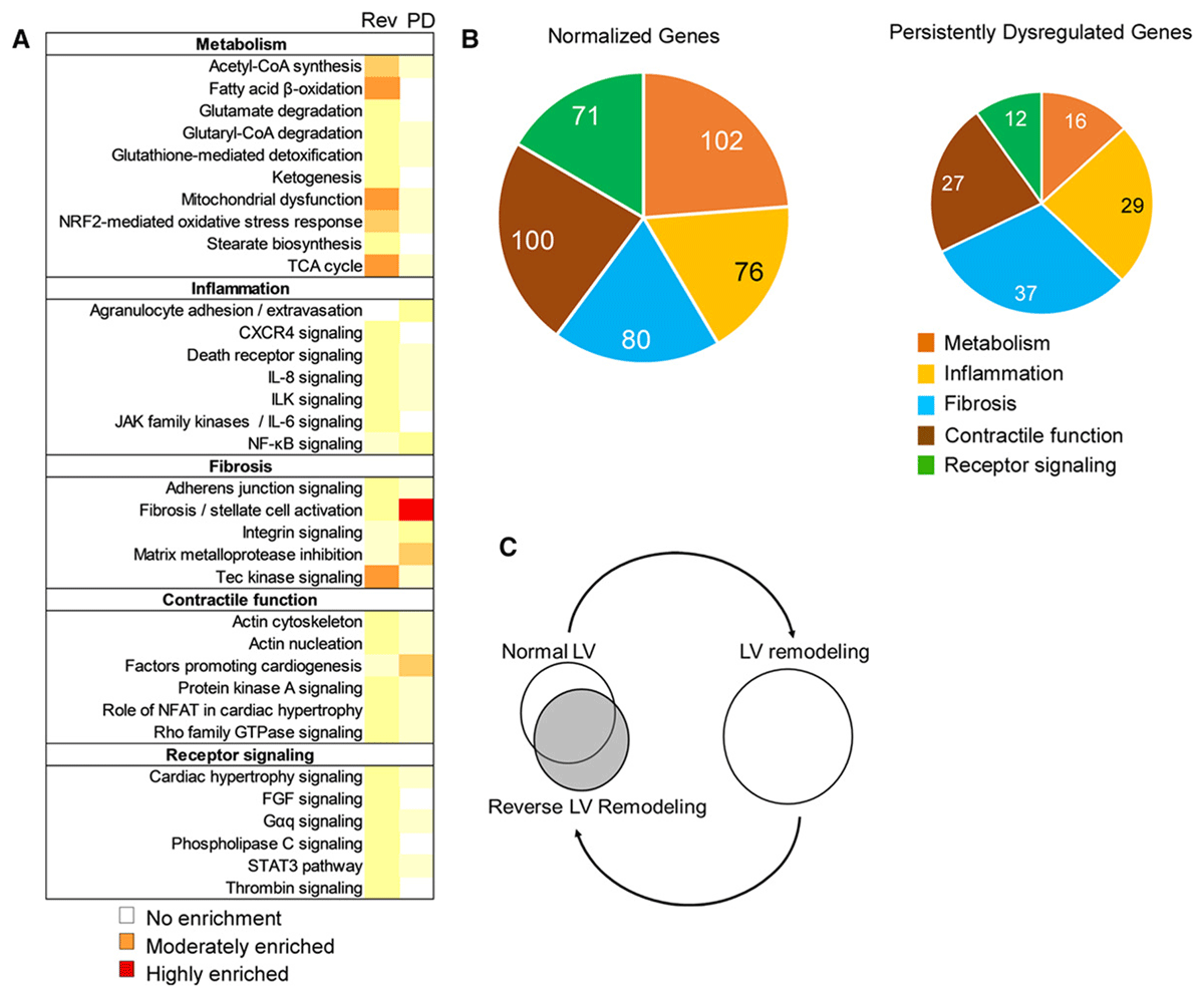
Figure 2
Proposed hypothetical model indicating that reverse left ventricular (LV) remodeling represents the summation of the complex interactions between multiple biological networks that adopt a novel nonpathological configuration, which only partly overlaps with the configuration present in normal hearts. Reproduced with permission from Weinheimer CJ et al.10
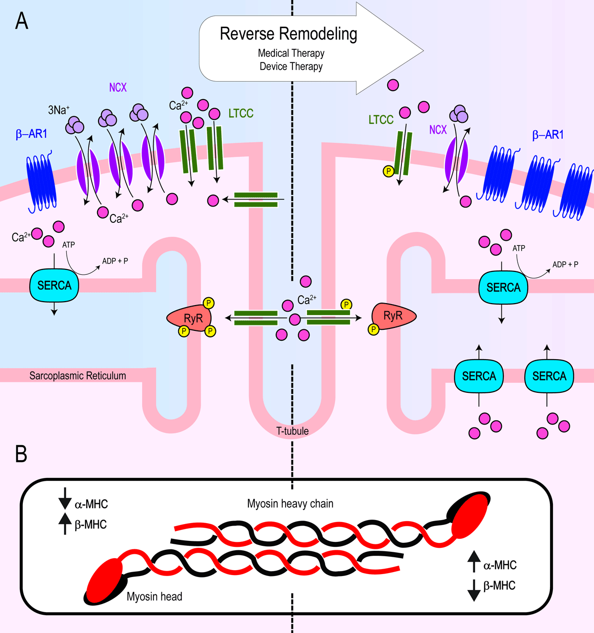
Figure 3
Summary schematic of key changes to cardiac myocyte biology from adverse remodeling (left) to reverse remodeling (right) after implementation of medical and device therapies. (A) Reverse remodeling leads to increased β-AR1 density and restoration of calcium handling and excitation-contraction coupling by way of decreased NCX density, decreased LTCC density albeit with increased activity (increased phosphorylation), increased SERCA density, and normalization of hyperphosphorylated RyR. (B) Reverse remodeling results in a shift in α-MHC and β-MHC ratios. α-MHC: alpha myosin heavy chain; β-AR1: beta adrenergic receptor 1; β-MHC: beta-myosin heavy chain; LTCC: L-type calcium channel; NCX: sodium calcium exchanger; SERCA: sarcoendoplasmic reticulum calcium ATPase; RyR: ryanodine receptor
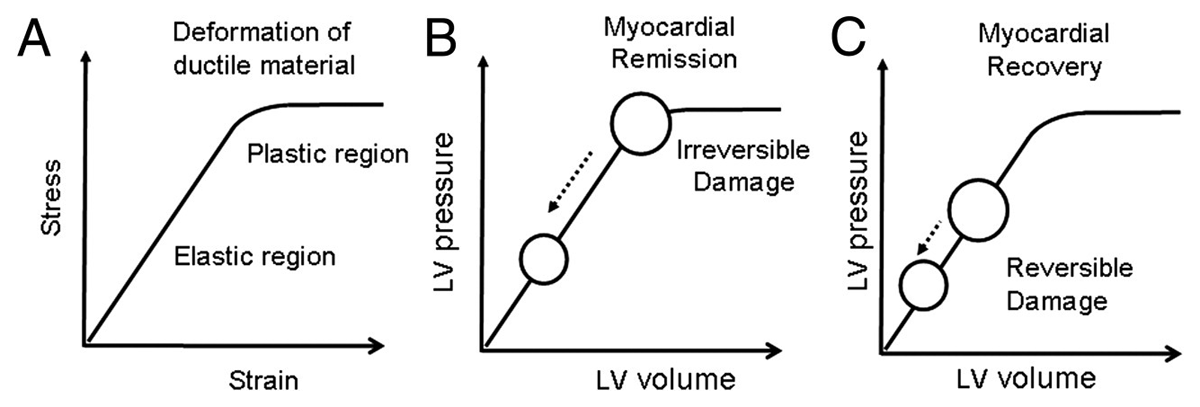
Figure 4
Mechanical engineering science and cardiac remodeling. (A) Diagram of a stress-strain curve of a ductile material illustrating the relationship between an applied force (stress) and deformation (strain). Deformation can lead to reversible changes in a material (elastic deformation) if the properties of the material are not changed, and irreversible changes in a material (plastic deformation). (B) Hypothetical model of reverse remodeling in a heart that has undergone irreversible damage (plastic deformation). (C) Hypothetical model of reverse remodeling with recovery in heart that has undergone reversible damage (elastic deformation). Reproduced with permission from Mann DL et al.2