
Introduction
Reverse left ventricular (LV) remodeling represents a coordinated multi-level process that allows the failing heart to adopt a new, less pathologic steady state associated with improved pump function and improved clinical prognosis. For some, etiologies of heart failure (HF) normalization of LV structure and function and recovery from HF symptomatology may occur spontaneously once the inciting event is resolved (eg, myocarditis). For other patients with chronic HF with reduced LV ejection fraction (HFrEF), improvements in LV structure and function may occur after initiating guideline-directed medical therapy (GDMT).1 Although utilization of GDMT may reverse LV remodeling with improved LV ejection fraction (LVEF) and improved HF symptoms, for the majority of HFrEF patients treated with GDMT, LVEF and LV structural abnormalities do not fully normalize. These patients remain at increased risk of redeveloping clinical HF despite maintenance of GDMT.1 Indeed, HF patients whose ventricles resume a more normal LV size and shape will experience one of two different clinical outcomes: freedom from HF events or recurrence of HF events. Previously the authors suggested that the term “myocardial remission” should refer to the normalization of the molecular, cellular, myocardial, and LV geometric changes that provoke cardiac remodeling but that are insufficient to prevent the recurrence of HF in the face of normal and/or perturbed hemodynamic loading conditions, whereas the term “myocardial recovery” should be used to describe the normalization of the molecular, cellular, myocardial, and LV geometric heart changes that provoke cardiac remodeling, allowing the heart to maintain preserved LV structure/function in the face of normal and/or perturbed hemodynamic loading conditions (Figure 1).2 Here we review the components of reverse LV remodeling in both experimental models and human studies. We also discuss the biological differences between myocardial recovery and remission with an emphasis on the need to maintain clinical remission from HF.
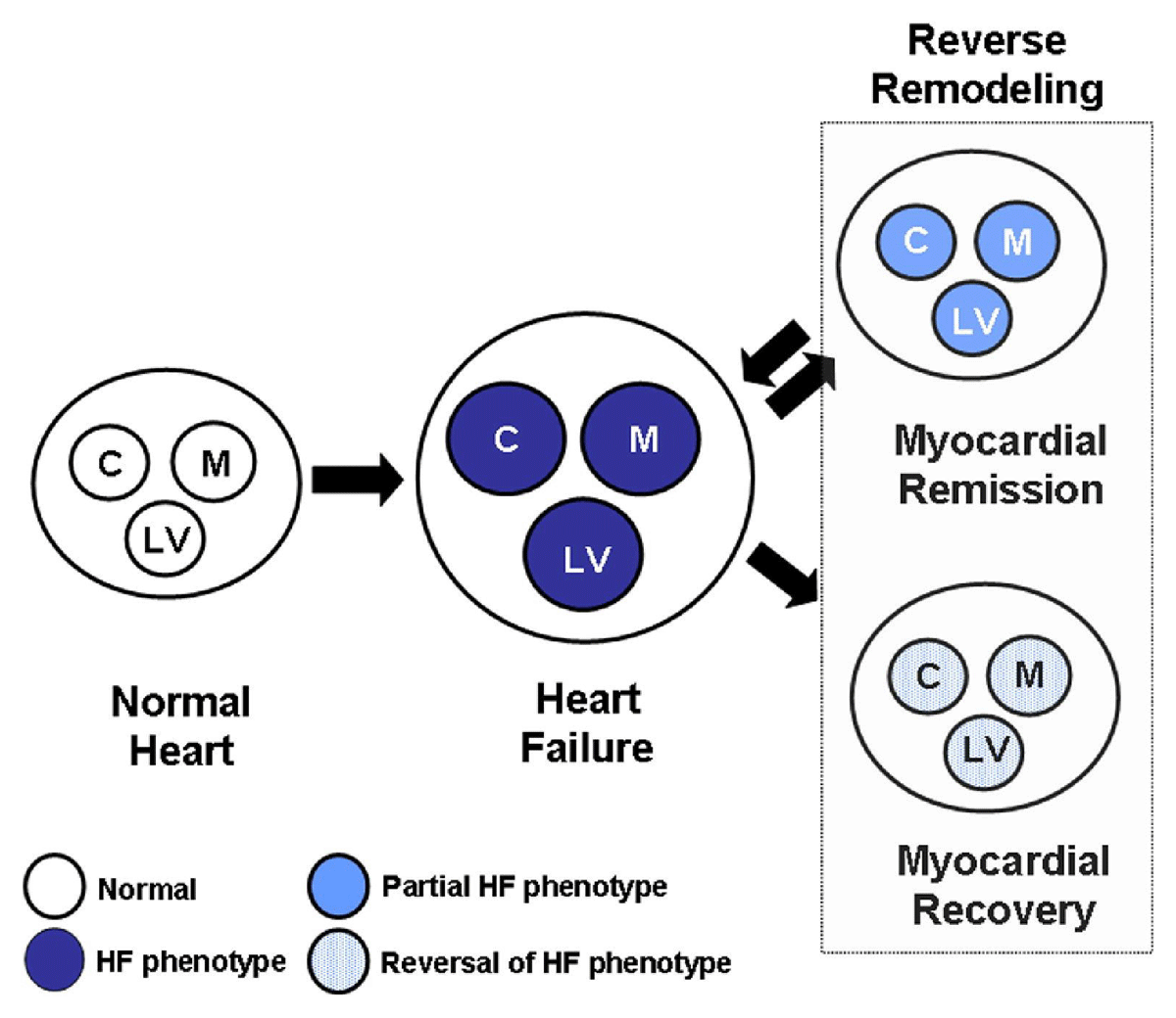
Figure 1
Reverse remodeling and myocardial recovery. Cardiac remodeling occurs secondary to abnormalities that arise in the biology of the cardiac myocyte (C), the myocardium (cardiocytes and extracellular matrix [M]), as well as LV geometry (LV), which have collectively been referred to as the HF phenotype. C: cardiac myocyte; M: myocardium; LV: left ventricular; HF: heart failure. Reproduced with permission from Mann DL et al.2
Reverse Left Ventricular Remodeling
Failing hearts undergo progressive LV dilation, increased sphericity, and LV wall thinning leading to ventricular-afterload mismatch that contributes to decreased stroke volume. Compounding this, the dilated spherical LV geometry results in restrictive tethering of the papillary muscles, which often leads to significant functional mitral regurgitation (MR), a product of leaflet malcoaptation during ventricular systole. MR further causes enlargement and remodeling of the left atrium, resulting in mitral annular dilation, propagating worse functional MR. Aside from reducing systemic output, significant MR causes further LV volume/pressure overload and remodeling. Collectively, these changes result in a rightward shift in the LV pressure-volume relationship, referred to as “adverse LV remodeling.” In post-infarction patients, remodeling changes are associated with worse clinical outcomes compared to other seemingly more obvious factors, including LVEF or severity of coronary arterial lesions.3, 4 Additional longitudinal studies of patients with LV dilation—but without myocardial infarctions, LV dimension, and sphericity—remain potent predictors of survival.5, 6
The term “reverse LV remodeling” arose from studies that observed a leftward shift in the LV end-diastolic pressure-volume relationship towards normal values in patients who had been supported with LV assist devices (LVAD) or with a cardiomyoplasty (a novel technique developed to improve LV function by wrapping the myocardium with latissimus dorsi muscle and then pacing the skeletal muscle).7, 8, 9 Importantly, while implemented, these devices allowed the heart to assume a more normal LV end-diastolic pressure-volume relationship and favorable LV elliptical geometry, which was preserved even when the LVAD was stopped/removed or pacing of the latissimus dorsi muscle was stopped. In the context of the present discussion, it is important to recognize that the calculation of LVEF incorporates LV end-diastolic volume in the denominator of the equation. Accordingly, in the clinical setting, improvements in LVEF are generally associated with a reciprocal decrease in LV end-diastolic volume rather than improved LV contractile function per se.
Biological Mechanisms of Reverse LV Remodeling
Remodeling events observed on the macroscopic scale, including distortion of LV geometry, deterioration of contractile function, and hemodynamic perturbations, are reflective of a dysfunctional and failing cardiac myocyte and its surrounding myocardium. Although first described as the normalization of cardiac function (LVEF > 50%) and left shift of the LV end-diastolic pressure-volume relationship, reverse LV remodeling represents a return to a less pathologic state rather than a complete normalization of HF biology (Figure 2).10
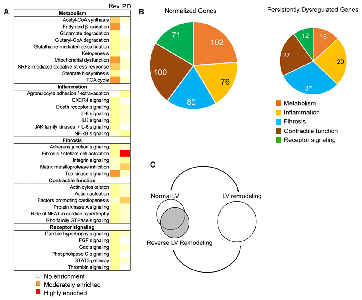
Figure 2
Proposed hypothetical model indicating that reverse left ventricular (LV) remodeling represents the summation of the complex interactions between multiple biological networks that adopt a novel nonpathological configuration, which only partly overlaps with the configuration present in normal hearts. Reproduced with permission from Weinheimer CJ et al.10
Remodeling and Recovery of Cardiac Myocyte Function
Histologic evaluation of failing hearts consistently shows that cardiac myocytes become hypertrophied in response to chronic ventricular overloading and increased diastolic wall stress. In the setting of volume overload, these changes in hemodynamic load lead to elongation of the cardiac myocyte and lengthwise addition of sarcomeres, resulting in eccentric LV remodeling, which is so named because of the eccentric position of the LV in the chest in patients with volume overload. A number of changes occur in cardiac myocyte biology in response to myocardial stretch and hemodynamic overload, neurohormonal activation (eg, norepinephrine, angiotensin II), and release of inflammatory mediators and oxidative stress. These changes in cardiac myocyte biology lead to this myocyte contractile dysfunction and increased myocyte cell death (Figure 3).
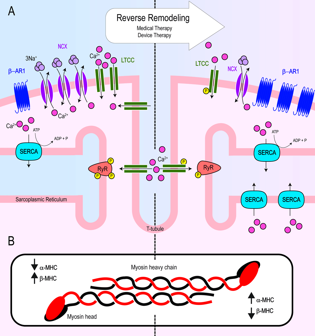
Figure 3
Summary schematic of key changes to cardiac myocyte biology from adverse remodeling (left) to reverse remodeling (right) after implementation of medical and device therapies. (A) Reverse remodeling leads to increased β-AR1 density and restoration of calcium handling and excitation-contraction coupling by way of decreased NCX density, decreased LTCC density albeit with increased activity (increased phosphorylation), increased SERCA density, and normalization of hyperphosphorylated RyR. (B) Reverse remodeling results in a shift in α-MHC and β-MHC ratios. α-MHC: alpha myosin heavy chain; β-AR1: beta adrenergic receptor 1; β-MHC: beta-myosin heavy chain; LTCC: L-type calcium channel; NCX: sodium calcium exchanger; SERCA: sarcoendoplasmic reticulum calcium ATPase; RyR: ryanodine receptor
Beta-Adrenergic Signaling
Ventricles obtained from HF patients demonstrate a marked reduction in beta-adrenergic receptor (β-AR) density, isoproterenol-mediated adenyl cyclase stimulation, and the contractile response to β-adrenergic agonists. The downregulation of β-ARs is likely mediated by increased levels of norepinephrine in the vicinity of the receptor. In patients with dilated cardiomyopathy, this reduction in receptor density involves primarily the β-AR1-receptor protein and mRNA and is proportional to the severity of HF. In contrast, the level of β-AR2-adrenergic receptor protein and mRNA are unchanged or increased.11 These changes are reversed in patients treated with medical and device therapies (Figure 3A). Patients treated with metoprolol compared to placebo had a significant upregulation in total β-receptor tissue density. Interestingly, there was no receptor upregulation in patients treated with carvedilol compared to control despite the fact that it was associated with a greater improvement in LVEF and stroke volume.12 It is possible that this is due to a difference in treatment length (6 months for metoprolol compared to 4 months for carvedilol). Changes in β-AR density may reflect a late effect of reverse LV remodeling effect.
In a more recent study of dilated cardiomyopathy, patients receiving carvedilol for 20 months had a significant increase in β-AR density, albeit measured indirectly and noninvasively by 11C-CGP12177 PET, which also predicted improved reverse remodeling events.13 Similarly, angiotensin-converting enzyme (ACE) inhibitors increased myocardial β-AR density compared to placebo.14 The application of device therapies, LVAD and cardiac resynchronization therapy (CRT), also has shown to enhance isoproterenol responsiveness as an indirect measure of β-AR desensitization.15, 16, 17, 18 In addition to changes in β-AR density, alterations in the topographic landscape of β-AR2-receptor protein is also likely to affect cardiomyocyte function. β-AR2-adrenergic receptors reside in transverse tubules (T-tubules) to produce localized cAMP signaling. However, in rat models of ischemic cardiomyopathy, β-AR2 receptors redistribute to the cell surface membranes and diffusely activate protein kinase A signaling.19, 20 Whether or not these topographic changes revert to the norm in reverse LV remodeling has yet to be determined.
Cardiac Myocyte Contractile and Structural Proteins
Myosin is a sarcomeric protein that plays a critical role in myocardial contractile function. Normal nonfailing ventricles predominantly consisted of β-myosin heavy chain (MHC) and up to 30% α-MHC (adult isoform of MHC), while failing hearts consistently downregulated α-MHC expression.21, 22 In a transgenic rabbit model where β-MHC is similarly the dominant ventricular isoform, α-MHC overexpression was found to be protective against tachycardia-induced cardiomyopathy and post-infarction remodeling.23 Additional rodent models of hypertensive and ischemic cardiomyopathy have shown that medical treatment with an ACE inhibitor or angiotensin receptor blocker conferred higher α-MHC levels.24, 25 In a trial of patients with idiopathic dilated cardiomyopathy, those that were treated with β-blocker therapy had restored transcript levels of α-MHC and sarcoplasmic-reticulum calcium ATPase (SERCA) and a decrease in β-MHC gene expression (Figure 3B).26
Similarly, patients undergoing unloading and reverse remodeling following LVAD support had a downregulation in β-MHC expression.27 Furthermore, patients who were treated with and responded to CRT consistently showed an increase in α-MHC and SERCA mRNA transcript levels.28, 29 These findings are consistent with those found in experimental dog models of ischemic cardiomyopathy and device unloading.30 Additionally biologic changes include an increase in other major sarcomeric proteins. This is evidenced by patients supported with LVAD who had increased expression of actin, myosin, tropomyosin, troponin C and T, and titin when comparing histologic tissue before and after LVAD treatment.31
Perhaps not unexpectedly, transduction of mechanical forces from a failing and dilated heart also leads to abnormalities in the cytoskeleton. Biopsies taken from patients with failing hearts had lower protein levels of vinculin and desmin and increased β-tubulin polymerization. These findings were reversed in patients mechanically supported with LVAD therapy.32 In a separate study, cytoskeleton transcript genes were evaluated in LVAD patients who underwent explant to transplant compared to those who underwent explant due to LV recovery. Those in the recovery group had significant changes in gene expression profiles of non-sarcomeric (lamin A/C, spectrin, and vinculin) and sarcomeric (β-actin, α-tropomyosin, a1-actinin, α-filamin A, and troponin T3) cytoskeleton markers.33 Other markers of cardiac remodeling, including natriuretic peptides, syndecan-4, vascular endothelial growth factor, and biomarkers of fibrosis (discussed below), are also dysregulated in HF.34 Specifically, a decrease in natriuretic peptides has been associated with reverse remodeling outcomes.35
Calcium Handling and Excitation-Contraction Coupling
Failing cardiac myocytes have impaired calcium handling, which leads to myocyte contractile dysfunction. Ryanodine receptors (RyR) regulate the release of calcium from the sarcoplasmic reticulum (SR) into the cytoplasm, which is required for muscle contraction. Interestingly, patients with HF have hyperphosphorylated levels of RyR via protein kinase A, resulting in calcium leak secondary to increased sensitivity of calcium-induced activation.36 RyR hyperphosphorylation was abrogated in failing hearts from patients treated with β-blocker therapy37 and in patients following LVAD support.36 However, this is controversial and likely an oversimplification as prior studies using mutant nonphosphorylatable RyR had no effect on cardiac function or sympathetic stimulation.38 On the other hand, SERCA, responsible for transporting cytosolic calcium back into the SR, is transcriptionally dysregulated in failing myocytes and also contributes to dysfunctional contraction. As described above, β-blocker and CRT therapy result in normalization of depressed SERCA transcript levels.26, 28, 29
More recent data demonstrated that despite decreased transcripts, SERCA protein levels remained unchanged in dilated and ischemic cardiomyopathies.39 What remains to be determined are SERCA protein levels in reverse remodeling. Lastly, failing cardiac myocytes have impaired excitation. L-type calcium channels (LTCC) exist as voltage sensors on the cell membrane, coupling membrane depolarization to SR calcium release through RyR. NCX is a sodium calcium exchanger responsible for extruding intracellular calcium to restore resting calcium levels. Together, LTCC and NCX are major regulators in membrane potential activation and are both dysregulated in failing cardiac myocytes. In a tropomodulin-overexpressing transgenic mouse model of HF, treatment with propranolol normalized protein levels and the density of SERCA, NCX, and LTCC.40 Similarly, mechanical LV unloading resulted in restoration of LTCC density,41 increased mRNA transcripts encoding SERCA, RyR, and NCX, and restored cardiac myocyte contractile force.42 Together, these data suggest that the dysregulation of excitation-contraction coupling found in failing cardiac myocytes is reversible after the implementation of medical and device therapies (Figure 3).
Myocyte Cell Death
Basic studies have suggested that progressive myocyte loss, through necrotic, apoptotic, or cell death pathways linked to autophagy, may contribute to progressive cardiac dysfunction and LV remodeling. Apoptosis, or programmed cell death, is an evolutionarily conserved process that allows multicellular organisms to selectively remove cells through a highly regulated program of cell suicide. Apoptosis is mediated by two pathways. The extrinsic pathway utilizes cell surface receptors, whereas the intrinsic pathway involves the mitochondria and endoplasmic reticulum (ER). Each of these pathways leads to caspase activation. The intrinsic pathway is responsible for transducing most apoptotic stimuli, including those caused by inadequate nutrients or survival factors, hypoxia, oxidative stress, nutrient stress, proteotoxic stress, DNA damage, and chemical and physical toxins. These stimuli ultimately converge on the mitochondria to trigger the release of apoptogenic proteins, such as cytochrome c, which leads to activation of proteolytic caspases, with subsequent DNA fragmentation.43, 44 In rat models of ischemic HF, inhibition of apoptosis partially protected against cardiac myocyte drop-out, replacement fibrosis, and myocardial dysfunction.45, 46
In healthy donor hearts, cytochrome c was localized in mitochondria, as expected under normal nonapoptotic conditions. In contrast, hearts obtained from patients with dilated cardiomyopathies had cytochrome c released into the sarcoplasmic space, a finding that was reversed after LVAD support.47 A separate study demonstrated that LVAD unloading significantly reduces TUNEL staining, a marker of cell death, although the overall incidence of apoptosis and its downstream mediators was quite low.48 However, when cardiac myocytes are closely scrutinized by electron microscopy, TUNEL-positive myocytes from dilated cardiomyopathy were not apoptotic but, rather, had increased DNA repair activity.49 This, in part, is likely due to the complex nature of cell death.
The exact mechanism of apoptosis in LV remodeling and volume overload is incompletely understood. Some insight can be gained from a study in cultured cardiac myocytes, which demonstrated activation of apoptotic pathways through mechanical stretch.50 Furthermore, common pathways associated with apoptosis (eg, NF-kβ and MAPK) are activated in failing hearts and are normalized with LVAD support.51, 52, 53 In addition to normalization of apoptotic factors, LVAD support also resulted in increased myocyte proliferation as measured by markers of cardiomyocyte mitosis and cytokinesis.54 In addition to LVAD support, CRT device implementation also has been shown to reduce apoptosis. Specifically, in a study where endomyocardial biopsies were obtained before and after CRT device implantation, patients receiving resynchronization had improved metrics LV remodeling and decreased histologic evidence of apoptosis.55 Viewed together, these data suggest that cell death pathways are activated in failing cardiac myocytes and that device therapies can attenuate this effect, but future studies are needed to further elucidate the mechanisms and biologic relevance of myocyte death.
Remodeling and Changes in the Extracellular Matrix
In addition to the biological changes that occur in cardiac myocyte during reverse LV remodeling, the extracellular matrix also undergoes important changes in organization and chemical composition.
Extracellular structure and function
One salient feature of the extracellular matrix is its network of fibrillar proteins that provide mechanical support to surrounding cardiac myocytes and translating myocyte contractile forces into LV ejection. Under normal homeostatic circumstances, type I and III collagens are the major fibrillar protein serving as a scaffold to transduce mechanical forces and maintain LV geometry. In transverse aortic constriction animal models of pressure overload, TGF-β, Smad 2/3 and Smad 1/5 pathways activate cardiac fibroblast to deposit collagens and other matrix proteins.56, 57 These changes result in myocardial stiffness akin to what is observed in patients with hypertensive heart disease.
In contrast, studies in patients with various cardiomyopathies are less conclusive and the data are mixed. This, in part, is likely due to the diverse etiologies and various stages of HF. For example, in ischemic cardiomyopathy, cardiac myocyte cell death is replaced by fibrinous scar and collagen deposition between capillaries and in the interstices surrounding cardiac myocytes. Correctively, patients with ischemic cardiomyopathy have a total increase in collagen deposition and decrease in the collagen I:III ratio, both of which were normalized with ACE inhibitor therapy.58 In a separate study, patients with dilated cardiomyopathy similarly had an increase in total collagen, but with an increase in the collage I:III ratio.59
Similar to ACE inhibitors, treatment with metoprolol also reduced myocardial fibrosis both in dog models of HF and in patients with dilated cardiomyopathies.60, 61 Further evidence of reverse remodeling was demonstrated in a substudy of the RALES (Randomized Aldactone Evaluation Study) trial, showing spironolactone therapy reduced procollagen type III amino-terminal peptide (PIIINP), a serum marker of collagen production and cardiac fibrosis.62 Additional studies show that patients receiving CRT device therapy have increased reverse LV remodeling, decreased fibrosis, and reduced circulating MMP-9 concentrations.55, 63 The data for LVAD support in reverse LV remodeling collagen deposition are conflicting and less clear. These discrepancies likely reflect the diverse and dynamic nature of cardiomyopathies and their medical treatment.
Collagen content is a highly regulated process that is not only influenced by production but also by turnover and post-translational modifications. Metalloproteases (MMPs) are collagenolytic enzymes commonly activated within a failing and dilated heart. Although the precise regulatory activators of MMPs are not known, tissue inhibitors of matrix metalloproteinases (TIMPs) can directly target and inhibit MMPs and their proteolytic function. Decreased TIMP-1 and TIMP-3 transcript and protein levels were found in failing hearts from patient hearts of both ischemic and dilated etiologies.64 In fact, TIMP3 deficient mice developed spontaneous LV dilation, cardiac myocyte hypertrophy, decreased collagen volume, and LV systolic dysfunction, mirroring remodeling events seen in human dilated cardiomyopathies.64
Matricellular Signaling Proteins and Myofibroblasts
Once thought to be a simple and inert network of mechanical structural proteins, the extracellular matrix is a complex milieu of diverse and dynamic signaling molecules. These previously underappreciated matricellular proteins can drive both inflammation and repair through “outside-in” signaling. In addition to the collagenolytic role of metalloproteases described above, MMPs can modulate inflammatory pathways through proteolytic processing of signaling molecules. Specifically, MMPs process cell membrane-bound tumor necrosis factor (TNF) to yield mature TNF, a proinflammatory cytokine and mediator of tissue repair mechanisms consistently elevated in patients with congestive HF.65, 66, 67 Additional studies have found that thrombospondin-1 (TSP-1), an inhibitor of angiogenesis and activator of TGFβ, is induced after tissue injury and is decreased in failing hearts.68, 69 In a pressure overload mouse model, TSP-1 expression was increased and a loss of TSP-1 function resulted in early hypertrophy and worse LV dilation without affecting collagen content.70 Other matricellular proteins, including tenascin-C, secreted protein acidic and rich in cysteine (SPARC), osteopontin, and periostin, have also been described in cardiac injury and remodeling.71 Cardiac fibroblasts are major effector cells responsible for secreting collagens’ matricellular proteins and can be activated by injury and neurohormonal activation.
In patients with dilated cardiomyopathy, elevated serum levels of carboxy-terminal propeptide of collagen type I (PICP) are associated with worse outcomes.72 Treatment with losartan or eplerenone can decrease concentrations of serum PICP, further suggesting a role of cardiac fibroblast in reverse remodeling.73, 74 Recent studies suggest a role for pirfenidone in reducing myocardial fibrosis in patients with HF with preserved ejection fraction.75 Wisper (Wisp2 super-enhancer-associated RNA) is a cardiac fibroblast-enriched lncRNA that regulates cardiac fibrosis after myocardial injury. Antisense oligonucleotides-mediated silencing of Wisper in vivo attenuated MI-induced fibrosis and cardiac dysfunction in a murine model of acute coronary ligation.76 Together, these data describe a complex network of extracellular matrix signaling molecules that drive biologic responses to cardiac stress. At the time of this writing, it is unclear how these matricellular proteins play a role in reverse LV remodeling.
Myocardial Microvasculature and Endothelial Dysfunction
Many of the aforementioned signaling molecules regulate endothelial cell survival and angiogenesis. This, in part, may explain why patients with dilated cardiomyopathies have alterations in capillary density. The concept of capillary rarefaction, or a reduced capillary density, in dilated cardiomyopathy suggests a mismatch between the oxygen supply and the demands of the myocardial tissue. This can contribute to the progression of HF due to inadequate perfusion and the resulting ischemia. In animal models of HF, medical therapy with β-blockers, ACE inhibitors, and aldosterone antagonists restored the loss of capillary density.60, 77, 78, 79 Additionally, patients mechanically unloaded with LVAD therapy underwent significant gene expression changes in vascular organization and migration signaling pathways.80 This is further supported by an increase in microvascular density with pulsatile LVAD unloading.81 Additional evidence of microvascular density recovery was demonstrated after 6 months of CRT.55 More recent data suggest proteins related to endothelial function are dysregulated. ANGPT2 (angiopoietin2) and VEGFR1 (vascular endothelial growth factor receptor 1) concentrations directly correlate with worse LV remodeling events, and a decrease in ANGPT2 is associated with LV reverse remodeling and outcomes.82 Although the data are consistent across animal and human models, further studies are needed to understand the primary triggers of microvascular density and its role in cardiomyopathies.
Myocardial Recovery Versus Myocardial Remission
At the beginning of this review, we noted that HF patients whose ventricles resume a more normal LV size and shape may experience freedom from or recurrence of HF events. Although we have discussed the various components of reverse LV remodeling, none of these studies directly addresses which of these changes is required to maintain clinical stability in ventricles that have undergone reverse LV remodeling.
Although the precise reasons for the lack of clinical stability in hearts that have undergone reverse LV remodeling are not known, one plausible explanation is that many of the multilevel molecular changes that occur during forward LV remodeling remain dysregulated in reverse LV remodeled hearts even though they may look phenotypically normal (Figure 2). As one example, transcriptional profiling studies of failing hearts have shown that only about 5% of genes that are dysregulated in failing hearts revert back to normal following LVAD support despite typical morphologic and functional responses to LVAD support.83 Second, although maximal calcium saturated force generation is improved in myocytes following LVAD support, force generation is still less than in myocytes from nonfailing controls despite reversal of cardiac myocyte hypertrophy.84 Third, most studies that have examined changes in the ECM following LVAD have suggested that the ECM does not revert to normal on its own and can actually be characterized by increased myocardial fibrosis. Moreover, our current understanding of changes in the ECM during LVAD support has focused on ECM content and not on the more fundamental issues of its 3D organization or the interactions between the collagen matrix and the resident cardiac myocytes, which are likely to be critically important. Fourth, although the LV end-diastolic pressure-volume relationship of LVAD-supported hearts are shifted leftward and overlap those found in nonfailing ventricles, the ratio of LV wall thickness to LV wall radius does not return to normal despite normalization of LV chamber geometry.85 This has important implications for the stability of LV function insofar as it implies that the LV wall stress elevated in reverse LV remodeled hearts.
Given that end-diastolic wall stress represents the load on the cardiac myocyte at the onset of systole, the observation that the r/h ratio is not normalized despite the normalization of LV global chamber properties suggests that the cardiac myocytes in reverse remodeled ventricles are still exposed to increased physiological stresses. Whether this represents the loss of functioning cardiac myocytes or failure of the 3D organization of the ECM to revert to normal is unknown. Thus, regression of the HF phenotype and the accompanying return towards a more normal cardiac phenotype during reverse remodeling does not, in and of itself, signify that the cellular/molecular biology and physiology of these hearts are normal, which may explain why reverse remodeling may be associated with different clinical outcomes.
Do Potential Biological Differences Explain Disparate Clinical Outcomes After Reverse Remodeling?
Although the potential biological differences between myocardial recovery and myocardial remission are not known, there are parallels in mechanical engineering science that may help to illuminate potential important differences and to frame future mechanistic discussions. In mechanics, deformation of a material refers to the change in the shape or size of an object due to an applied force. Figure 4A shows a representative one-dimensional stress versus strain diagram of a material that is exposed to an increased load.2 With increasing stress, there is an increase in the length of the material up until the point when no further changes in length are possible without the material breaking. Importantly, if the material returns to its original state when the load is removed, this is referred to as “elastic deformation.” In contrast, if the mechanical properties of the material are changed irreversibly when applying stress, such that the object will return only partially to its original properties when the stress is removed, this is referred to as “plastic deformation.” It is sometimes the case that elastic deformation occurs under a certain level of stress and plastic deformation occurs when that stress level is exceeded. Regardless, the important distinction is whether or not the material returns to its original state when the stress is removed. Although precise parallels between cardiac remodeling in HF and deformation of solid materials following loading are not appropriate, there could be a heuristic parallel between reverse remodeling and plastic deformation insofar as the reverse remodeled heart does not revert completely to normal after cessation of hemodynamic overloading (Figure 4B). Although speculative, it is possible that myocardial recovery is more analogous to elastic deformation in that the recovered heart reverts back to normal after hemodynamic overloading is removed (Figure 4C).
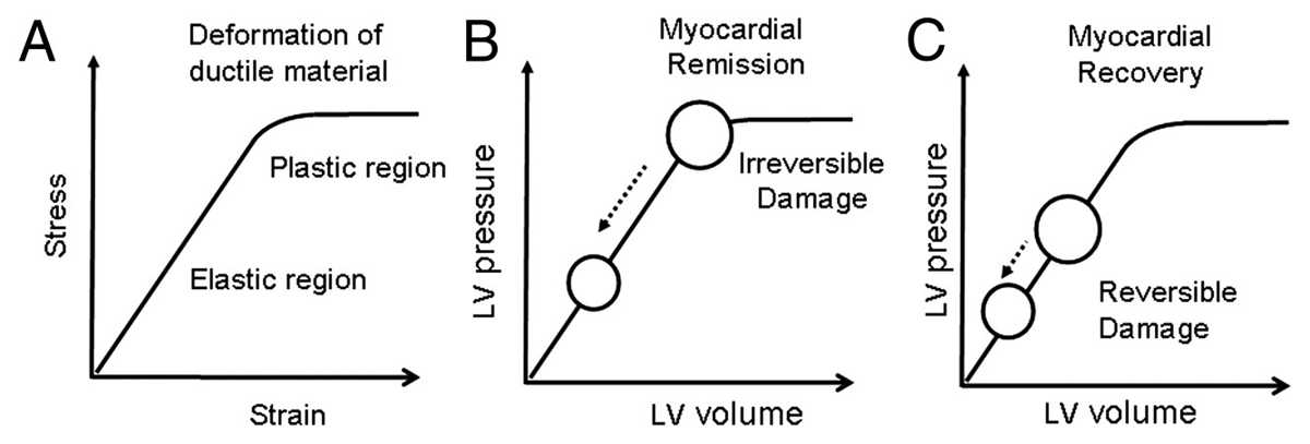
Figure 4
Mechanical engineering science and cardiac remodeling. (A) Diagram of a stress-strain curve of a ductile material illustrating the relationship between an applied force (stress) and deformation (strain). Deformation can lead to reversible changes in a material (elastic deformation) if the properties of the material are not changed, and irreversible changes in a material (plastic deformation). (B) Hypothetical model of reverse remodeling in a heart that has undergone irreversible damage (plastic deformation). (C) Hypothetical model of reverse remodeling with recovery in heart that has undergone reversible damage (elastic deformation). Reproduced with permission from Mann DL et al.2
Human Models of Reverse LV Remodeling
Reverse LV remodeling and normalization of LVEF have been observed in numerous clinical scenarios wherein discontinuation of the inciting event, including cardiotoxic agents, peripartum cardiomyopathy, stress-induced cardiomyopathy, and viral myocarditis, often results in significant improvement in LV size, shape, and function. However, rates of myocardial recovery have been increasing in more recent clinical studies, which is most likely attributable to advances in and widespread application of GDMT and device therapies.
Medical Therapies
Several studies show that treatment with enalapril compared to placebo prevent progression of LV remodeling and result in fewer HF-related deaths,86, 87 and a substudy of the Val-HeFT (Valsartan Heart Failure) trial demonstrated that the addition of valsartan to an existing ACE inhibitor or β-blocker therapy further reduced LV diameter.88 Additional therapies such as carvedilol and spironolactone have been shown to reduce LV volumes when added to ACE inhibitors alone and with β-blockers, respectively, and have been longstanding cornerstone therapies in reducing mortality.89, 90 Interestingly, patients on equivalent drug therapy (β-blocker, ACE inhibitor or ARB, and aldosterone receptor blocker) and with LVEF between 40% to 50% had significantly improved clinical outcomes if they recovered from a worse LV function (LVEF < 40%) compared to either unchanged (LVEF 40-50%) or deteriorated function (LVEF > 50%).91 Lastly, SGLT2 inhibitors are the most recent of adopted medical therapies to demonstrate a reduction primarily in HF hospitalizations when added to standard therapy and also have been shown to reduce LV volumes in some patients.92
Device Therapies
Device therapies also have shown to reduce ventricular volumes and contribute towards reverse remodeling. Both pulsatile and continuous-flow LVADs and axial or centrifugal pumps provided equivalent LV pressure unloading, albeit pulsatile devices provided a greater unloading to LV volume due to higher pump output. Despite these differences, both pulsatile and continuous-flow therapies were equivalent in hemodynamic measures of LV reverse remodeling, although there may be serum biomarkers to suggest greater biological remodeling with the use of pulsatile pumps.93, 94, 95 Cardiac resynchronization therapy has been widely implemented in patients with moderate to severe systolic HF and LV desynchrony as evidenced by a prolonged QRS interval, portending poorer outcomes. Patients treated with CRT had improved contractile function, smaller LV volumes, and less mitral regurgitation at both short- and long-term follow-up.96, 97
Durability of HF with Reverse LV Remodeling and Improved LVEF
Although applications of medical and device therapies have reshaped the landscape and management of HF, it remains a remarkably morbid and mortal disease with rates of death comparable to those of aggressive malignancies. Additionally, the efficacy of current HF therapeutics is highly variable and often dependent on the etiology of the cardiomyopathy as well as duration and severity. Even with reverse LV remodeling that results in normalization of LV structure and function, the vast majority of HF patients remain vulnerable to future HF events. One recent study evaluated the durability of clinical stability in patients with dilated cardiomyopathy who were started on GDMT and followed for 10 years. Of the 408 enrolled patients with dilated cardiomyopathy, only 15% normalized their LV structure and function on GDMT (defined as an LVEF > 50% and LV end-diastolic dimension index < 33 mm/m2). Remarkably, only 60% of the patient cohort with normalized LV structure and function on GDMT were able to maintain clinical stability over the ensuing decade.98 In the TRED-HF (Therapy Withdrawal in Recovered DCM) study, patients with dilated cardiomyopathy and normal LV size and function were randomized to continued treatment with GDMT or phased withdrawal. The investigators found that 44% of patients who discontinued medical therapy had HF relapse within 6 months, which was not observed in the patients who continued GDMT and remained clinically stable.99 As noted by the authors of the TRED-HF study, their findings suggest that “for many patients, improvement in cardiac function following treatment does not reflect full and sustained recovery but rather reflects remission, which requires at least some treatment to be maintained.”99 Viewed together, these data not only highlight the heterogenous response to conventional medical therapies but also the fragility of maintaining clinical stability despite normalization of LV structure and function.
Conclusion
Although reverse LV remodeling occurs in the vast majority of patients who have prolonged LVAD support—and occurs in a significant proportion of HFrEF patients treated with GDMT—reverse LV remodeling with an improved LVEF does not necessarily signify that the patient is cured from HF. Indeed, many HF patients whose ventricles resume a more normal LV size and shape will experience recurrent HF events despite being maintained on state-of-the-art GDMT and receiving excellent clinical care. Further, the results of the TRED-HF study suggest that GDMT is required to maintain clinical stability even in patients who have normalization of their LV structure and function. Collectively, these studies suggest that while achieving normalization of their LV structure and function should be an important goal of HF therapy, we also need to think more holistically about the totality of the HF patient experience and concentrate our future research efforts on identifying the optimal therapeutic approach(es) that will maintain patients in remission, free from future HF events.
Key Points
Reverse remodeling is the process by which a failing heart undergoes efforts towards normalization of geometric, hemodynamic, and biologic changes associated with improved left ventricular ejection fraction and clinical outcomes.
Patients and animal models with heart failure can achieve reverse remodeling through the implementation of medical and device therapies.
The biological underpinnings of myocardial reverse remodeling are complex and associated with a reversion towards a more normal cardiac myocyte and extracellular biology.
Reverse remodeling and myocardial recovery represent improved yet fragile new steady states continuously at risk for future adverse remodeling events. A more complete mechanistic understanding is required to leverage the full therapeutic potential of novel medical and device treatments.
Competing Interests
The authors have no competing interests to declare.