
1. Introduction
Hamstring muscles play a fundamental role in lower-limb function, particularly in hip extension and knee flexion during functional activities such as sitting, walking, and running (Timmins et al., 2020). Hamstring tightness can reduce range of motion and alter normal kinetic linkages, potentially impairing movement efficiency and increasing the risk of musculoskeletal injury (Gajdosik, Hatcher, & Whitsell, 1992; Kotteeswaran, Snigdha, & Alagesan, 2014). While overuse is a well-recognized contributor, hamstring tightness is also commonly associated with underuse, particularly among individuals with sedentary lifestyles (Chaphekar et al., 2021). University students represent a population at high risk due to prolonged sitting during academic activities, with a previous study reporting a prevalence as high as 45% among female students (Thakur & Rose, 2016). Furthermore, prolonged sitting and insufficient physical activity have been identified as important factors associated with hamstring tightness in university students (Shukla & Patel, 2021; Thakur & Rose, 2016). Given that the present study included female university students with sedentary behaviors and prolonged sitting, these epidemiological findings are relevant to the study sample and support the rationale for investigating hamstring flexibility in this population.
From a myofascial perspective, the hamstrings are part of the superficial back line (SBL), a continuous connective tissue network extending from the plantar fascia to the epicranial fascia (Myers, 2014). This myofascial chain contributes to force transmission and movement coordination across multiple body segments, particularly in the sagittal plane. Dysfunction or increased tension in one region of the SBL may influence mechanical behavior and movement in other interconnected regions. For example, hamstring tightness has been associated with altered lumbopelvic mechanics and may contribute to movement dysfunction and low back pain (Kotteeswaran, Snigdha, & Alagesan, 2014).
Based on the concept of myofascial continuity, interventions targeting one structure within the SBL may produce remote effects on other structures along the chain. Static hamstring stretching is commonly used to improve flexibility and reduce muscle tightness (Medeiros et al., 2016; Page, 2012). Additionally, self-myofascial release techniques, such as foam rolling, have been shown to improve hamstring flexibility when applied directly to the hamstrings (Junker & Stöggl, 2015). Interestingly, previous studies have demonstrated that applying self-myofascial release to distant structures within the same myofascial chain, such as the plantar fascia, can also improve hamstring flexibility and functional performance, including sit and reach scores (Do et al., 2018; Grieve et al., 2015; Shetty & Dsouza, 2018). Furthermore, combining hamstring stretching with plantar fascia mobilization (PFM) has been shown to produce greater improvements in hamstring flexibility compared with stretching alone (Izhar et al., 2023). These findings support the concept that releasing tension in one region of the SBL may influence flexibility and function in other interconnected regions.
Despite these findings, most previous studies have primarily focused on isolated flexibility outcomes while providing limited information regarding body segment kinematics during functional movement tasks. Evaluating body segment angles during the sit and reach task may provide additional insight into multi-segment movement patterns beyond flexibility alone. Because the sit and reach task primarily involves sagittal plane forward bending, two-dimensional assessment of pelvic, hip, lumbar, thoracic, and shoulder angles can characterize the relative contribution of different body segments during the movement. Such information may improve understanding of how posterior chain interventions influence movement patterns and functional performance beyond isolated flexibility outcomes. Furthermore, evidence regarding the combined effects of PFM and active posterior chain stretching (APCS) applied over consecutive sessions remains limited. In addition, most previous studies, primarily focused on immediate responses and isolated flexibility outcomes, with limited investigation of cumulative short-term effects and body segment kinematics during functional movement tasks. Understanding these combined and multi-segment responses is important for optimizing intervention strategies targeting the posterior myofascial chain.
Therefore, this study aimed to compare the effects of APCS alone and APCS combined with PFM (APCS+PFM) on hamstring flexibility, body segment kinematics, and sit and reach performance in individuals with hamstring tightness over consecutive sessions. It was hypothesized that APCS+PFM would produce greater improvements in hamstring flexibility, body segment angles, and sit and reach scores than APCS alone.
2. Methods
2.1 Study design
This study employed a 2 × 3 mixed factorial design. Participants who met the inclusion criteria were randomly allocated to the APCS group or the APCS+PFM group using a computer-generated random sequence. Group assignments were prepared before participant enrollment and implemented by an investigator who was not involved in outcome assessment. Popliteal angles, body segment angles during the sit and reach test, and sit and reach scores were measured at baseline (before intervention; T0), immediately post-intervention (T1), and after three consecutive days (T2). The primary outcome measure was the popliteal angle, which was selected as an indicator of hamstring flexibility. Secondary outcomes included body segment angles during the sit and reach task and sit and reach performance.
2.2 Participants
A total of 18 female university students aged 18–25 years were recruited and allocated to two groups: APCS (N = 9) and APCS+PFM (N = 9). Eligibility criteria included hamstring muscle tightness (popliteal angle > 20°), a body mass index (BMI) of 18.5–22.9 kg/m² (Asian-Pacific classification), sitting for more than 6 h/day, and engaging in less than 150–300 min/week of moderate-intensity or 75–150 min/week of vigorous-intensity aerobic physical activity in accordance with the World Health Organization (WHO) 2020 physical activity guidelines (Bull et al., 2020; Fatima et al., 2017; Lim et al., 2017). Exclusion criteria included generalized joint hypermobility (Beighton score ≥ 4), abnormal lower extremity alignment (hallux valgus, high arch, flat feet, knee hyperextension, knock knees, bowlegs, femoral anteversion, femoral retroversion, and excessive anterior or posterior pelvic tilt), abnormal spinal alignment (scoliosis, thoracic hypokyphosis, thoracic hyperkyphosis, lumbar hypolordosis, and lumbar hyperlordosis), limb length discrepancy, musculoskeletal injuries within the past three months, and a history of surgery or fracture (Cooper et al., 2018; Engelbert & Rombaut, 2022; Joshi et al., 2018). Written informed consent, approved by the Human Research Ethics Committee of Chulabhorn Royal Academy (EC 098/2566), was obtained from all participants prior to participation.
The sample size was determined based on a previous study (Williams & Selkow, 2019), using sit and reach distance for the sample size estimation because this outcome was the most comparable variable available from studies investigating similar interventions during study planning. An estimated effect size (partial eta squared) of 0.12, indicating a moderate effect, was used. An alpha level of 0.05 and a statistical power of 0.80 were set, and the calculation was performed using G*Power version 3.1.9.4. The initial calculation indicated a total sample size of 14 participants. After accounting for a 20% dropout rate, the final sample size was adjusted to 18 participants, with nine participants allocated to each group.
2.3 Popliteal angle measurement
The popliteal angle was measured in the dominant limb, as determined by a ball-kicking test (Hoffman et al., 1998). All participants were identified as right-limb dominant. Digital photogrammetry was conducted to assess the popliteal angle. Non-reflective spherical markers (15 mm in diameter) were placed on the lateral epicondyle of the femur and the lateral malleolus of each participant. A camera mounted on a tripod was positioned 40 cm above the floor and 200 cm from the participant. The test limb was positioned with the hip and knee flexed to 90° and stabilized using a plyometric box (75 × 90 × 30 cm), while the contralateral limb remained parallel to the floor in the supine position (Waseem et al., 2009). In addition, the pelvis was secured with a strap during testing to control accessory movements and maintain lumbopelvic stability throughout the assessment. Participants were instructed to gradually extend the test knee to the maximal range while maintaining the hip at 90° of flexion without compensatory posterior pelvic tilt or lumbar flexion, and the contralateral knee was kept fully extended (Waseem et al., 2009). The final position was held for 5 seconds to allow image capture (Chaphekar et al., 2021). The procedure was performed for three repetitions, with a 30-second rest between trials. The mean value of the three trials was used for statistical analysis. The popliteal angle was analyzed from the images using Kinovea software (version 0.9.5). It was defined as the angle formed by the intersection of an imaginary vertical line passing through the marker placed on the lateral epicondyle of the femur and a line connecting the markers on the lateral epicondyle of the femur and the lateral malleolus (Herrington, 2013). The popliteal angle was measured by a blinded investigator. The intra-rater reliability of the measurement was excellent (ICC(3,3) = 0.90, 95% CI: 0.78–0.93), calculated from repeated measurements performed as part of the present study.
2.4 Sit and reach measurement
The sit and reach test was performed using a Digital Front Softening Tester (T.K.K., Japan), adapted as a digital alternative to the standard sit and reach box to measure sit and reach distance and the flexibility of the posterior kinetic chain, including the lower back, torso, and shoulder girdle. Participants started in a long sitting position on the floor with full knee extension, the plantar surfaces of the feet in contact with the testing apparatus in a neutral ankle position, and both arms extended forward with the shoulders flexed, parallel to the lower limbs, in a relaxed position (Kawano et al., 2010). The starting position (score) was recorded directly from the measuring scale of the apparatus. Participants were instructed to gradually reach forward by sliding their hands along the apparatus as far as possible while maintaining knee extension and avoiding lateral trunk flexion. At the end range, participants were instructed to maintain the position for 2 seconds to allow image capture (Lemmink et al., 2003). The final sit and reach score was recorded from the measuring scale. The sit and reach score represented the excursion achieved during the sit and reach test and was calculated as the difference between the final and starting scores. The test was performed for three repetitions with a 30-second rest between trials. The mean value of the three trials was used for statistical analysis. All measurements were conducted by a blinded investigator who demonstrated excellent intra-rater reliability (ICC(3,3) = 0.95, 95% CI: 0.87–0.98), calculated from repeated measurements performed as part of the present study.
2.5 The body segment angle measurement
Body segment angles, including the shoulder, thoracic, lumbar, pelvic, and hip angles, were assessed during the sit and reach test using photogrammetry. Non-reflective spherical markers were placed on the following anatomical landmarks of each participant: the spinous processes of C7, T1, T12, and L5; the glenohumeral joint (GH); the anterior superior iliac spine (ASIS); and the greater trochanter of the femur (GT) (Figure 1A). During the assessment, participants were instructed to maintain a forward-bending movement pattern and to avoid trunk rotation and lateral flexion throughout testing. Marker visibility was verified by the investigator before and during image capture to ensure that all anatomical landmarks remained clearly identifiable. A camera mounted on the tripod was positioned at a height of 48 cm and at a distance of 200 cm from the participant on the dominant right side. All photographs were obtained using a standardized setup with a fixed camera position, distance, height, lens setting, and focus, while all participants were positioned consistently throughout the assessment. These procedures were implemented to minimize measurement error associated with two-dimensional photogrammetry. Marker positions were captured at the starting and ending positions during the sit and reach test following the standardized test protocol. Joint angles were analyzed from the images using Kinovea software (version 0.9.5). The shoulder angle was defined as the angle between an imaginary horizontal line passing through C7 and a line connecting C7 to the GH joint (Sohrabi et al., 2020). The thoracic angle in the sagittal plane was defined as the angle formed by the intersection of two straight lines drawn through T1 and T12, each parallel to the longitudinal axis of the trunk. The lumbar angle was defined as the angle formed by the intersection of two straight lines drawn through T12 and L5, parallel to the longitudinal axis of the trunk. The pelvic angle was defined as the angle between a horizontal line parallel to the floor and a straight line passing through L5, representing the longitudinal axis of the trunk (Mier & Shapiro, 2013). The hip joint angle was defined as the angle formed by the intersection of a line connecting the ASIS to the GT and a horizontal line passing through the GT (Kawano et al., 2010) (Figure 1B). Body segment angular excursions were calculated as the difference between the angles measured at the starting and ending positions. The mean value of the three trials was used for statistical analysis. All measurements were performed by a blinded investigator who demonstrated excellent intra-rater reliability (ICC(3,3) = 0.90, 95% CI: 0.80–0.95), calculated from repeated measurements performed as part of the present study.
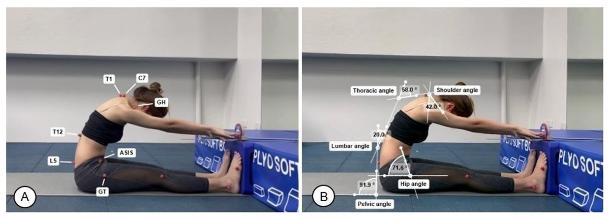
Figure 1
Body segment angle measurement during the sit and reach test. (A) Anatomical marker positions showing C7, T1, T12, L5, GH, ASIS, and GT. (B) Calculation of shoulder, thoracic, lumbar, pelvic, and hip angles using Kinovea software.
2.6 Interventions
Two intervention programs were implemented: APCS and APCS+PFM. Participants in the APCS group performed active hamstring and gastrocnemius stretching exercises. The stretching exercises were performed in a standardized sequence, with the hamstring stretch performed first, followed by the gastrocnemius stretch. For the active hamstring stretch, participants stood on one leg while placing the heel of the stretching leg on a chair, with the knee fully extended and the ankle in dorsiflexion. From this starting position, participants were instructed to flex the trunk forward and reach toward the toes as far as possible until a stretch sensation was perceived in the hamstring muscles. The stretch was held for 30 seconds before returning to the starting position (Shetty & Dsouza, 2018) (Figure 2A). Three repetitions were performed on each side, with a 30-second rest interval between repetitions. The procedure was then repeated on the contralateral leg. For the active gastrocnemius stretch, participants stood facing a wall with both hands placed on the wall at shoulder height and shoulder-width apart. They then simultaneously stepped one leg posteriorly and leaned the trunk forward, maintaining full extension of the stretching leg while the contralateral leg remained anterior with the knee flexed, until a stretch sensation was perceived in the gastrocnemius muscle of the stretching leg (Figure 2B). The stretch was held for 30 seconds, followed by a 30-second rest interval between repetitions. Three repetitions were performed on each side. The starting leg was randomly assigned for each session. Both stretching exercises were performed for three consecutive days.
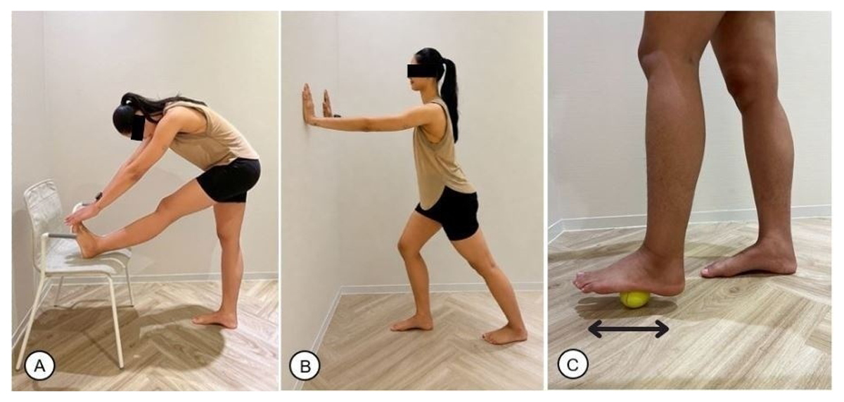
Figure 2
Intervention procedures. (A) Active hamstring stretching performed with the knee fully extended and the ankle dorsiflexed while the heel was placed on a chair. (B) Active gastrocnemius stretching performed against a wall with the stretching knee maintained in full extension. (C) Plantar fascia mobilization performed using a tennis ball rolled along the plantar surface of the foot from the heel toward the metatarsal heads with emphasis on the medial longitudinal arch.
The APCS+PFM program consisted of the APCS program combined with PFM. Participants in this group performed the same stretching protocol as described for the APCS group. Following completion of the stretching exercises on each leg, PFM was performed on the same side before proceeding to the contralateral leg. During the PFM intervention, participants rolled a tennis ball under the plantar surface of the foot from the heel to the metatarsal heads, with emphasis on the medial longitudinal arch (Grieve et al., 2015; Izhar et al., 2023; Shetty & Dsouza, 2018) (Figure 2C). Participants were instructed to apply pressure sufficient to produce mild discomfort without pain, corresponding to a Numeric Rating Scale (NRS) score of 5–7, for 2 minutes (Cheatham et al., 2015; Russo et al., 2023; Shetty & Dsouza, 2018; Takeuchi, Akizuki, & Nakamura, 2021). All participants used the same tennis ball throughout all intervention sessions and received standardized verbal instructions regarding rolling technique, movement direction, intervention duration, and pressure application. In addition, all intervention procedures were supervised by the investigator throughout the study period. This program was conducted for three consecutive days. The intervention programs were evaluated by experts using the Item-Objective Congruence (IOC) index, yielding a score of 0.81, indicating high content validity.
2.7 Statistical analysis
All statistical analyses were performed using SPSS version 26.0 (IBM Corp., Armonk, NY, USA). Data normality was assessed using the Shapiro–Wilk test, and all outcomes were normally distributed. Differences in baseline participant characteristics between the two groups were evaluated using independent samples t tests. A 2 × 3 (group × time) mixed-design repeated-measures ANOVA was conducted to examine the effects of group and time on the dependent variables, including the popliteal angle, body segment angles, and sit and reach score. The assumption of sphericity was evaluated using Mauchly’s test, and Greenhouse-Geisser corrections were applied when the assumption was violated. Bonferroni-adjusted pairwise comparisons were performed for post hoc analyses when significant main effects or interactions were identified. Statistical significance was set at p < 0.05 with a 95% confidence level. Effect sizes for the ANOVA were reported as partial eta squared and interpreted as follows: 0.01 = small effect, 0.06 = moderate effect, and 0.14 = large effect. Effect sizes for post hoc comparisons were calculated using Cohen’s d and interpreted as small (0.2), medium (0.5), or large (0.8).
3. Results
3.1 Characteristics of participants
There were no significant differences between the groups of the general characteristics of the participants, including BMI, height, weight, sitting hours, and popliteal angle. These results indicated homogeneity of participants (Table 1).
Table 1
General characteristics of participants (Mean ± SD).
| CHARACTERISTICS | APCS+PFM (N = 9) | APCS (N = 9) | P |
|---|---|---|---|
| BMI (kg/m²) | 19.76 ± 1.12 | 19.64 ± 0.84 | 0.501 |
| Height (cm) | 159.33 ± 3.74 | 163.67 ± 5.02 | 0.417 |
| Weight (kg) | 50.22 ± 4.18 | 52.67 ± 3.39 | 0.664 |
| Sitting hours (h) | 7.67 ± 2.18 | 8.89 ± 2.26 | 0.260 |
[i] APCS+PFM: active posterior chain stretching combined with plantar fascia mobilization, APCS: active posterior chain stretching, BMI: body mass index.
3.2 Body segment angles
For the repeated-measures ANOVA analyses, Mauchly’s test indicated that the assumption of sphericity was satisfied for the analyses of the hip angle, pelvic angle, thoracic angle, and popliteal angle. Greenhouse–Geisser corrected degrees of freedom and p values were applied for the analyses of the lumbar angle, shoulder angle, and sit and reach score because the assumption of sphericity was violated.
No significant interaction between group and time was observed. There were no significant main effects of time on the shoulder, thoracic, or lumbar angles. In contrast, a significant main effect of time was observed for the pelvic and hip angles across the three measurement times (p = 0.028; Effect size = 0.079, and p = 0.048; Effect size = 0.281, respectively) (Table 2). Post hoc analyses revealed that the pelvic angle at T2 was significantly larger than at T0 (p = 0.009; Effect size = 0.722) and T1 (p = 0.001; Effect size = 0.910). Similarly, the hip angle at T2 was significantly greater than at T0 (p = 0.006; Effect size = 0.883) and T1 (p = 0.016; Effect size = 0.880). These findings indicate progressive improvements in pelvic and hip alignment over time.
Table 2
Body segment angular excursions during the sit and reach test across time and between groups (mean ± SD).
| BODY SEGMENT ANGLES (°) | GROUP | INTERACTION EFFECTS | TIME | GROUP | ||||||||
|---|---|---|---|---|---|---|---|---|---|---|---|---|
| APCS+PFM (T0) | APCS+PFM(T1) | APCS+PFM(T2) | APCS(T0) | APCS(T1) | APCS(T2) | p | ES | p | ES | p | ES | |
| Shoulder | 31.74 ± 12.48 | 34.49 ± 14.11 | 37.88 ± 14.66 | 31.96 ± 10.92 | 27.88 ± 12.07 | 33.73 ± 11.36 | 0.14 | 0.05 | 0.08 | 0.12 | 0.53 | 0.20 |
| Thoracic | 24.14 ± 10.22 | 24.09 ± 11.51 | 22.77 ± 10.63 | 16.16 ± 6.36 | 14.74 ± 9.18 | 16.92 ± 8.62 | 0.26 | 0.09 | 0.42 | 0.03 | 0.07 | 0.04 |
| Lumbar | 11.20 ± 4.78 | 12.06 ± 4.99 | 12.86 ± 6.19 | 6.70 ± 4.67 | 8.53 ± 5.75 | 8.65 ± 5.04 | 0.32 | 0.42 | 0.61 | 0.09 | 0.09 | 0.11 |
| Pelvic | 14.57 ± 6.49 | 14.92 ± 3.87 | 19.34 ± 4.50*,** | 18.74 ± 5.99 | 19.92 ± 7.57 | 22.47 ± 7.79*,** | 0.09 | 0.12 | 0.02 | 0.07 | 0.08 | 0.14 |
| Hip | 9.13 ± 2.31 | 9.73 ± 1.61 | 13.49 ± 4.86*,** | 11.16 ± 4.46 | 11.64 ± 4.40 | 13.57 ± 5.14*,** | 0.15 | 0.02 | 0.04 | 0.28 | 0.34 | 0.08 |
[i] APCS+PFM: active posterior chain stretching combined with plantar fascia mobilization, APCS: active posterior chain stretching, T0: baseline, T1: immediately post-intervention, T2: after three consecutive days, ES: effect size. *p < 0.05, within-group comparison between T0 and T2. **p < 0.05, within-group comparison between T1 and T2.
3.3 Sit and reach score
No significant interaction between group and time was observed. A significant main effect of time was observed across the three measurement times (p = 0.001; Effect size = 0.405). The main effect of time showed that the sit and reach score excursion at T1 was significantly higher than that at T0 (p = 0.028; Effect size = 0.588), the score at T2 was significantly greater than that at T0 (p = 0.001; Effect size = 0.925), and the score at T2 was significantly greater than that at T1 (p = 0.014; Effect size = 0.641). The mean ± SD values of sit and reach score excursion in the APCS+PFM and APCS groups were as follows: at T0, the scores were 22.18 ± 6.77 cm and 24.83 ± 8.15 cm; at T1, 24.41 ± 7.12 cm and 26.60 ± 9.24 cm; and at T2, 27.72 ± 8.36 cm and 27.91 ± 8.21 cm (Figure 3).
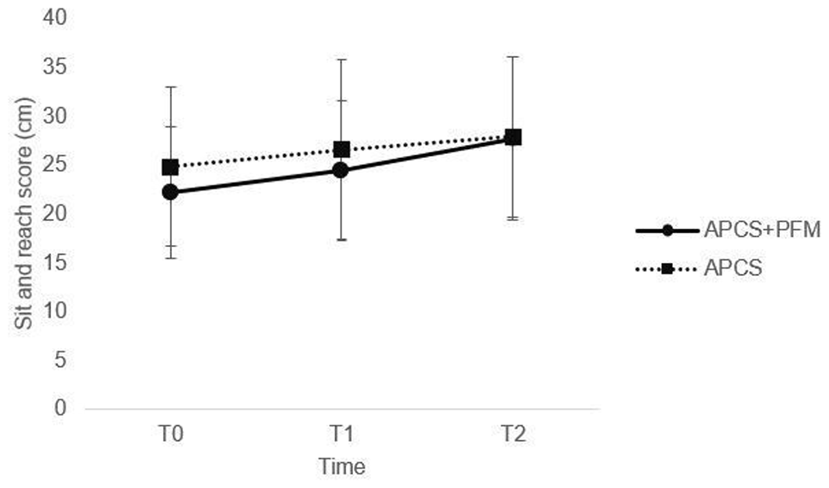
Figure 3
Sit and reach score excursion for the APCS and APCS+PFM groups at T0, T1, and T2. APCS: active posterior chain stretching, APCS+PFM: active posterior chain stretching combined with plantar fascia mobilization.
3.4 Popliteal angle
A significant group × time interaction was observed for the popliteal angle (F(2,32) = 2.215, p = 0.046; Effect size = 0.122). In the APCS+PFM group, the popliteal angle at T1 was significantly lower than that at T0 (mean difference = –4.57°, 95% CI: –6.30 to –2.84; p = 0.004; Effect size = 1.330), the angle at T2 was smaller than that at T0 (mean difference = –8.85°, 95% CI: –12.80 to –4.90; p = 0.003; Effect size = 1.424), and the angle at T2 was also smaller than that at T1 (mean difference = –4.28°, 95% CI: –6.70 to –1.86; p = 0.010; Effect size = 1.115). The mean ± SD of popliteal angles were 37.27 ± 8.28° at T0, 32.70 ± 7.18° at T1, and 28.42 ± 7.57° at T2. In the APCS group, the popliteal angle at T1 was significantly smaller than that at T0 (mean difference = –5.63°, 95% CI: –10.08 to –1.18; p = 0.002; Effect size = 1.516), and the angle at T2 was smaller than that at T0 (mean difference = –5.79°, 95% CI: –6.99 to –4.59; p = 0.004; Effect size = 1.342). The mean ± SD of popliteal angles at T0, T1, and T2 were 45.53 ± 8.39°, 39.90 ± 9.36°, and 39.74 ± 8.89°, respectively. Additionally, the popliteal angle in the APCS+PFM group was significantly lower than that in the APCS group at T2 (p = 0.030; Effect size = 0.263) (Figure 4). This significant reduction in popliteal angle indicates an improvement in hamstring flexibility, as a smaller popliteal angle reflects greater knee extension range of motion, indicating reduced hamstring tightness.
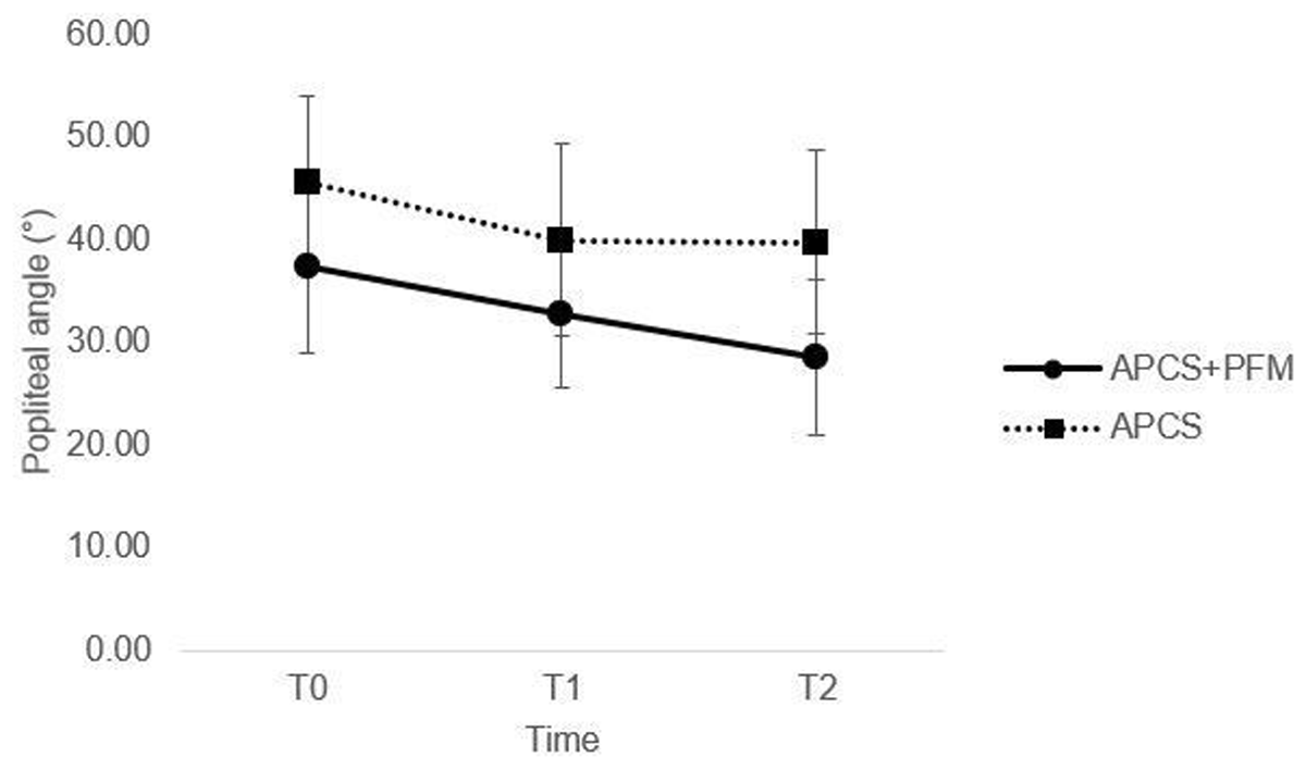
Figure 4
Popliteal angle for the APCS and APCS+PFM groups at T0, T1, and T2.
APCS: active posterior chain stretching, APCS+PFM: active posterior chain stretching combined with plantar fascia mobilization.
4. Discussion
The present study demonstrated that APCS+PFM produced greater improvements in hamstring flexibility, as reflected by a greater reduction in popliteal angle, compared with APCS alone. In addition, both intervention groups showed improvements in sit and reach performance and associated body segment kinematics, particularly pelvic and hip angles, over the three-day intervention period. These findings support the hypothesis that incorporating PFM into posterior chain stretching enhances short-term hamstring flexibility. The observed improvements in both groups suggest that repeated posterior chain interventions may influence movement patterns during the sit and reach task regardless of the inclusion of PFM. In our study, although matched-pair grouping based on age, BMI, and sitting hours was not applied, the independent t-test results showed no significant differences in these characteristics between the groups. Therefore, the observed changes in the dependent variables were less likely to be influenced by participant characteristics and may be partly attributable to the intervention program.
Active hamstring stretching, either alone or in combination with gastrocnemius stretching, can increase hamstring flexibility through both mechanical and neurophysiological mechanisms (Weerapong et al., 2004). Mechanically, stretching promotes viscoelastic deformation of the musculotendinous unit, which may reduce passive stiffness (Weerapong et al., 2004). Neurophysiologically, stretching may facilitate reciprocal inhibition, whereby activation of the antagonist muscle contributes to relaxation of the target muscle (Sharman et al., 2006; Weerapong et al., 2004). In addition, previous studies have reported that both hamstring stretching alone and combined hamstring and gastrocnemius stretching improve hamstring flexibility compared with a control group, with the combined stretching producing greater improvements (Russell et al., 2010). It is possible that addressing multiple structures along the same myofascial chain, rather than focusing solely on the target muscle, may contribute to greater improvements in flexibility.
Furthermore, a previous study demonstrated that stretching the hamstring muscles during ankle dorsiflexion, which involves concurrent stretching of the gastrocnemius, increased cervical flexion and extension range of motion (Hyong & Kang, 2013). This observation indicates that stretching one region of the body may influence flexibility in distant regions. Such remote effects may be explained by mechanical interactions within connective tissues that link different body segments (Hyong & Kang, 2013). Therefore, stretching structures that are mechanically associated with the target muscle may enhance flexibility through force transmission along connective tissue pathways. These mechanisms may explain the remote flexibility improvements observed in our study.
Our study focused on the PFM technique, which involves repeatedly applying mechanical pressure through a rolling friction method using a tennis ball under the plantar surface of the foot with body weight. This approach has been proposed to stimulate fascial mechanoreceptors, including Golgi tendon organs and Ruffini endings, thereby reducing local muscle and fascial tension (Do et al., 2018; Hyong & Kang, 2013; Remvig et al., 2008; Schleip, 2003; Tozzi, 2012). In addition, mechanical loading during rolling may increase local tissue temperature and blood flow, enhance fascial hydration, improve viscoelastic properties, and reduce passive tissue stiffness (Bradbury-Squires et al., 2015; Izhar et al., 2023; Kim et al., 2020; MacDonald et al., 2013; Russo et al., 2023; Sefton, 2004). These local mechanical and physiological responses may be transmitted to more proximal structures through anatomically continuous fascial connections, consistent with the concept of myofascial force transmission (Cruz-Montecinos et al., 2015; Grieve et al., 2015; Krause et al., 2016), and may partially explain the greater reduction in hamstring tightness observed in the APCS+PFM group compared with the APCS group.
These mechanisms may explain the immediate reduction in hamstring tightness observed in our study. This finding is consistent with a previous study that also reported immediate improvements in sit and reach scores and dorsiflexion angles following SMR using a foam roller applied to the plantar fascia. Notably, flexibility increased by up to 70% within 2 minutes after the intervention (Fauris et al., 2021). Based on the concept of the SBL, in which the plantar fascia is anatomically connected to the calf muscles and further linked to the hamstrings, applying PFM to the plantar region may help immediately reduce tension along the fascial chain, including the hamstrings. Moreover, several studies have reported that PFM produces acute effects on improving hamstring flexibility, as evidenced by improvements in sit and reach scores, toe-touch tests, and passive straight leg raise tests (Do et al., 2018; Grieve et al., 2015). These findings support the notion that PFM can immediately reduce hamstring tightness through remote effects mediated by the fascial system.
However, the present study differs from these previous studies in several important aspects. Most previous investigations primarily examined the immediate effects of isolated plantar fascia interventions in healthy individuals, with outcomes largely limited to flexibility measures assessed immediately after a single treatment session. In contrast, the present study evaluated the combined effects of APCS and PFM over three consecutive days in sedentary female university students with hamstring tightness. Furthermore, in addition to hamstring flexibility and sit and reach performance, body segment kinematics were assessed during a functional movement task at multiple time points (T0, T1, and T2), providing insight into both immediate and short-term responses to the intervention.
Moreover, the cumulative short-term effects observed after three consecutive days of intervention may be explained by mechanisms similar to those underlying the immediate responses. Repeated mechanical loading through the combination of PFM and active stretching may induce viscoelastic creep, increase tissue compliance, and facilitate the loosening of collagen cross-links within connective tissue (Burk et al., 2019; Gabriel et al., 2022; Kalichman & David, 2017; Wilke et al., 2018). These adaptations may enhance stretch tolerance and contribute to progressive improvements in target muscle flexibility over the intervention period. In addition, the observed improvements may also reflect perceptual factors associated with stretch-related discomfort rather than changes in tissue extensibility alone.
While the continued reduction in popliteal angle from T1 to T2 in the APCS+PFM group may suggest an additive or cumulative effect of PFM when combined with APCS, alternative explanations for the observed between-intervention differences cannot be excluded. Moreover, the APCS+PFM group demonstrated lower baseline popliteal angles than the APCS group, although this difference was not statistically significant. Therefore, these findings should be interpreted cautiously.
In addition, the APCS+PFM group demonstrated an approximately 8.85° reduction in popliteal angle, indicating improved hamstring flexibility after three consecutive days of intervention. Although a clinically meaningful threshold for change in popliteal angle has not been established in individuals with hamstring tightness, the magnitude of improvement observed in the present study may be considered practically relevant. A previous study reported improvements in hamstring flexibility ranging from approximately 5° to 12° following stretching interventions, and an improvement of approximately 8.9° in active straight leg raise range of motion has been reported after an acute stretching intervention (López-Miñarro et al., 2012). Although different flexibility assessments were used, the magnitude of change observed in the present study falls within the range previously reported for stretching interventions and may therefore reflect a substantial improvement in hamstring flexibility. These findings suggest that APCS+PFM produced substantial short-term improvements in hamstring flexibility.
Importantly, the present study also evaluated body segment kinematics during a functional movement task. Significant improvements were observed in pelvic and hip angles, whereas shoulder, thoracic, and lumbar angles did not significantly change across time. These findings suggest that improved sit and reach performance may be associated with greater pelvic rotation and hip flexion rather than increased spinal flexion during forward bending. From a practical perspective, greater pelvic rotation and hip flexion during forward bending may reflect improved lumbopelvic movement coordination and a more efficient distribution of motion along the posterior chain during sagittal plane movements. Although these changes were observed during the sit and reach test, they may have implications for movement quality in activities requiring coordinated lumbopelvic motion. Therefore, sit and reach performance should be interpreted as an integrated functional outcome reflecting the combined contribution of multiple movement components rather than a measure of a single anatomical structure or flexibility characteristic.
From a biomechanical perspective, this movement pattern may reflect improved lumbopelvic contribution associated with enhanced hamstring extensibility, as reduced passive tension in the hamstrings may facilitate greater anterior pelvic rotation and hip flexion during forward bending. Previous studies have reported that greater hamstring extensibility is associated with increased anterior pelvic tilt and hip flexion during sit and reach performance, whereas reduced hamstring extensibility is associated with increased thoracic flexion compensation during the sit and reach task (López-Miñarro & Rodríguez-García, 2010; Mier & Shapiro, 2013). Therefore, these findings provide additional insight beyond isolated flexibility measurements and support the concept that interventions targeting the posterior chain may influence multi-segment movement patterns and functional performance.
This study has several limitations. First, the findings should be interpreted with caution, as they reflect only the immediate and short-term cumulative effects of the intervention rather than long-term adaptations. In addition, the absence of a no-intervention, sham, or PFM-only comparison group limits the ability to completely exclude potential influences of familiarization or repeated testing effects. Accordingly, these results should be considered preliminary, and future longitudinal studies incorporating additional control conditions are warranted to determine whether the observed improvements are sustained over time and to further distinguish intervention-specific effects from potential testing-related effects. Second, our sample consisted of sedentary individuals with hamstring tightness, which may limit the generalizability of the findings. Other populations, such as males, athletes, and individuals with musculoskeletal or clinical conditions, may demonstrate different tissue properties, movement characteristics, and neuromuscular responses that could influence the effectiveness of the intervention. Therefore, further research is needed to examine the effects of this program in males, athletic populations, and individuals with musculoskeletal or clinical conditions. Third, pressure application during PFM was based on subjective tolerance criteria rather than objective quantification methods. Future studies may incorporate objective quantification methods to improve intervention standardization and consistency. In addition, body segment kinematics were assessed using two-dimensional photogrammetry during the sit and reach task. However, this method may not have fully captured subtle multi-planar movements and could have introduced projection error. Future studies may incorporate three-dimensional motion analysis to further clarify multi-planar movement characteristics during sit and reach performance. Finally, the present study did not include direct assessments of tissue stiffness or neuromuscular activity, such as shear wave elastography or surface electromyography. Future studies incorporating these measures may help clarify whether the observed improvements are primarily related to mechanical, neurophysiological, or perceptual mechanisms associated with stretch tolerance.
5. Conclusion
This study extends previous evidence by moving beyond earlier studies that primarily examined the immediate effects of isolated PFM on flexibility outcomes after a single intervention session. In contrast, the present study evaluated the combined effects of APCS along the lower limb segment of the SBL and PFM across three consecutive intervention sessions while simultaneously assessing body segment kinematics during a functional movement task. This broader approach provides additional insight into both short-term cumulative changes in hamstring flexibility and movement patterns. Our findings indicate that incorporating PFM into stretching of the whole lower limb may contribute to greater short-term improvements in hamstring flexibility compared with APCS alone. However, both interventions also improved sit and reach performance and pelvic and hip angular excursions over time. These findings suggest that repeated posterior chain interventions may influence movement patterns during the sit and reach task regardless of the inclusion of PFM. Collectively, these findings provide preliminary evidence supporting the integration of PFM as an adjunct to posterior chain stretching in sedentary female university students with hamstring tightness and suggest that addressing multiple interconnected structures along the posterior chain may enhance immediate and short-term flexibility and functional movement outcomes.
Ethics and consent
This study was conducted in accordance with the Declaration of Helsinki. Ethical approval was obtained from the Human Research Ethics Committee of Chulabhorn Royal Academy (EC No. 098/2566). Written informed consent was obtained from all participants prior to participation in the study.
Acknowledgements
The authors would like to thank the Laboratory of the School of Human Kinetics and Health, Chulabhorn Royal Academy, for providing the facilities and venue for data collection. The authors also thank Pandech C. and Hinsungnoen S. for their assistance in conducting the study.
Data Accessibility Statement
The data are not publicly available due to privacy and ethical restrictions but are available from the corresponding author on reasonable request.
Author Contributions
All authors contributed substantially to the manuscript. Pongserm S. was responsible for investigation, methodology, formal analysis, and writing the original draft. Kongtong W. was responsible for conceptualization, supervision, review and editing, and served as the corresponding author. All authors have read and agreed to the published version of the manuscript.