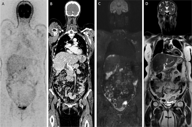
Have a personal or library account? Click to login
Whole Body Diffusion-Weighted MRI in Ovarian Cancer Staging Cover

Whole Body Diffusion-Weighted MRI in Ovarian Cancer Staging

Open Access
|Nov 2019

Figures & Tables

jbsr-103-1-1948-g1.png
Figure 1

Patient with diagnosis of ovarian cancer: (A, B) Time-of-flight FDG-PET/CT depicts limited number of peritoneal metastases. (C, D) Contrary, WB-DWI/MRI shows diffuse nodular and confluent carcinomatosis visible as b1000 hyperintense lesions on the (C) DWI-image.

DOI: https://doi.org/10.5334/jbsr.1948 | Journal eISSN: 2514-8281
Language: English
Submitted on: Sep 14, 2019
Accepted on: Sep 20, 2019
Published on: Nov 18, 2019
Published by: Ubiquity Press
In partnership with: Paradigm Publishing Services
Publication frequency: 1 issue per year

© 2019 Vincent Vandecaveye, Raphaëla Dresen, published by Ubiquity Press
This work is licensed under the Creative Commons Attribution 4.0 License.