
Introduction
The genus Alcea (Malvaceae) comprises approximately 70–75 species worldwide, primarily distributed in Southwest Asia (Escobar et al., 2012), with 34 species recorded in Iran (Pakravan 2008). Linnaeus (1753), following Tournefort (1700), was the first to distinguish between the genera Alcea and Althaea. However, later botanists such as Willdenow (1800), De’Candolle (1828), and Baker (1890) merged these two genera under the name Althaea. Alefeld (1862) moved some Althaea species to Alcea later. Subsequent taxonomic studies on Alcea were conducted by Boissier (1867) and Iljin (1949).
More comprehensive taxonomic investigations were carried out by Zohary (1963a, 1963b) in Southwest Asia, and by Riedl (1976) and Pakravan (2008) within the Iranian flora. Given the high degree of polymorphism in morphological traits (Pakravan, 2001), various biosystematic approaches have been employed to aid in the taxonomy of Alcea, including analyses of seed coat characteristics (Ozkan & Uzunhisarciklic, 2007), seed protein electrophoresis (Pakravan, 2025), palynology (Cabi et al., 2009), and anatomical studies (Mohammad et al., 2018). The most significant phylogenetic research on Alcea was conducted by Escobar et al. (2012), who confirmed its monophyly and its distinction from Althaea.
Anatomical studies are considered valuable tools in plant classification (Metcalfe & Chalk, 1979). Numerous researchers have utilized anatomical traits of stems, leaves, and petioles across various plant families to aid in classification (Ozcan & Demiralay, 2018; Azizian & Cutler, 1982). Notable studies on Alcea and Althaea include those by Ozkan & Uzunhisarciklic (2007) in Turkey and Shaheen et al. (2010) in Pakistan, both of which provided important insights into species delimitation.
Given that petiole anatomy plays a significant role in defining species and genus boundaries in vascular plants (Thacker et al., 2021), and considering the lack of comprehensive anatomical studies on Alcea, this research aims to investigate the anatomical characteristics of several Alcea taxa to contribute to their taxonomic classification and species positioning within the genus.
Materials and methods
In this study, samples from 15 taxa (11 species and 4 varieties) of the genus Alcea were examined: Alcea kurdica (Schlecht.) Alef var. kurdica, A. kurdica var. leiocarpa Zohary, A. kurdica var. laxiflora (I. Riedl) Pakravan, A. rechingeri (Zohary) I. Riedl, A. wilhelminae I. Riedl var. wilhelminae, A. sulphurea (Boiss. & Hohen.) Aef., A. tarica Pakravan, A. glabrata var. glabrata, A. glabrata var. microcarpa (Zohary) Pakravan & Ghahreman, A. angulata Freyn & Sint., A. hyrcana (Grossh.) Grossh., A. schirazana Alef., A. hohenackeri (Boiss. & Huet.) Boiss., and A. wilhelminae var. lineariloba (I. Riedl) Pakravan, A. sacsachanica Iljin (Table 1). For anatomical analysis, free hand sectioning from herbarium specimens were carried out. For each taxon one to four populations, with at least four individuals in each population and three petioles from each individual were examined. The samples were preserved in the Alzahra University herbarium (ALUH) (Thiers, 2025). In all cases, leaves from the lower inflorescence were selected.
Table 1:
Studied taxa with voucher number and collection information.
Tabela 1: Preučevani taksoni s številko vavčerja in podatki o nabirkih.
| Taxon | Herbarium sheet ID | Collector | Location |
|---|---|---|---|
| A. angulata | ALUH-26379 | Pakravan & Darrehshury | Tehran: Firuzkuh |
| A. angulata | ALUH-26391 | Pakravan & Darrehshury | Robat-karim Tehran: |
| A. angulata | ALUH-26393 | Pakravan & Darrehshury | Tehran: Qom Road |
| A. glabrata var. glabrata | ALUH-26436 | Pakravan & H. Moghadam | Tehran: 50 km from Bueen Zahra to Eshtehard |
| A. glabrata var. microcarpa | ALUH-26372 | Pakravan & Darrehshury | Tehran: 30 km from Karai to Challus |
| A. hohenackeri | ALUH-26411 | Pakravan & Darrehshury | Azarbaijan: 10 km from Oshnavieh to Urmia |
| A. sachsachanica | ALUH-26390 | Pakravan & H. Moghadam | Azarbaijan: Ghushchi pass. |
| A. kurdica var. kurdica | ALUH-26410 | Pakravan & H. Moghadam | Kurdestan: 13 km from Kamyaran to Sanandaj |
| A. kurdica var. kurdica | ALUH-26426 | Pakravan & H. Moghadam | Azarbaijan: Tabriz, Bostan-abad to Mianeh |
| A. kurdica var. kurdica | ALUH-26428 | Pakravan & Darrehshury | Fars: Estahban to Shiraz, Mian Kotal |
| A. kurdica var. kurdica | ALUH-26395 | Pakravan & H. Moghadam | Hamedan: 3 km from Avaj to Razan |
| A. kurdica var. laxiflora | ALUH-26434 | Pakravan & H. Moghadam | Tehran: 50 km from Bueen Zahra to Eshtehard |
| A. kurdica var. laxiflora | ALUH-26443 | Pakravan & H. Moghadam | Kermanshah: Between Khamseh ane Bisotun |
| A. kurdica var. leiocarpa | ALUH-26400- | Pakravan & H. Moghadam | Hamedan: Lalehjin |
| A. rechingeri | ALUH-26430 | Pakravan & Darrehshury | Fars: Estahban to Shiraz |
| A. schirazana | ALUH-26394 | Pakravan & Darrehshury | Fars: Betweeen Ardakan and Komehr |
| A. hyrcana | ALUH-12440 | Pakravan | Gilan: Talesh to Kallkhal |
| A. sulphurea | ALUH-26378 | Pakravan & Darrehshury | Tehran: 3 km from Darbandsar to Fasham |
| A. sulphurea | ALUH-14645 | Darzi | Tehran: Shemiran |
| A. tarica | ALUH-26380 | Pakravan | Tehran: Damavand, Tar Lake. |
| A. wilhelminae var. lineariloba | ALUH-26437 | Pakravan & H. Moghadam | Azarbaijan:15 km from Mianeh to Bostan-abad |
| A. wilhelminae var. lineariloba | ALUH-26419 | Pakravan & H. Moghadam | Azarbaijan: Ghushchi pass. |
| A. wilhelminae var. wilhelminae | ALUH-26416 | Pakravan & H. Moghadam | Azarbaijan: Ghotur valley |
| A. wilhelminae var. wilhelminae | ALUH-12435 | Pakravan | Azarbaijan: Ardebil to Khalkhal |
The samples were first boiled in water and then stored in 70% alcohol solution for one week. Free hand sectioning was manually prepared from the middle portion of the petiole using razor blades. The sections were stained using a double-staining method with methyl green and carmine aluminize (Nobarinezhad et al., 2018) and subsequently photographed (at least five microscopic slides) with an Olympus BX51 light microscope equipped with an Olympus DP-12 camera.
Morphometry and clustering analysis
In the analysis, 13 anatomical traits were examined (Table 2), including three qualitative and ten quantitative traits, derived from both the petiole surfaces and their cross-sections. For the quantitative traits, each characteristic was measured, and the mean along with the standard deviation was calculated. Cluster analysis based on Euclidean distance by Ward method was performed in SPSS ver. 25 (2017) software.
Results
Anatomical traits of petiole were examined, including the overall shape of the petiole in cross-section, epidermal structure, cortex, and vascular bundles.
Shape of the petiole: In Alcea kurdica var. kurdica, A. kurdica var. leiocarpa, A. rechingeri, A. wilhelminae var. wilhelminae, A. sulphurea, A. tarica, A. glabrata var. glabrata, and two populations from Firouzkuh and Robat Karim of A. angulata., the petiole shape is circular, whereas in other species, it is triangular (Figures 3 & 4A, B, C).
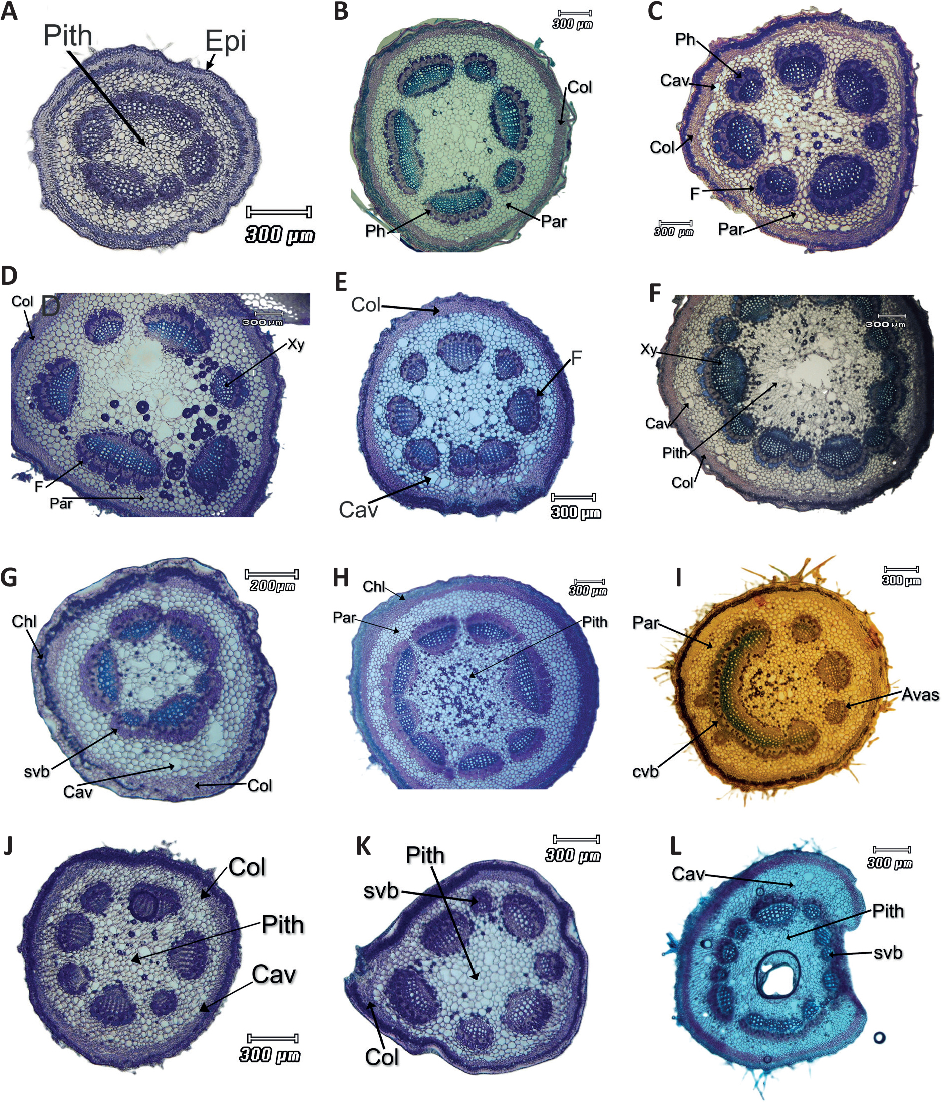
Figure 3:
Cross sections of Alcea petioles: A: A. hohenackeri, B: A. kurdica var. leiocarpa, C: A. kurdica var. laxiflora, D: A. sachsachanica, E: A. tarica, F: A. rechingeri, G: A. wilhelminae var. lineariloba, H: A. angulata, I: A. sulphurea, J: A. glabrata var. glabrata, K: A. glabrata var. microcarpa, L: A. hyrcana. Scale bars: 300 µm. Avas: additional vascular bundle, cav: cavity, Chl: chlorenchyma, Col: collenchyma, Cut: cuticle, Cvb: continuous vascular bundles, Epi: epidermis, F: phloem fibre, Par: parenchyma, Ph: phloem, svb: small vascular bundle, Xy: xylem.
Slika 3: Prečni prerezi pecljev vrst v rodu Alcea: A: A. hohenackeri, B: A. kurdica var. leiocarpa, C: A. kurdica var. laxiflora, D: A. sachsachanica, E: A. tarica, F: A. rechingeri, G: A. wilhelminae var. lineariloba, H: A. angulata, I: A. sulphurea, J: A. glabrata var. glabrata, K: A. glabrata var. microcarpa, L: A. hyrcana. Merila: 300 µm. Avas: dodatni žilni snop, cav: votlina, Chl: klorenhim, Col: kolenhim, Cut: kutikula, Cvb: neprekinjeni žilni snopi, Epi: povrhnjica, F: floemsko vlakno, Par: parenhim, Ph: floem, svb: majhen žilni snop, Xy: ksilem.
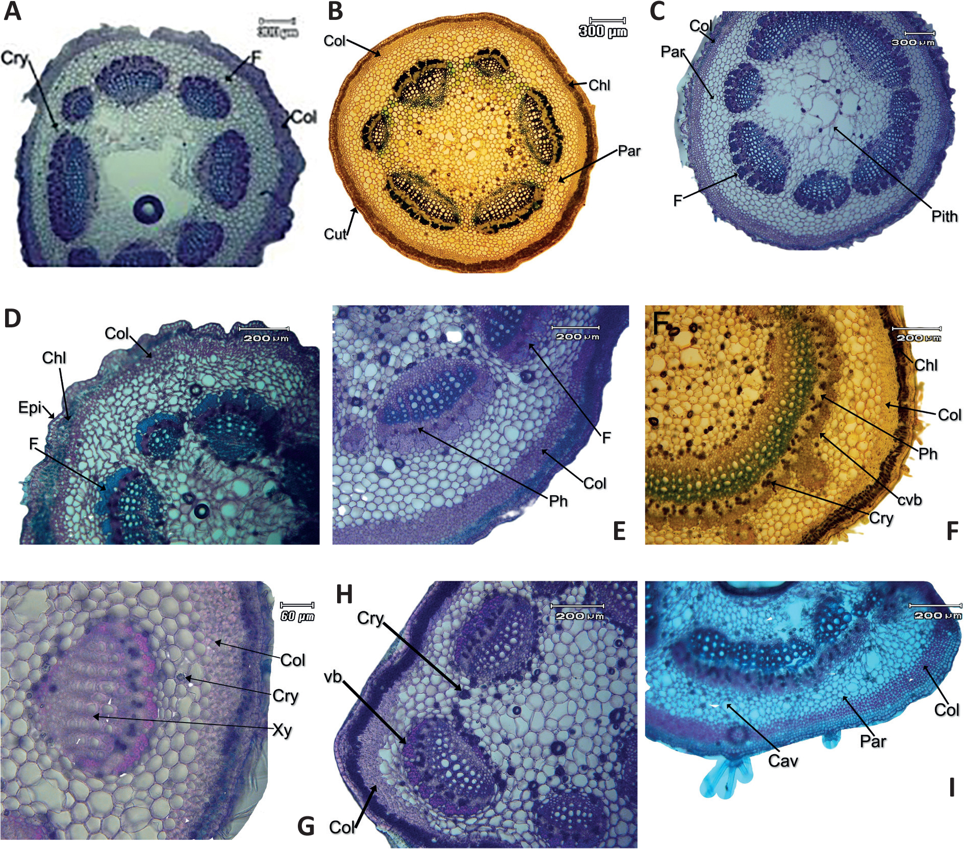
Figure 4:
Cross section of Alcea petioles: A: A. schirazana, B: A. wilhelminae var. wilhelminae, C: A. kurdica var. kurdica, D: A. wilhelminae var. lineariloba, E: A. angulata, F: A. sulphurea, G: A. glabrata var. glabrata, H: A. glabrata var. microcarpa, I: A. hyrcana. Scale bars: 300 µm (A, B, C,), 200 µm (D, E, F, H, J), 60 µm (G). cav: cavity, Chl: chlorenchyma, Col: collenchyma, cry: crystal, Cut: cuticle, cvb: continuous vascular bundle, Epi: epidermis, F: phloem fibre, L.vb: lateral vascular bundle, Par: parenchyma, Ph: phloem, svb: small vascular bundle, Xy: xylem.
Slika 4: Prečni prerezi pecljev vrst v rodu Alcea: A: A. schirazana, B: A. wilhelminae var. wilhelminae, C: A. kurdica var. kurdica, D: A. wilhelminae var. lineariloba, E: A. angulata, F: A. sulphurea, G: A. glabrata var. glabrata, H: A. glabrata var. microcarpa, I: A. hyrcana. Merilo: 300 µm (A, B, C,), 200 µm (D, E, F, H, J), 60 µm (G). cav: votlina, Chl: klorenhim, Col: kolenhim, crystal: kristal, Cut: kutikula, cvb: neprekinjen snop. Epi: povrhnjica, F: floemsko vlakno, L.vb: lateralni žilni snop, Par: parenhim, Ph: floem, svb: majhen žilni snop, Xy: ksilem.
Epidermis: In most taxa, the epidermis consists of one-layer except in A. glabrata. var glabrata, A. schirazana and A. angulata that have two layers of epidermis cells. The epidermis cells are round to cuboidal in shape and covered by a thin cuticle, with stomata interrupting the surface in some areas. Except for A. schirazana Alef. and A. wilhelminae var. wilhelminae, which lack trichomes, all other species exhibit combination of various types of trichomes, including simple, star-shaped, bifurcate, three radiate and glandular (Figure 2).
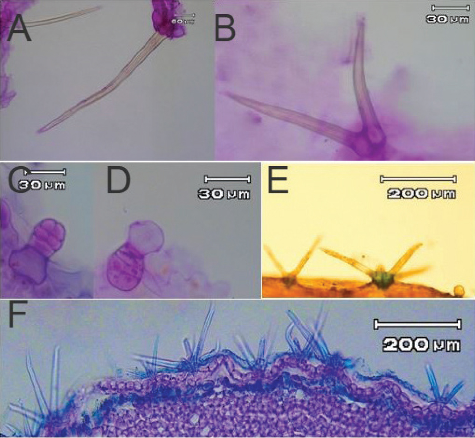
Figure 2:
Different types of trichomes on the petiole of Alcea species. A: simple, B: bifurcate, C & D: glandular, E & F: stellate.
Slika 2: Različne vrste trihomov na listnem peclju vrst v rodu Alcea. A: navadni, B: dvokraki, C in D: žlezni, E in F: zvezdasti.
Cortex: Beneath the epidermis, several layers of chlorenchyma and collenchyma cells form the cortex. Below the epidermis, chlorenchyma layers containing storage material were observed, with 2 to 5 layers of cells. The thickest chlorenchyma layers were found in A. wilhelminae var. wilhelminae and the thinnest in A. hohenackeri. Beneath the chlorenchyma, collenchyma layers consisting of 3 to 10 rows of cells are observed, which were angular in all samples. The minimum number of collenchyma layers were observed in A. wilhelminae var. wilhelminae, and the most were in A. sulphurea. The thickest collenchyma layers were found in A. sulphurea, and the thinnest in A. hyrcana (Grossh.) Grossh. The thickest parenchyma was found in A. rechingeri, and the thinnest in A. glabrata var. microcarpa. This region also contains secretory cavities (Figures 3 & 4).
Beneath the collenchyma layers, parenchyma cells are found, which consist of 4 to 10 rows of round, oval, or polygonal cells, with intercellular spaces (Figures 4 & 5).
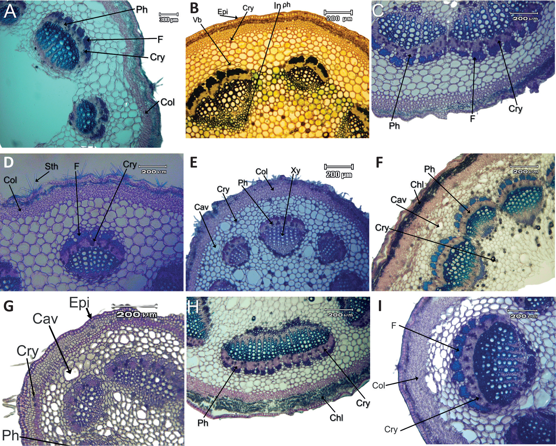
Figure 5:
Cross section of Alcea petioles: (A): A. schirazana, (B): A. wilhelminae var. wilhelminae, (C): A. kurdica var. kurdica, (D): A. sachsachanica, (E): A. tarica, (F): A. rechingeri, (G): A. hohenackeri, (H): A. kurdica var. leiocarpa, (I): A. kurdica var. laxiflora. Scale bars: 200 µm. cav: cavity, Chl: chlorenchyma, Col: collenchyma, cry: crystal, Cut: cuticle, Epi: epidermis, F: phloem fibre, In ph: internal phloem, L.vb: lateral vascular bundle, Par: parenchyma, Ph: phloem, svb: small vascular bundle, Sth: stellate trichome, Xy: xylem.
Slika 5: Prečni prerezi pecljev vrst v rodu Alcea: (A): A. schirazana, (B): A. wilhelminae var. wilhelminae, (C): A. kurdica var. kurdica, (D): A. sachsachanica, (E): A. tarica, (F): A. rechingeri, (G): A. hohenackeri, (H): A. kurdica var. leiocarpa, (I): A. kurdica var. laxiflora. Lestvice: 200 µm. cav: votlina, Chl: klorenhim, Col: kolenhim, Cut: kutikula, cry: kristal, Epi: povrhnjica, F: floemsko vlakno, In ph: notranji floem, L.vb: lateralni žilni snop, Par: parenhim, Ph: floem, Sth: zvezdasti trihom, svb: majhen žilni snop, Xy: ksilem,
Vascular bundles: Vascular bundles are bicollateral except in A. angulata. Arrangement had a great stability and systematic concept. It included sclerenchyma fibre cells, phloem bundles, vascular cambium, xylem, inner phloem, inner xylem and pith. The petiole vascular bundles were divided into two abaxial and adaxial, and unlike in some other members of the Malvaceae family, medullary vascular bundles were not observed in these species. The vascular cylinder consisted of xylem and phloem vascular bundles, which in all samples, had a free and crescent shape, often elliptical, with varying sizes in a ring. A significant variation in the number of vascular bundles was observed in different Alcea taxa. Some taxa had large vascular bundles, while others had small vascular bundles, with anywhere from 6 to 8 bundles observed among different species. The minimum number of vascular bundles were found in A. sulphurea and A. hohenackei (5), and the maximum number of vascular bundles were observed in A. rechingeri (16). Additional vascular bundles were observed only in A. sulphurea and A. angulata (Figures 4 & 5).
Among the studied taxa, most of the them had separate vascular bundles. In some taxa, the bundles were independent and very close to each other, while in the following taxa, continuous vascular bundles were observed:
A. angulata (3 continuous bundles), A. hyrcana (3 to 4 continuous bundles), A. rechingeri (9 fused bundles), A. kurdica var. kurdica (4 continuous bundles). U-shaped continuously vascular bundles were observed only in A. sulphurea (Figure 3I).
There were one to two rows of cambium cells between the xylem and phloem bundles, but due to the high density of crystals in this area, they are not clearly visible in the images (Figure 4G). Above each large vascular bundle and some small ones, a set of fiber cells with thick walls is located, known as the phloem fiber cap. The number of phloem fiber cap layers varies among the taxa. Based on the height of the cap in the cellular layers, the phloem cap can be classified as large (more than 80 µm), medium (between 50 to 75 µm), or small (between 25 to 45 µm).
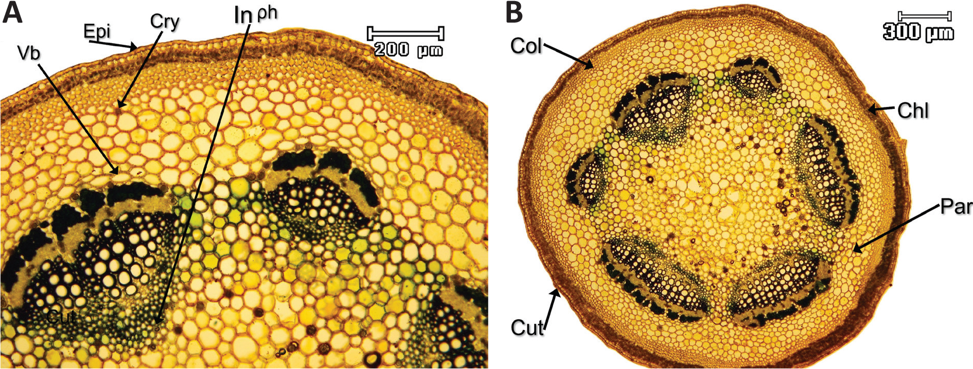
Figure 1.
Details of petiole structure in Alcea wilhelminae var. wilhelminae. Chl: chlorenchyma, Col: collenchyma, Cry: crystal, Cut: cuticle, Epi: epidermis, In ph: internal phloem, Par: parenchyma, V.b: vascular bundle.
Slika 1: Podrobnosti v strukturi listnega peclja pri vrsti Alcea wilhelminae var. wilhelminae. Chl: klorenhim, Col: kolenhim, Cry: kristal, Cut: kutikula, Epi: povrhnjica, In ph: notranji floem, Par: parenhim, V.b: žilni snop.
Species such as A. sulphurea, A. sachsachanica, A. tarica, and A. angulata had large fiber caps. The taxa, like A. hohenackeri, A. hyrcana, A. glabrata var. glabrata, A. kurdica var. leiocarpa and A. wilhelminae var. lineariloba had small fiber caps, while the remaining taxa had medium-sized fiber caps (Figures 4 & 5, Table 2).
Phloem bundles with the widest diameter were found in A. kurdica var. kurdica (Figure 4C), while the narrowest phloem was found in A. angulata (Figure 4E, Table 2).
The xylem bundles with the widest diameter were observed in A. kurdica var. laxifolia (Figure 5I), while the narrowest were found in A. hyrcana (Figure 4I). In all taxa, both xylem and phloem are present internally (except in A. angulata). In all taxa, the petiole contains a large area of ground tissue of the pith, which consists of parenchyma cells with thin walls and appears in various shapes such as round, oval, or polygonal, except in A. hyrcana, A. rechingeri and A. sacsachanica, which had hollow pith. Additionally, secondary phloem fibers were observed at the edges of the pith, except in A. angulata, where they were sparse and minimal (Figures 4 & 5).
Crystals were also found in various parts of the petiole, including the cortex, phloem vessels, and pith (Figures 4 & 5).
Table 2:
Anatomical features of petiole in Alcea species.
Tabela 2: Anatomske značilnosti pecljev vrst v rodu Alcea.
| characters | Parench thick μm | No. Collen | Collen thick (μm) | T. fib scl. | Phlo thick (μm) | Xylem thick (μm) | Chlorench thick (μm) | T. V. B. | Add. V, B | M.V.B | Vas. B. pattern | Epi. Th. | Pet sh |
|---|---|---|---|---|---|---|---|---|---|---|---|---|---|
| Taxa | |||||||||||||
| A. angulata | 182.3–187.7 | 5–7 | 91.1–115.2 | 74.4–78.3 | 37.5–46.1 | 116.8–125.2 | 51.2–53.7 | 12 | + | 10 | Free bundle Ring | 25.8–32.6 | C |
| A. glabrata var. glabrata | 170.1–175.8 | 4–6 | 81.5–93.4 | 32.–34.2 | 41.9–45.5 | 132.2–135.3 | 36.4–38.2 | 7 | - | 4 | Free bundle Ring | 30.1–32.6 | C |
| A. glabrata var. microcarpa | 144.3–146.8 | 4–5 | 82.7–90.3 | 54.9–63.7 | 43.4–45.9 | 134.2–136.1 | 46.9–48 | 7 | - | 5 | Free bundle Ring | 21.1–23.6 | T |
| A. hohenackeri | 204.2–207.1 | 2–3 | 56.1–59.4 | 43.3–44.51 | 47.5–52.5 | 171.9–174.5 | 30.4–31.9 | 5 | - | 4 | Free bundle Ring | 20.6–23.7 | T |
| A. hyrcana | 186.9–187.2 | 3–7 | 58.9–110.8 | 35.6–37.7 | 50.7–55.4 | 99.8–103.3 | 36.9–37.5 | 12 | - | 2 | Free bundle Ring | 26.7–29.8 | T |
| A. kurdica var. kurdica | 200.3–238.3 | 4–5 | 89.5–94.4 | 52.3–80.65 | 56.4–70.7 | 121.9–199.6 | 35.6–49.5 | 7 | - | 5 | Free bundle Ring | 28.5–32 | C |
| A. kurdica var. laxiflora | 243.9–250 | 5–6 | 97.5–100.2 | 63.1–63.2 | 63.8–65.7 | 256.6–270.5 | 42.5–42.6 | 7 | - | 4 | Free bundle Ring | 31.7–34.3 | T |
| A. kurdica var. leiocarpa | 256.3–270.8 | 4 | 87.3–87.7 | 45.8–48.1 | 63.7–65.7 | 129.9–133.7 | 46.2–50.1 | 6 | - | 4 | Free bundle Ring | 22.6–24.7 | C |
| A. rechingeri | 309.8–330.4 | 5–6 | 99.7–105.4 | 66.2–67.4 | 63.5–64.2 | 186.9–187.6 | 48.8–51.7 | 16 | - | 10 | Fused bundle Ring | 27.8–30.3 | C |
| A. schirazana | 257.6–278.4 | 8–9 | 103.9–124.9 | 74.7–76.1 | 60.2–64.2 | 180.8–182 | 43.7–47.3 | 8 | - | 6 | Free bundle Ring | 30.7–32.2 | T |
| A. sachsachanica | 297.10 | 6–7 | 107.3–109.6 | 76–85.23 | 60.8–63.1 | 195.8–198.5 | 43.8–44.8 | 6 | - | 4 | Free bundle Ring | 28.6–31.5 | T |
| A. sulphurea | 229.6–258.8 | 5–6 | 100–103.9 | 60.8–82.4 | 60.9–63.9 | 160–167.3 | 44.5–46.1 | 5 | + | 1 | U-shape Free bundle | 25.8–25.9 | C |
| A. tarica | 201.9–215.7 | 5–6 | 104.6–108.2 | 87.9–93.3 | 51.2–54.3 | 166.6–168.5 | 31.2–34.5 | 8 | - | 5 | Free bundle Ring | 22.8–25.5 | C |
| A. wilhelminae var. lineariloba | 190.7–192.4 | 3–4 | 60.9–81.6 | 47.2–47.4 | 39.8–39.9 | 107.1–118.8 | 61.6–63.7 | 6 | - | 4 | Free bundle Ring | 31.3–37.0 | T |
| A. wilhelminae var. wilhelminae | 294.6–297.6 | 4–5 | 87.9–102.6 | 61.2–66.75 | 55.2–58.2 | 219.7–227.74 | 46.2–87.2 | 6 | - | 4 | Free bundle Ring | 36.8–37.2 | C |
[i] Parench thick: Parenchyma thickness, No. Collen: Number of collenchyma layers, No. Chloren: Layer number of Chlorenchyma, T. fi. Scl.: Thickness of sclerenchyma fibers, Phlo thick: Phloem thickness, Xylem thick: Xylem thickness, Collen thick: collenchyma thickness, T. V. B.: Total Number of vascular bundles, Add. V, B: Additional vascular bundles, M.V.B: Major vascular bundle, Vas. B. pattern: vascular bundles pattern, Epi. Th.: Epiderm thickness, C: circular, T: triangle, Pet. Sh: Petiole shape, - & +: Presence or absence of additional vascular bundles. The highest value of each trait state is in bold.
Clustering analysis
Cluster analysis was conducted based on Euclidean distance by using the Ward method in SPSS software. 25 (2017) based on 11 qualitative petiole anatomical traits. The resulting dendrogram (Figure 6) grouped 15 taxa of the genus Alcea into two main clusters (A and B) at an average taxonomic distance of 25 units. The first major cluster (A) is further divided into two sub-cluster, C and D, which separate at a taxonomic distance of 6. Cluster C comprises A. tarica, A. hohenackeri, A. sulphurea, A. wilhelminae var. lineariloba, which are further split into two smaller sub-clusters, F and G. Cluster D, also separated at a taxonomic distance of 6, includes A. glabrata and A. hyrcana. The second major cluster (B) consists of A kurdica var. laxiflora, A. kurdica var. kurdica, A. wilhelminae and A. sachsachanica.
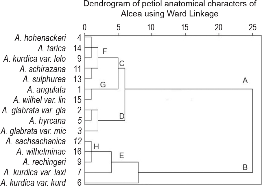
Figure 6:
Dendrogram based on quantitative petiole anatomical characters of Alcea species.
Slika 6: Dendrogram, ki temelji na kvantitativnih anatomskih značilnostih pecljev vrst v rodu Alcea.
Discussion
The findings of this study align with those of Mohammad et al. (2018), who conducted a cladistic analysis based on anatomical traits between different genera of the Malvaceae family. Their research also confirmed that key anatomical features of the petiole, such as sclerenchyma fibres, the structure of vascular elements (including their size, shape, and distribution), the overall vascular pattern, and the presence of crystals, play a crucial role in classification and phylogenetic analysis within the Malvaceae family.
Metcalfe and Chalak (1979) stated that the arrangement of the vascular structures of the petiole changes with the age of the leaf. However, many previous scientists showed that the arrangement of vascular structures in the petiole and midvein of the leaf has a systematic meaning (Moon et al., 2009; Schweingruber et al., 2014; Talip et al., 2017; Thacker et al., 2021; Silva et al., 2018; Marasek-Ciolakowska et al., 2021.
The convergence of A. schiraziana to A. kurdica var. leiocarpa is attributed to their similar characteristics in the thickness of collenchyma, parenchyma, phloem, and chlorenchyma. Similarly, the close positioning of A. kurdica var. laxiflora, A. kurdica var. kurdica and A. rechingeri is due to shared traits in collenchyma thickness, phloem structure, petiole shape, and the number of vascular bundles. In contrast, A. wilhelminae var. wilhelminae and var. lineariloba have separated into distinct clusters due to dissimilarity in sclerenchyma and collenchyma width, phloem thickness, and petiole shape, and thus did not group together.
The statistical analysis in this study clearly demonstrates that, while the delimitation of some taxa aligns with previous classifications based on morphological traits, others do not conform to these groupings. In traditional classification systems based on morphology, species such as A. wilhelminae, A. glabrata, and A. sachsachanica are placed in the same group (Zohary, 1963), while A. kurdica and A. rechingeri are classified together in another. However, in the dendrogram based on petiole anatomical traits (Figure 6), only closely related accessions appear in adjacent branches. Such as, A. sulphurea is positioned next to A. angulata, as both share mericarps without wings, leaves with shallow or no indentations, and densely velvety pubescence. Similarly, A. kurdica var. laxiflora is clustered with A. kurdica and A. rechingeri, also A. glabrata var. microcarpa is clustered with A. glabrata, and A. wilhelminae with A. sachsachanica, which confirmed their close taxonomic relationships, while A. wilhelminae and A. lineariloba are found in a convergent group cluster. These results are completely consistent with the results of phylogenetic analysis based on molecular markers on Alcea species in Irano-turanian region (Escobar et. al., 2012).
In contrast, the placement of some other taxa does not correspond to their expected taxonomic relationships (Figure 6). For example, it was beyond our expectation that A. tarica was placed beside the A. kurdica or A. wilhelminae var. lineariloba was placed beside the A. angulata. It is due to taxonomic relationships being based mostly on morphological characters. They do not always correspond to genetic relationships, just as in molecular systematics, some taxa are not placed in their taxonomic positions based on morphological traits.
Conclusion
Petiole anatomy of 15 taxa of the genus Alcea was studied using hand section and double staining, and some for the first time. Some species have identified by their anatomical characters. Clustering analysis based on petiole anatomical traits indicated that while some anatomical traits of the petiole could aid in species identification, they are not particularly useful for grouping closely related species.
Acknowledgements
We wish to thank the Faculty of Plant Sciences, Alzahra University for providing us with many facilities to conduct this study. Also, the authors wish to thanks of Dr. Mahhvash Seifali for helping in statistical analysis.