Figure 1:
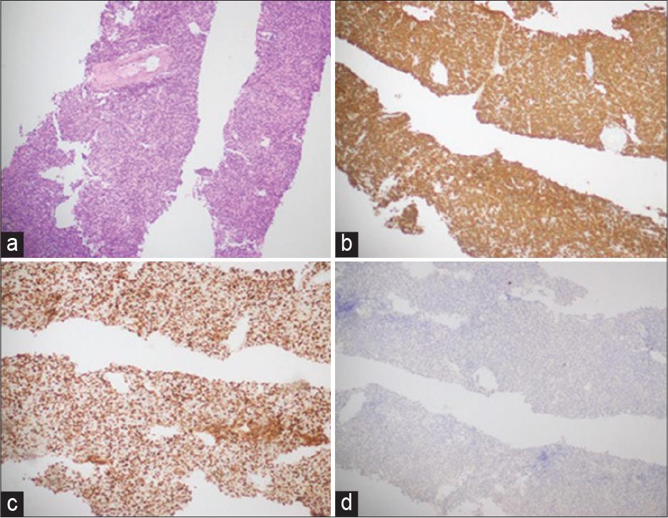
Figure 2:
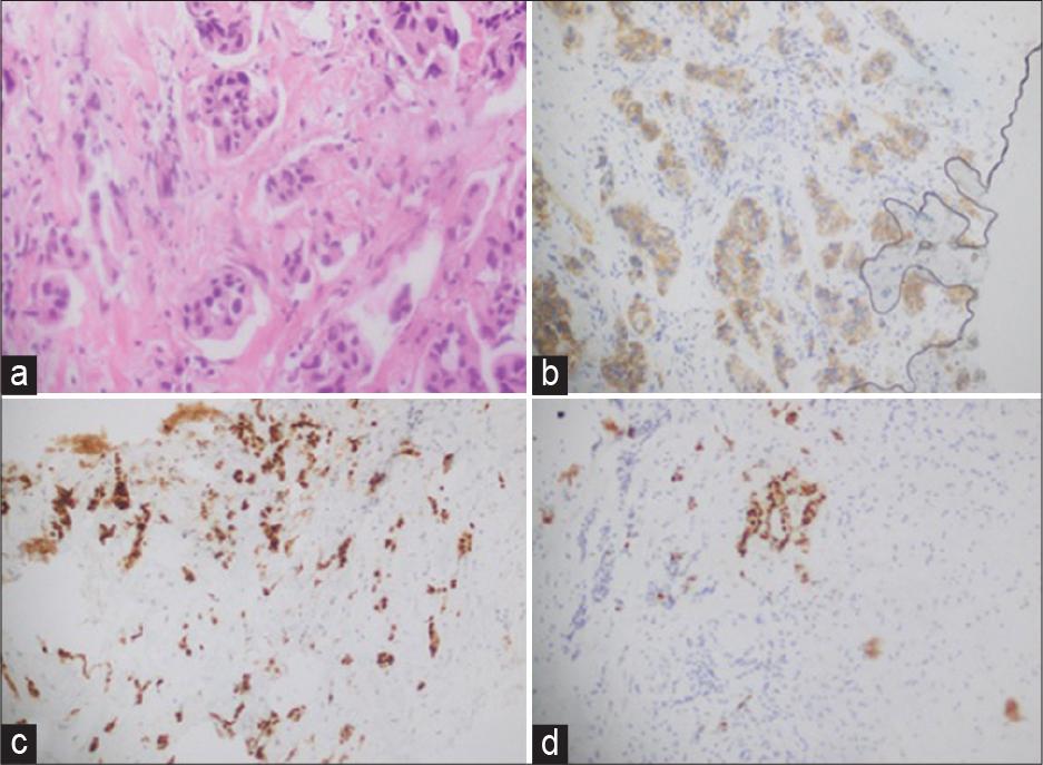
Figure 3:
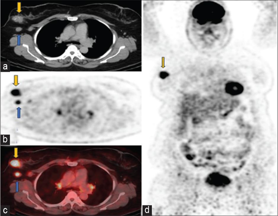
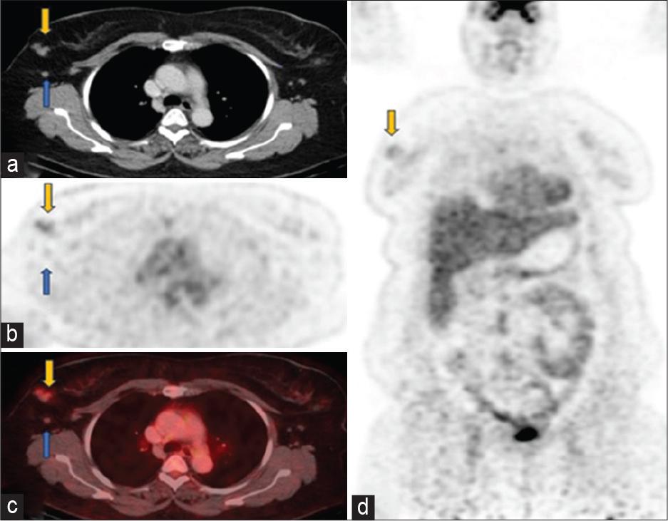
Figure 4:
© 2024 Tahira Yasmeen, Sobia Umar, Mariah Mairah Razi, published by Shakuat Khanum Memorial Cancer Hospital and Research Centre
This work is licensed under the Creative Commons Attribution-NonCommercial-ShareAlike 4.0 License.