
Introduction
As the third most common malignant tumor globally, colorectal cancer (CRC) poses a serious public health challenge due to its high morbidity and mortality. Among males, CRC exhibits a higher incidence and mortality rate compared to females, with cancer statistics showing that between 2015 and 2019, the average yearly CRC incidence rate was 33% higher in males, and their overall mortality rate was 43% higher than that of females. Notably, CRC has emerged as the leading cause of cancer-related death in males under the age of 50 and as the second most common in females of the same age group (Tsokkou et al. 2025). Although conventional treatments—including radical surgical resection, systemic chemotherapy, and precision radiotherapy—have demonstrated definite efficacy in patients with localized CRC, their effectiveness remains limited in cases of metastatic or recurrent refractory disease. Notably, rapid advances in tumor immunology have led to breakthrough treatments represented by immune checkpoint inhibitors (ICIs), which have not only significantly improved clinical outcomes in a subset of CRC patients but also fundamentally reshaped the treatment landscape. ICIs have become an integral part of modern multidisciplinary comprehensive cancer care (Weng et al. 2022). These monoclonal antibodies target immunosuppressive receptors by specifically blocking key immune checkpoint molecules on T cells—such as programmed cell death protein 1 (PD-1), programmed death-ligand 1 (PD-L1), and cytotoxic T-lymphocyte-associated protein 4 (CTLA-4)—thereby reversing tumor-mediated immunosuppression and reactivating T-cell-mediated anti-tumor immune responses. Over the past decade, ICIs have shown transformative clinical efficacy across multiple malignancies, significantly improving survival outcomes—including in CRC—and enabling long-term survival benefits in some patients (Alsaafeen et al. 2025). The quest for reliable biomarkers to predict ICIs efficacy and safety is a critical and active area of research across multiple cancer types. For instance, in melanoma, circulating inflammatory cytokines have been investigated as potential dynamic predictors of response to anti-PD-1 therapy (Mirjačić Martinović et al. 2023). Currently, microsatellite status represents one of the most established biomarkers for predicting response to immunotherapy. However, gastrointestinal cancer patients with microsatellite instability-high (MSI-H) or mismatch repair-deficient (dMMR) characteristics exhibit considerable heterogeneity in treatment response. Approximately 30% of MSI-H patients demonstrate primary resistance to ICIs, and about 17% develop acquired resistance after two years of treatment—a rate that tends to increase over time (André et al. 2020). Despite immunotherapy being a hotspot in contemporary anti-cancer research, the application of ICIs in CRC remains exploratory, and validated predictive biomarkers for treatment efficacy are still lacking.
The gut microbiota is a community of symbiotic microorganisms residing in the human intestinal tract. Disruption of its homeostasis can alter microbial community structure, impair host physiological functions, and contribute to the development of various diseases. Accumulating evidence indicates that the gut microbiota plays a regulatory role in tumor immunotherapy, with certain bacteria influencing the efficacy of immune checkpoint inhibitors (ICIs) through immune modulation or metabolic pathways. In mouse models, depletion of endogenous flora using broad-spectrum antibiotics markedly reduced the antitumor effect of PD-1 inhibitors in colorectal cancer (CRC), underscoring the necessity of an intact microbiota for ICI activity (Zhang SL et al. 2021). Subsequent studies revealed that responders exhibited higher abundance of Lactobacillus, and specifically Lactobacillus paracasei was shown to enhance CD8+ T-cell recruitment to tumor sites via induction of CXCL10 expression (Zhang SL et al. 2022). A clinical cohort of 74 patients with advanced gastrointestinal tumors receiving PD-1/PD-L1 inhibitors demonstrated that Eubacterium, Lactobacillus, and Streptococcus were associated with clinical benefit, whereas Bacteroides and Coprococcus were enriched in non-responders (Peng et al. 2020). Akkermansia muciniphila has repeatedly been identified as a beneficial commensal. Transplantation of this bacterium into germ-free or antibiotic-treated mice via fecal microbiota transplantation (FMT) restored PD-1 inhibitor efficacy through IL-12-dependent recruitment of CCR9+CXCR3+CD4+ T cells (Routy et al. 2018). Further studies indicate that A. muciniphila sustains anti-PD-1 response by modulating glycerophospholipid metabolism, whereas Bacteroides-induced chronic inflammation may suppress immune activation (Xu X et al. 2020). Bifidobacterium pseudolongum enhances ICI response by secreting inosine, which activates Th1 cells via the adenosine A2A receptor in T cells (Mager et al. 2020). Similarly, Bifidobacterium breve combined with anti-PD-1 treatment increased the CD8+/Treg ratio and elevated IFN-γ and IL-2 levels in CRC mouse models (Yoon et al. 2021). Clostridium complex strain CC4 was shown to significantly augment CD8+ T-cell infiltration; monotherapy or combination with PD-1 blockade nearly eradicated tumors, indicating strong synergistic potential (Montalban-Arques et al. 2021). Beyond CRC, Helicobacter pylori infection was correlated with reduced survival in non-small cell lung cancer (NSCLC) patients treated with PD-1 inhibitors (Oster et al. 2022). In intrahepatic cholangiocarcinoma, non-responders to anti-PD-1 therapy exhibited declining microbial diversity over time. Bacteroides was enriched in responders, while Proteobacteria dominated in non-responders (Mao et al. 2021). The efficacy of antigen presentation by dendritic cells and sustained activation of effector T cells in the tumor microenvironment are closely linked to the gut microbiota and its metabolites. Microbial products such as short-chain fatty acids (SCFAs) and secondary bile acids can modulate host gene expression via ligandreceptor interactions and influence immune cell fate (Ocvirk and O’Keefe 2021). Trimethylamine N-oxide (TMAO), derived from microbial metabolism of dietary choline, promotes inflammation and immune activation (Wu K et al. 2020). In models of pancreatic ductal adenocarcinoma and triple-negative breast cancer, TMAO potentiated ICI response by activating the dendritic cell-CD8+ T-cell axis and M1 macrophages (Mirji et al. 2022; Wang H et al. 2022). Extracellular polysaccharides secreted by lactic acid bacteria have been shown to enhance ICI efficacy in tumor-bearing mice (Kawanabe-Matsuda et al. 2022). Additionally, synergistic production of indole-3-propionic acid by Lactobacillus johnsonii and Clostridium sporogenes promotes H3K27 acetylation in the Tcf7 super-enhancer region, enhancing stem-like properties in CD8+ T cells (Tpex) and improving response to immune checkpoint blockade across cancer types (Jia D et al. 2024). In summary, specific bacterial species and their metabolites modulate ICI efficacy through immune and metabolic mechanisms. The composition of the gut microbiota may serve as a predictive biomarker for treatment response and offers promising avenues for therapeutic intervention.
In recent years, advances in high-throughput metabolomics have provided unprecedented opportunities to systematically characterize the human gut metabolome. Under homeostatic conditions, intestinal metabolites and microbial communities work synergistically to maintain host physiological functions and promote health. In contrast, dysbiosis in their composition and function can contribute to the development of various diseases, including carcinogenesis. Studies have shown that the accumulation of oncometabolites and pathogenic microorganisms is closely associated with colorectal cancer (CRC) progression, while certain probiotics or beneficial symbiotic bacteria may exert anti-tumor effects through the production of protective metabolites. Given this functional significance, a growing number of studies are exploring fecal, serum, and plasma metabolites as clinical biomarkers for CRC diagnosis and prognosis. Combinations of microbial and metabolic biomarkers have demonstrated improved diagnostic accuracy (Chen F et al. 2022). Currently, predictive biomarkers for immunotherapy response in CRC remain limited. Although tumor mutational burden, neoantigen load, tumor-infiltrating lymphocytes, and immunoglobulins represent promising candidates, the gut microbiota is increasingly recognized as a key regulator of immunotherapy outcomes. This is not only due to its close association with CRC pathogenesis, but also because microbial sequencing technologies have become highly robust and standardized. Therefore, both intestinal flora and their metabolites hold great promise as predictive biomarkers for immunotherapy efficacy in CRC. They may provide a novel molecular foundation for future personalized treatment strategies.
In summary, the host-microbiota-metabolite network is widely recognized as a key determinant of immune checkpoint inhibitor (ICI) efficacy. Variations in microbial composition and metabolic profiles may substantially influence treatment outcomes. Therefore, pre-treatment metagenomic sequencing of fecal samples, combined with quantitative analysis of key bacterial strains and relevant metabolic markers associated with either beneficial or adverse effects, holds promise for predicting treatment response and informing clinical decision-making and individualized therapeutic optimization. This study aims to characterize the gut microbiome and metabolome profiles of MSI-H CRC patients undergoing immunotherapy and to systematically evaluate their role in anti-tumor responses. These findings will provide a theoretical and data-driven foundation for future microbiota-based prognostic assessment and precision interventions.
Experimental
Materials and Methods
Study subjects
Based on the guidelines of the Union for International Cancer Control (UICC), a total of 20 patients with stage III–IV colorectal adenocarcinoma were retrospectively enrolled in this study. This retrospective cohort study was conducted after obtaining approval from the Ethics Committee of Zhengzhou Central Hospital (Approval No. ZXYY2025033). All research-specific procedures, including patient identification, data collection, and biospecimen sampling for this study, were initiated only after ethical approval. We retrospectively screened the medical records of patients with stage III–IV colorectal adenocarcinoma who were treated with immune checkpoint inhibitors at Zhengzhou Central Hospital Affiliated to Zhengzhou University. Clinical immunotherapy for these patients was initiated as part of standard care between November 2024 and January 2025. From this treated pool, we identified and included 20 eligible patients into our analytical cohort between January and March 2025. The inclusion of patients and collection of their baseline fecal samples were performed concurrently during this period (January–March 2025), specifically within 1–7 days after the patients had completed at least 2–3 cycles of their ongoing immunotherapy. Inclusion Criteria: (1) Histologically or cytologically confirmed stage III–IV colorectal adenocarcinoma with microsatellite instability test results of MSI-H/dMMR; (2) Completion of at least two cycles of immunotherapy (e.g., anti-PD-1, anti-PD-L1, or anti-CTLA-4 agents); (3) Availability of complete medical records, including diagnostic pathology reports and imaging evaluations with at least one measurable lesion; (4) Ability to provide at least one fecal sample after initiation of treatment. Exclusion Criteria: (1) Presence of additional malignant tumors; (2) Diagnosis of autoimmune disorders; (3) Use of probiotics, antibiotics, or acid-suppressing medications within the preceding 6 months; (4) Pre-existing intestinal diseases such as immune enteritis, ulcerative colitis, or Crohn’s disease.
Clinical data collection
Clinical and biochemical data, including patient age, gender, tumor location, TNM stage, metastatic sites, genetic expression profiles, and pretreatment serum lactate dehydrogenase (LDH) levels, were retrospectively collected from electronic medical records. These data were generated as part of routine clinical care prior to immunotherapy initiation. All biochemical analyses, including LDH measurement, were performed in the Central Laboratory of Zhengzhou Central Hospital Affiliated to Zhengzhou University using standardized automated clinical analyzers following established protocols and internal quality control procedures. Genomic profiling was performed using a validated next-generation sequencing (NGS) panel targeting cancer-related genes. Microsatellite instability (MSI) status was determined by analyzing microsatellite loci, with MSI-H defined as instability at ≥ 30% of analyzed loci. Mismatch repair (MMR) status was evaluated based on the mutational signatures of MMR deficiency. BRAF mutation status, including the V600E mutation, was assessed from the NGS data.
Treatment regimens
Patients received one of the following original (non-biosimilar) immune-checkpoint inhibitors, all administered intravenously according to the China-approved package inserts: Tislelizumab (PD-1): 200 mg flat dose, q3w (BeiGene, China), Sintilimab (PD-1): 200 mg flat dose, q3w (Innovent, China), Camrelizumab (PD-1): 200 mg flat dose, q3w (Hengrui, China), Adebrelimab (PD-L1): 1200 mg flat dose, q3w (Heng-rui, China), Atezolizumab (PD-L1): 1200 mg flat dose, q3w (Roche, Switzerland). Treatment continued until disease progression, unacceptable toxicity, or patient withdrawal; each patient completed ≥ 2 cycles. No biosimilars were used.
Response evaluation
Tumor response was assessed every 6 to 8 weeks using computed tomography (CT) and/or magnetic resonance imaging (MRI) according to Response Evaluation Criteria in Solid Tumors version 1.1 (RECIST 1.1) (Eisenhauer et al. 2009). Efficacy was evaluated based on the following criteria: Complete Response (CR): Disappearance of all target lesions, with any pathological lymph nodes having a short axis diameter reduced to < 10 mm. Partial Response (PR): At least a 30% decrease in the sum of the longest diameters of target lesions, taking as reference the baseline sum. Progressive Disease (PD): At least a 20% increase in the sum of the longest diameters of target lesions (with an absolute increase of ≥ 5 mm), or the appearance of one or more new lesions. Stable Disease (SD): Neither sufficient shrinkage to qualify for PR nor sufficient increase to qualify for PD. Patients were classified into two groups based on their best overall response per RECIST 1.1: Clinical Benefit Response (CBR, the responder group): Patients who achieved CR, PR, or SD lasting ≥ 3 months. Non-Clinical Benefit (NCB, the non-responder group): Patients with SD lasting < 3 months or PD.
Experimental workflow
Stool sample collection
Morning stool samples were collected from CRC patients 1–7 days after they had completed at least 2–3 cycles of immunotherapy. Approximately 1 g of fresh stool was collected under sterile conditions, aliquoted into cryogenic tubes, and immediately frozen at –80°C. All eligible fecal samples underwent simultaneous metagenomic sequencing and metabolomic analysis to minimize batch effects.
Fecal microbiome analysis
Genomic DNA was extracted using a magnetic bead-based stool DNA extraction kit (AU46111-96, BioTeke). Qualified DNA (200 ng) was sheared to 200–500 bp using a Bioruptor™ Pico sonicator. A sequencing library was constructed with the TruSeq Nano DNA LT Library Preparation Kit (Illumina). Paired-end sequencing (PE150) was performed on an Illumina NovaSeq 6000 platform (LC-Bio, China). Raw reads were quality-controlled using fastp (v0.23.4), and host-derived reads were removed by alignment to the human genome with Bowtie2 (v2.2.0). Quality-filtered reads were assembled using MEGAHIT (v1.2.9). Contigs > 500 bp were used for CDS prediction with MetaGeneMark (v3.26). Redundant CDS were clustered (95% identity, 90% coverage) using MMseqs2 to generate a non-redundant Unigene set. Abundance was quantified by aligning reads back to this set. Taxonomic and functional (KEGG, GO) annotations were assigned using DIAMOND (v0.9.14) against NCBI NR, KEGG, and GO databases.
Fecal metabolomic analysis
Approximately 50 mg of fecal sample was homogenized in 500 μl of 80% icecold methanol. After protein precipitation and centrifugation, the supernatant was lyophilized, reconstituted in 50% methanol, and centrifuged again. Chromatographic separation was performed on a Thermo Vanquish Flex UPLC system with an ACQUITY UPLC T3 column (100 mm × 2.1 mm, 1.8 μm). The mobile phase consisted of 5 mmol/l ammonium acetate with 5 mmol/l acetic acid in water (A) and acetonitrile (B). Mass spectrometry was conducted on a Q-Exactive Plus instrument in both positive and negative ionization modes using data-dependent acquisition. Quality control (QC) samples were analyzed and interspersed throughout the batch. Raw data were processed using XCMS in R for peak detection and alignment. Metabolites were annotated by matching accurate mass (*m/z* error < 10 ppm) against PubChem, HMDB, and an inhouse database.
Statistical Analysis
Statistical analysis of clinical data
Clinical and demographic data were analyzed using SPSS (v25.0) and R (v4.0). Normally distributed continuous variables are presented as mean ± SD and compared using Student’s t-test; non-normally distributed variables as median (IQR) and compared with the Mann-Whitney U test. Categorical variables were compared using the chi-square or Fisher’s exact test. A two-sided P < 0.05 was considered significant.
Statistical analysis of microbiome data
Diversity analysis: Alpha diversity indices (Chao1, Shannon, Simpson) were calculated using QIIME1 and compared with the Mann-Whitney U test. Beta diversity based on Bray–Curtis dissimilarity was visualized via PCoA and tested using PERMANOVA. Differential abundance: Differentially abundant taxa were identified using LEfSe (LDA > 3.0, P < 0.05). The Mann-Whitney U test was applied for differential analysis of functional pathways (P < 0.05, |log2FC| > 1). To account for multiple comparisons, Benjamini-Hochberg false discovery rate (FDR) correction was applied, and the key findings remained consistent after correction (adjusted P < 0.10). Species-level correlation networks were constructed based on Spearman correlations (|ρ| > 0.8).
Statistical analysis of metabolomic data
Data pre-processing: Metabolites with >80% missingness were removed. Missing values were imputed using KNN, and data were normalized by PQN. Differential metabolite identification: Differential metabolites were identified by combining univariate (Student’s t-test, P < 0.05) and multivariate (VIP ≥ 1 from PLS-DA) criteria, with fold change threshold > 1.2. FDR correction (Benjamini-Hochberg) was applied for multiple testing, and the major findings remained robust after correction (adjusted P < 0.10). Pathway enrichment: Functional enrichment of KEGG pathways was assessed using a hypergeometric test (P < 0.05) and MSEA (|NES| > 1, FDR < 0.25).
Results
Comparison of baseline characteristics
The baseline clinical features of patients in the non-clinical benefit (NCB) and clinical benefit response (CBR) groups were analyzed, including demographics (age, gender), tumor characteristics (location, TNM stage, metastatic sites), and genomic profiles. No statistically significant differences were observed in these conventional clinicopathological features between the two groups (all P ≥ 0.05), as detailed in Table I. In contrast, analysis of laboratory parameters revealed a notable difference. Serum levels of lactate dehydrogenase (LDH), a key enzyme in tumor glycolysis and a biomarker associated with tumor burden, were significantly elevated in the NCB group compared to the CBR group (282.13 ± 67.76 U/L vs. 201.96 ± 23.46 U/L; t = 3.535, P = 0.005). The mean difference was 80.17 U/L (95% CI: 30.33 to 130.01). This finding indicates that while the groups were balanced in terms of standard clinical and pathological variables, patients with poorer response to immunotherapy exhibited higher baseline systemic tumor metabolic activity, as reflected by elevated LDH.
Table I
Baseline comparison between NCB and CBR groups.
| Element | CBR (n=10) | NCB (n=10) | t | P |
|---|---|---|---|---|
| Age | 45.10±11.82 | 56.20±13.44 | -1.961 | 0.065 |
| Gender | 0.628 | |||
| Male | 6 (60.0) | 8 (80.0) | ||
| Female | 4 (40.0) | 2 (20.0) | ||
| Tumor location | 1.000a | |||
| Right | 3 (30.0) | 2 (20.0) | ||
| Center | 7 (70.0) | 8 (80.0) | ||
| T | 1.000a | |||
| T2 | 1 (10.0) | 0 (0.0) | ||
| T3 | 7 (70.0) | 8 (80.0) | ||
| T4 | 2 (20.0) | 2 (20.0) | ||
| N | 1.000a | |||
| N0 | 3 (30.0) | 3 (30.0) | ||
| N1 | 5 (50.0) | 5 (50.0) | ||
| N2 | 2 (20.0) | 1 (10.0) | ||
| N3 | 0 (0.0) | 1 (10.0) | ||
| Ml | 10 (100.0) | 10 (100.0) | ||
| Transfer site | 1.000a | |||
| Liver | 8 (80.0) | 7 (70.0) | ||
| Others | 2 (20.0) | 3 (30.0) | ||
| Gene expression | 1.000a | |||
| RAS or BRAF are mutant | 6 (60.0) | 5 (50.0) | ||
| RAS and BRAF are wild type | 4 (40.0) | 5 (50.0) | ||
| Whether the primary lesion has been resected | 0.303a | |||
| Yes | 9 (90.0) | 6 (60.0) | ||
| No | 1 (10.0) | 4 (40.0) | ||
| LDH | 201.96 ± 23.46 | 282.13 ± 67.76 | 3.535 | 0.005 |
Overall Gut Microbial Community Structure
Analysis of the overall gut microbial community structure revealed no significant differences in alpha diversity (assessed by Chao1, Shannon, and Simpson indices) or beta diversity (assessed by Bray-Curtis dissimilarity) between the NCB and CBR groups (all P ≥ 0.05; detailed results are provided in Supplementary Fig. S1 and the PERMANOVA/Adonis test results are shown in Supplementary Table SI). This indicates that the differential response to immunotherapy was not driven by broad ecological shifts in the microbial community, prompting a focused investigation into specific taxonomic and functional features.
Analysis of microbial taxon abundance between groups
Figure 1 illustrates the relative abundance of the gut microbiota at different taxonomic levels in the non-clinical benefit (NCB) and clinical benefit response (CBR) groups. Superkingdom Level (Fig. 1A): Bacteria were the dominant superkingdom in both groups, with Viruses and Archaea accounting for only a minimal proportion, confirming that the bacterial component is the central focus of the gut microbial community. Phylum Level (Fig. 1B): Bacillota and Bacteroidota were the two most dominant phyla in both groups. However, the NCB group exhibited a higher relative abundance of Bacteroidota, while the CBR group showed a slightly greater proportion of Bacillota. Class Level (Fig. 1C): The NCB group showed a higher abundance of Bacteroidia, whereas the CBR group was enriched with Clostridia. Order Level (Fig. 1D): Consistent with class-level findings, the order Bacteroidales was more prevalent in the NCB group. Family Level (Fig. 1E): The NCB group showed a higher relative abundance of Bacteroidaceae, while Lachnospiraceae was more enriched in the CBR group. (Fig. 1F): Genera such as Bacteroides, Phocaeicola, and Segatella demonstrated higher relative abundance in the NCB group compared to the CBR group. Species Level (Fig. 1G): Segatella copri was more enriched in the NCB group, whereas Escherichia coli was more prevalent in the CBR group. These species-level differences suggest a potential microbiological basis for the observed divergence in clinical response phenotypes. Together, these results indicate that although both groups share a similar overall microbial structure, specific taxa at multiple taxonomic levels exhibit abundance profiles that distinguish clinical responders from non-responders.
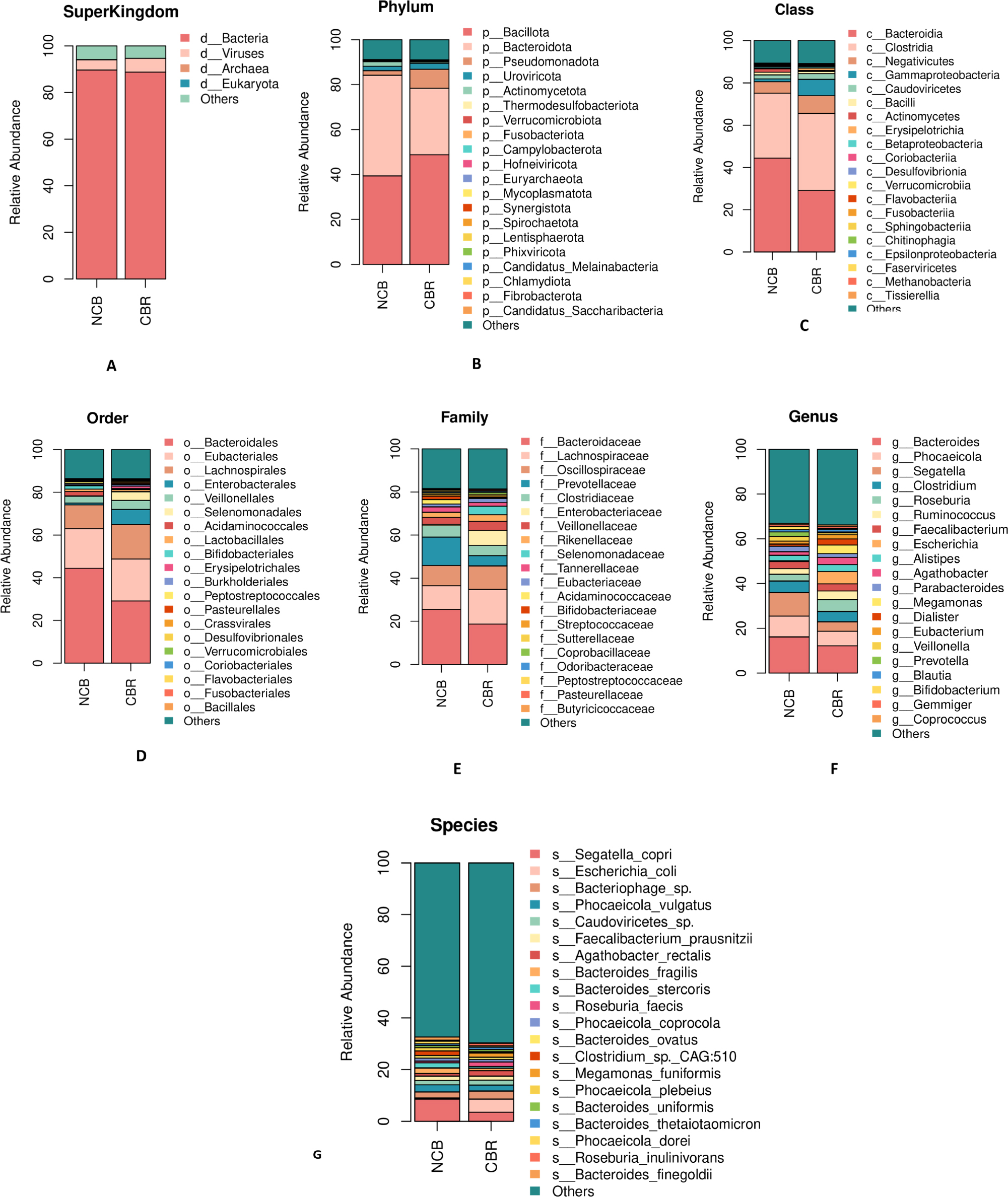
Fig. 1.
Relative abundance of gut microbiota at different taxonomic levels in the NCB and CBR groups. Stacked bar plots show the mean relative abundance of taxa in each group. This figure is intended to illustrate compositional patterns; statistical analyses of differential abundance are presented separately in subsequent sections. (A) Superkingdom level: Bacteria constituted the dominant domain in both groups. (B) Phylum level: Bacillota and Bacteroidota were the dominant phyla shared by both groups, with variations in their relative abundances between groups. (C) Class level: Bacteroidia showed higher relative abundance in the NCB group, whereas Clostridia showed higher relative abundance in the CBR group. (D) Order level, (E) Family level, and (F) Genus level: These levels further reflected the trend of Bacteroidota-related taxa being more enriched in the NCB group, and Lachnospiraceae, among others, being enriched in the CBR group. (G) Species level: Analysis precisely identified Segatella_copri and Escherichia_coli as characteristic species showing differential abundance patterns for the NCB and CBR groups, respectively. Together, these observations reveal compositional differences in gut microbiota structure between the groups, providing a microbiological basis for phenotype associations.
Intramicrobial correlation network analysis
To further elucidate microbial interactions and their potential implications for host health and ecology, Spearman correlation analysis was conducted on the top 30 genera based on relative abundance within both the CBR and NCB groups. The results are summarized using heatmaps and network diagrams. In the CBR group (Fig. 2A): Significant positive correlations were observed among genera such as Lachnospira, Agathobacter, and Coprococcus, which were widely correlated with multiple beneficial taxa. In contrast, Escherichia and Haemophilus exhibited significant negative correlations with most other genera. In the NCB group (Fig. 2B): Faecalibacterium showed strong positive correlations with Blautia and Dorea, whereas Bacteroides was negatively correlated with numerous other genera. Network Analysis of Strong Correlations: Pairwise interactions with |rho| > 0.8 were selected to construct correlation networks (Fig. 2C for CBR, Fig. 2D for NCB). These networks visualize robust genus-level associations, highlighting potential cooperative or competitive ecological relationships that may influence microbial community stability and function. Functional Relevance: The distinct correlation patterns between groups suggest differing microbial ecological structures that may contribute to variations in host immune modulation and treatment outcomes. These interactions offer valuable targets for further mechanistic investigation into microbiota-mediated regulation of host health. This analysis provides a comprehensive view of genus-level microbial associations and their possible role in shaping a microbiota environment conducive to clinical response in colorectal cancer immunotherapy.
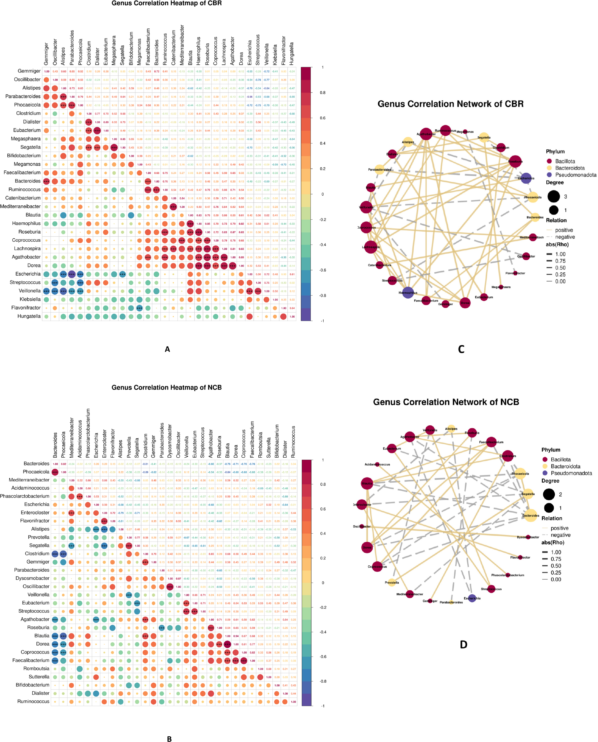
Fig. 2.
Intramicrobial Correlation Network Analysis: (A) and (B): Heatmaps displaying Spearman correlation coefficients among the top 30 most abundant genera in the CBR and NCB groups, respectively. Dot size and color intensity represent the strength and direction of correlations: larger and redder dots indicate stronger positive correlations, while larger and bluer dots indicate stronger negative correlations. Asterisks (*P < 0.05, **P < 0.01, ***P < 0.001) are marked within the heatmap cells to indicate statistical significance. (C) and (D): Correlation network diagrams for the CBR and NCB groups, respectively. Nodes represent bacterial genera, and edges represent strong correlations (|rho| > 0.8). Edge color indicates the type of correlation: red for positive and blue for negative associations.
Functional annotation based on KEGG database
Functional annotation of the fecal metagenomes was performed using the Kyoto Encyclopedia of Genes and Genomes (KEGG) database to compare metabolic potential between the CBR and NCB groups. Level 1 Functional Categories (Fig. 3A): Metabolism was the most dominant functional category in both groups. The CBR group exhibited a slightly higher relative abundance of metabolic functions compared to the NCB group, followed by Genetic Information Processing and Environmental Information Processing. Categories such as Cellular Processes, Human Diseases, and Organismal Systems were low in abundance in both groups. The increased metabolic activity in the CBR group suggests a more active microbial community with enhanced carbohydrate and energy metabolism. Level 2 Functional Modules (Fig. 3B): Significant differences were observed in Level 2 pathways. The CBR group was enriched in: Carbohydrate metabolism, Metabolism of cofactors and vitamins, Xenobiotics biodegradation and metabolism, Cell motility, Membrane transport; The NCB group showed higher abundance in: Amino acid metabolism, Metabolism of terpenoids and polyketides. Level 3 Pathway Analysis (Fig. 3C): Further refinement at Level 3 indicated that the CBR group was enriched in: Biosynthesis of secondary metabolites, Two-component system and ABC transporters. The NCB group demonstrated higher activity in: Amino sugar and nucleotide sugar metabolism. Key KEGG Orthologs (KOs) (Fig. 3D): Specific KOs significantly enriched in the CBR group included genes related to the type IV secretion system, D-alanyl-D-alanine carboxypeptidase, and several hypothetical proteins. The NCB group was enriched in KOs such as DNA ligase (ligA), conserved hypothetical proteins, and beta-galactosidase. These results reveal distinct functional profiles between the two groups, with the CBR group exhibiting enhanced metabolic activities linked to microbial cooperation, nutrient transport, and xenobiotic degradation, potentially supporting a favorable microenvironment for immunotherapy response.
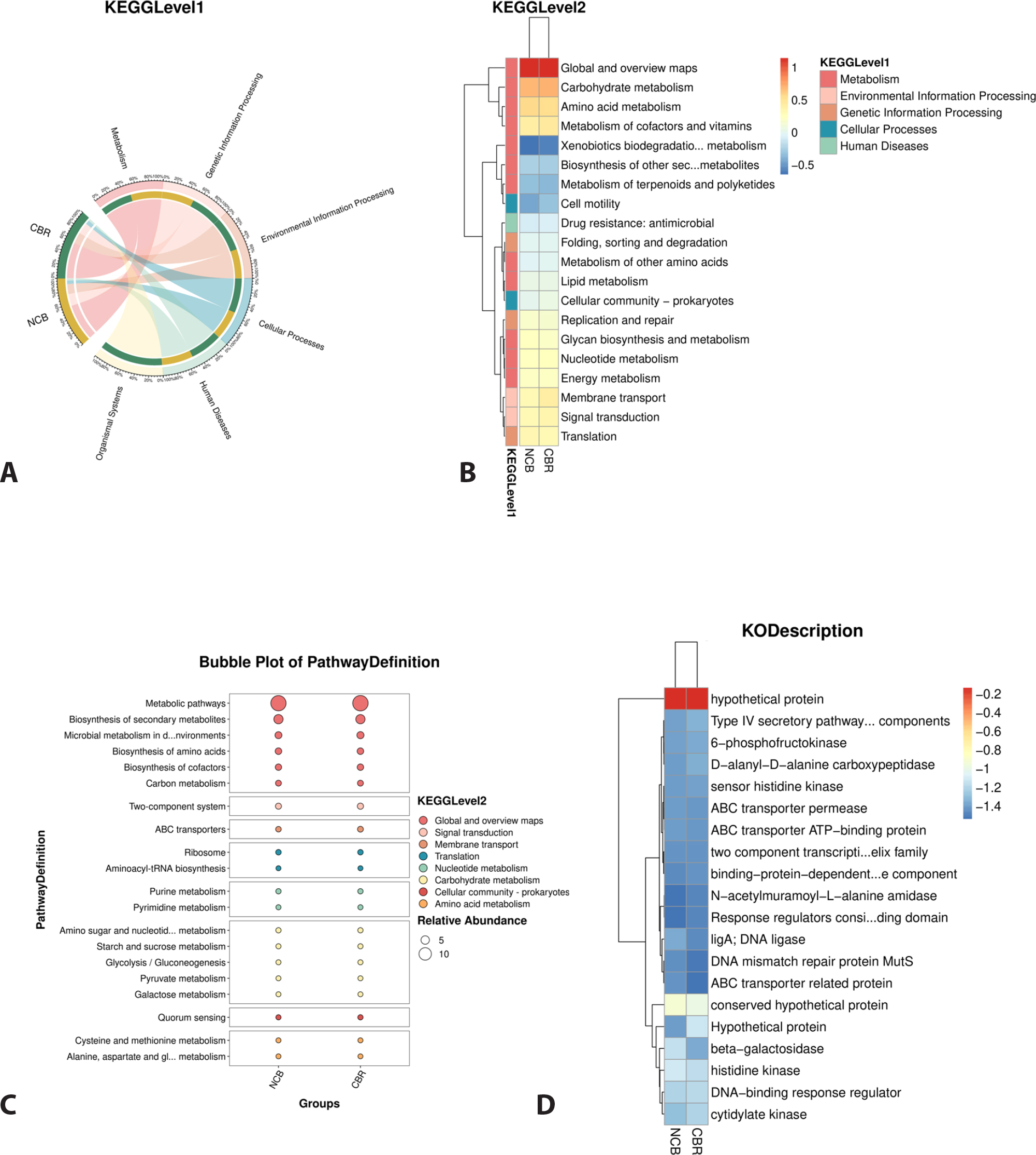
Fig. 3.
KEGG Database Functional Notes: (A) Level 1 functional distribution shows that Metabolism was the predominant category in both groups, with a slightly higher proportion in the CBR group. (B) Level 2 functional module analysis revealed group-specific enriched pathways: the CBR group was enriched in pathways such as Carbohydrate metabolism and Metabolism of cofactors and vitamins, whereas the NCB group showed higher activity in pathways like Amino acid metabolism. (C) Comparison of specific metabolic pathways further confirmed that the CBR group was more active in pathways including Biosynthesis of secondary metabolites and the Two-component system. (D) Pathway Definition level analysis precisely identified key differential genes between groups; for instance, the CBR group was enriched in Type IV secretory pathway components, and the NCB group was enriched in ligA. These results indicate that the gut microbiota in the CBR group possesses a greater potential for active substance transport and energy metabolism.
Differential analysis of species and functional entries
To identify microbial taxa and functional pathways significantly associated with treatment response, we performed comparative analyses using the Mann-Whitney U test on species abundance and KEGG pathway data, with significance thresholds set at |log2FC| > 1 and P < 0.05. The top 20 most abundant and significantly different species showed s_Clostridium_unclassified was enriched in the CBR group, while s_Roseburia_hominis dominated in the NCB group (Fig. 4A). Functional analysis revealed significantly higher abundance of limonene and pinene degradation and caprolactam degradation pathways in the CBR group, whereas prodigiosin biosynthesis showed higher abundance in the NCB group despite its overall low expression level (Fig. 4B). LEfSe analysis (LDA Score > 3, P < 0.05) further confirmed the strong association of s_Clostridium_unclassified with the CBR group, and significant enrichment of s_Marseilla_massiliensis and s_Roseburia_hominis in the NCB group across taxonomic hierarchies (Fig. 4C), highlighting potential microbial and functional biomarkers for predicting immunotherapy response in colorectal cancer.
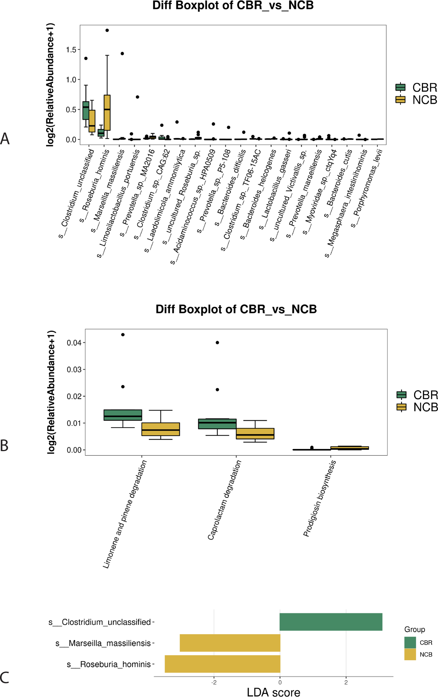
Fig. 4.
Differential Analysis of Species and Functional Entries: (A) Species difference boxplot based on the Mann-Whitney U test (|log2FC| > 1, p < 0.05), displaying the top 20 most abundant significant differential species. The results show that s_Clostridium_unclassified was significantly enriched in the CBR group, whereas s_Roseburia_hominis was more predominant in the NCB group. (B) KEGG PathwayDefinition functional difference boxplot shows that the CBR group was significantly enriched in Limonene and pinene degradation and Caprolactam degradation, while the NCB group had a higher abundance of Prodigiosin biosynthesis. (C) LEfSe analysis (LDA Score > 3, P < 0.05) identified differential taxa with discriminative power between groups, further validating s_Clostridium_unclassified as a biomarker for the CBR group, and s_Marseilla_massiliensis and s_Roseburia_hominis as marker species for the NCB group.
Analysis of species and functional contribution
Based on the abundance of species and their correspondence to the top five most abundant KEGG pathways, we assessed the contribution of microbial taxa in the NCB and CBR groups to these metabolic processes. Analysis of the ko01100 (Metabolic Pathways), ko01110 (Biosynthetic pathway of secondary metabolites), ko01120 (Microbial metabolic pathway in different environments), ko01230 (Biosynthesis of Amino Acids), and ko01240 (Biosynthesis pathway of cofactors) pathways revealed that s_Bacteroides_unclassified was the dominant species in both groups. However, significant differences in species abundance were observed between groups: s_Bacteroides_unclassified and s_Segatella_copri were more abundant in the NCB group, while s_Escherichia_coli and s_Megamonas_unclassified showed higher abundance in the CBR group (Fig. 5A–E). These findings suggest that although certain core species are shared between groups, their differential abundance may drive functional variations in key metabolic pathways, potentially influencing host response to immunotherapy.
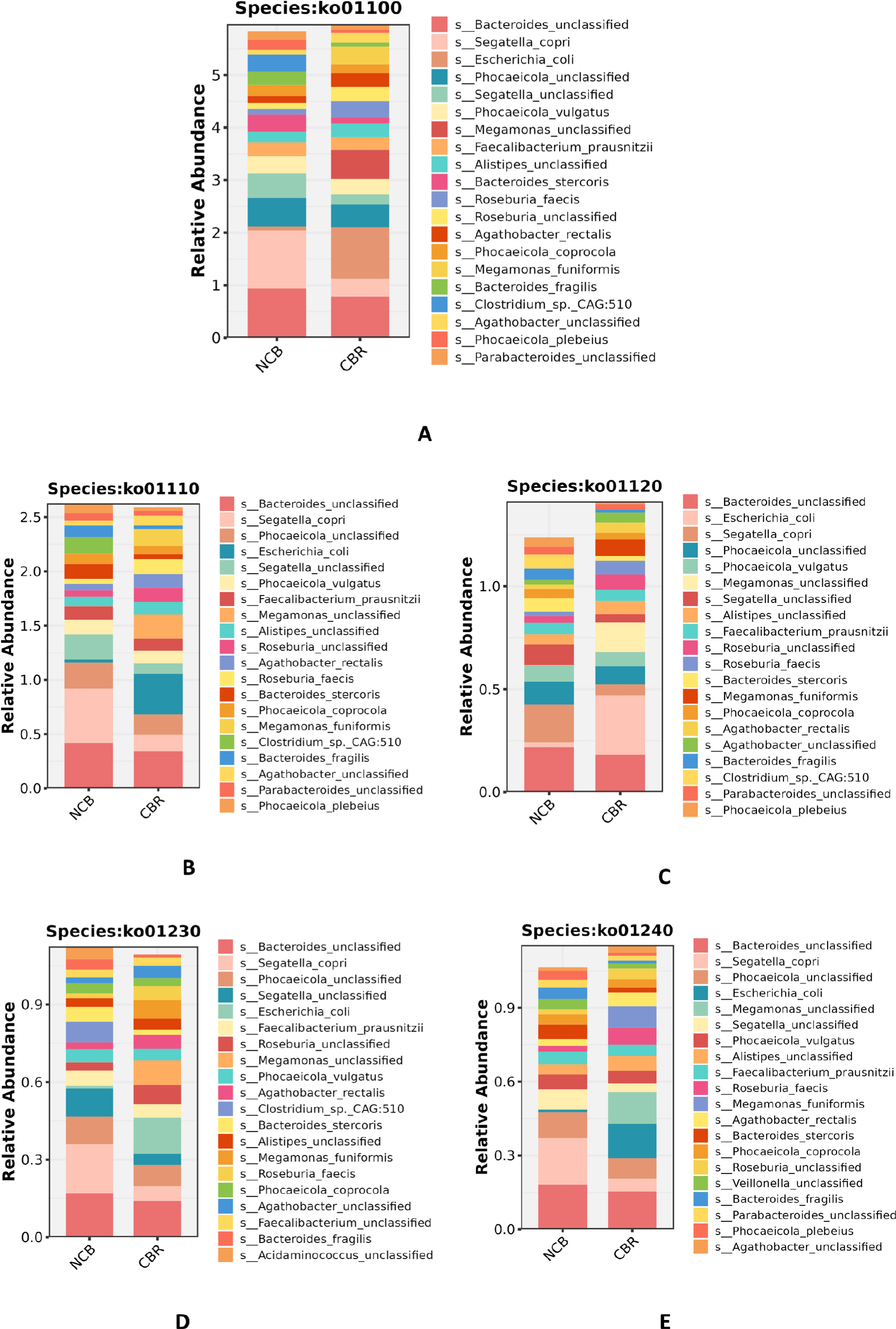
Fig. 5.
Analysis of Species and Functional Contribution: Panels A to E illustrate the relative contributions of species to the top five most abundant KEGG pathways: ko01100 (Metabolic pathways), ko01110 (Biosynthesis of secondary metabolites), ko01120 (Microbial metabolism in diverse environments), ko01230 (Biosynthesis of amino acids), and ko01240 (Biosynthesis of cofactors), respectively. The analysis reveals that s_Bacteroides_unclassified was the dominant contributing species shared by both groups. However, significant intergroup differences were observed: the contribution of s_Bacteroides_unclassified and s_Segatella_copri was higher in the NCB group, whereas s_Escherichia_coli and s_Megamonas_unclassified were enriched in the CBR group.
Screening and identification of differential metabolites
The PLS-DA results (Fig. 6A) revealed a clear separation between the two groups along the first principal component (PC1, 7.34% explained variance) and the second principal component (PC2, 5.62% explained variance), indicating significant differences in their metabolic profiles. To validate model robustness, a permutation test with 200 iterations was performed (Fig. 6B). The results demonstrated that all randomly permuted Q2 values were substantially lower than those of the original model, which had an R2-intercept of 0.9607 and a Q2-intercept of −0.2785, confirming that the model was not overfitted and exhibited high predictive reliability and reproducibility. A total of 78 significantly differential metabolites were identified (Fig. 6C) based on the criteria: fold change (FC) ≥ 1.2 or FC ≤ 1/1.2, P < 0.05, and variable importance in projection (VIP) ≥ 1. Among these, 35 metabolites were up-regulated and 43 were down-regulated in the CBR group compared to the NCB group, while the remaining 1,463 metabolites did not show significant differences. Visualization of the results included a heatmap of differential metabolites (Fig. 6D) and boxplots illustrating abundance variations of key metabolites (Fig. 6E). These findings underscore distinct metabolic alterations between clinical responders and non-responders, providing a metabolic perspective on potential mechanisms influencing immunotherapy outcomes in colorectal cancer.
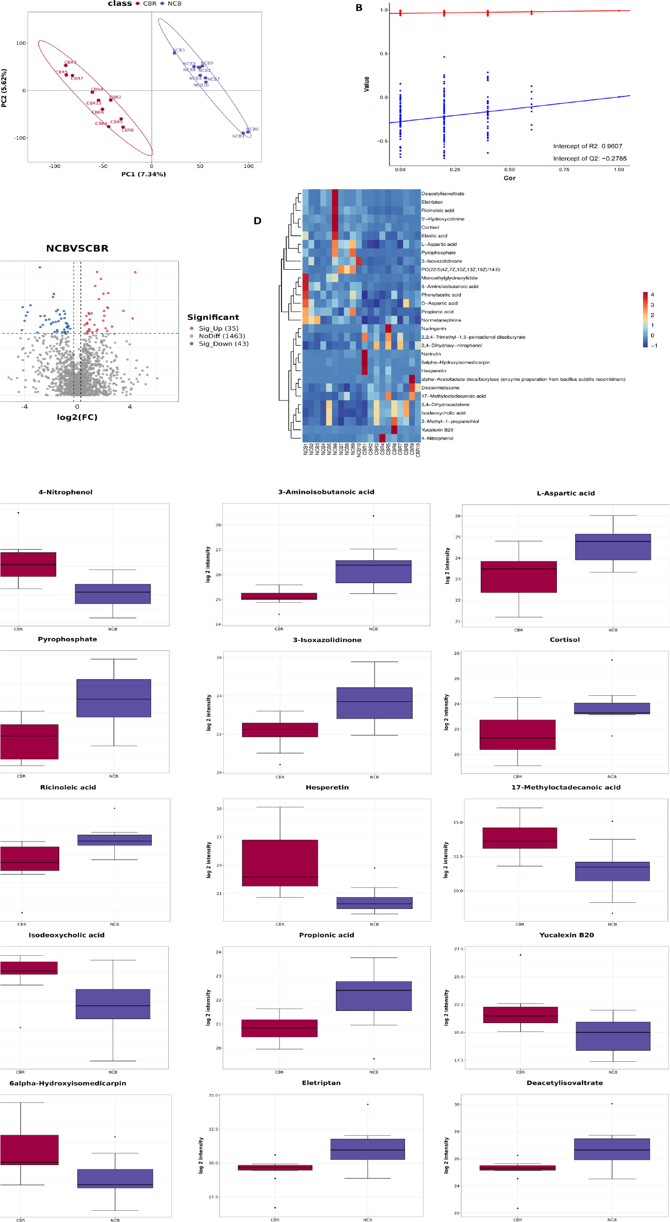
Fig. 6.
Screening and Identification of Differential Metabolites: (A) PLS-DA score plot demonstrates distinct separation between CBR and NCB groups along the first two principal components, reflecting significant intergroup metabolic differences. (B) Permutation test (200 iterations) confirmed model robustness, with original Q2 and R2 values significantly higher than permuted results, indicating no overfitting. (C) Volcano plot visualizes differential metabolites using log2(fold change) and -log10(P-value), highlighting 78 significant metabolites (35 upregulated, 43 downregulated) meeting thresholds of |log2FC| ≥ 0.26, P < 0.05, and VIP ≥ 1. (D) Heatmap displays the top 30 most significant metabolites (lowest P-values), showing clear group-specific clustering. (E) Boxplots illustrate the top 15 most significantly altered metabolites (P ≤ 0.01), with 4-Nitrophenol, Hesperetin, 17-Methyloctadecanoic acid, Isodeoxycholic acid, Yucalexin B20, and 6alpha-Hydroxyisomedicarpin significantly elevated in the CBR group, while 3-Aminoisobutanoic acid, L-Aspartic acid, Pyrophosphate, 3-Isoxazolidinone, Cortisol, Ricinoleic acid, Propionic acid, Eletriptan, and Deacetylisovaltrate were more abundant in the NCB group.
KEGG enrichment and correlation analysis of metabolites
KEGG enrichment analysis of significantly differential metabolites revealed that metabolic pathways—particularly Metabolic pathways, Microbial metabolism in diverse environments, and Biosynthesis of secondary metabolites—contained the highest number of altered metabolites, indicating substantial metabolic disruption may influence immunotherapy efficacy (Fig. 7A). Bubble plot analysis (Fig. 7B) further demonstrated significant enrichment in pathways including D-amino acid metabolism, Alanine, aspartate and glutamate metabolism, ABC transporters, and Protein digestion and absorption. A metabolite-pathway interaction network (Fig. 7C) exhibited a core-periphery structure, with key functional modules such as Metabolic pathways and Microbial metabolism in diverse environments at the core, and pivotal metabolite nodes including L-Aspartic acid, L-Alanine, Propionic acid, and Cortisol driving group differences. Gene Set Enrichment Analysis (GSEA) indicated that Protein digestion and absorption and D-amino acid metabolism were significantly activated in the NCB group (high NES, low P-value), whereas Nucleotide metabolism and Purine metabolism were suppressed (NES < 0, P < 0.05), suggesting altered nutrient metabolism and cell proliferation regulation (Fig. 7D). Correlation analysis of the top 30 most significant metabolites (Fig. 9E) and a network of high-confidence associations (|rho| > 0.7, Fig. 7F) identified Deacetylisovaltrate, Cortisol, Ricinoleic acid, Eletriptan, and Valgaciclovir as highly connected hub metabolites, indicating their potential role as regulators of metabolic flux and phenotypic divergence between response groups.
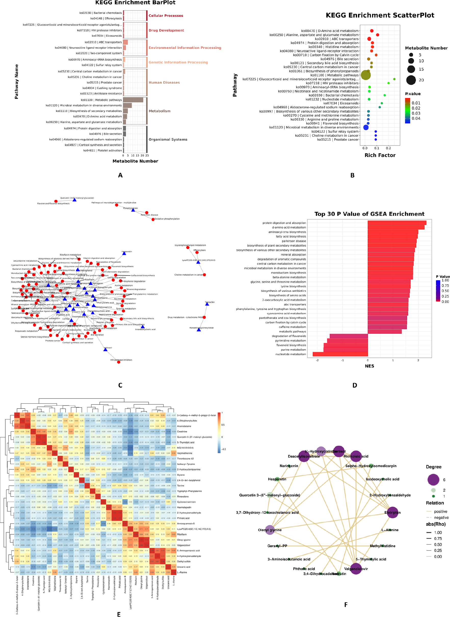
Fig. 7.
KEGG Enrichment and Correlation Analysis of Metabolites: (A) Hierarchical bar chart showing KEGG pathway enrichment, with horizontal axis indicating the number of differential metabolites per pathway, vertical axis listing pathway names, and color representing KEGG Level 1 classification. (B) Bubble plot of the top 28 enriched pathways (P ≤ 0.05), with Rich Factor (differential metabolites/total metabolites in pathway) on the horizontal axis, pathway names on the vertical axis, point size showing metabolite count, and color indicating enrichment P-value. (C) Regulatory network between metabolites (triangles, top 30 by P-value) and pathways (circles), where connectivity reflects functional importance. (D) Enrichment bar chart displaying pathway names versus Normalized Enrichment Score (NES), with bar color ranging from blue (P=1.0) to red (P=0.0) to show significance. (E) Correlation heatmap with red indicating positive and blue negative metabolite relationships. (F) Correlation network diagram where nodes represent metabolites and edges (|rho| > 0.7) reflect associations, with highly connected nodes indicating functional importance in metabolic flux and phenotypic differences.
Correlation analysis between differential microbes and metabolites
Spearman’s correlation analysis was conducted between significantly differential metabolites (P < 0.05) and differential microorganisms, with results visualized in a heatmap (Fig. 8A). Given the exploratory nature of this analysis, correlations with |ρ| ≥ 0.60 and nominal P < 0.05 were considered for interpretation. For additional quality control, FDR correction was applied, and the key correlation patterns remained consistent after correction (adjusted P < 0.10). The analysis revealed distinct association patterns: s_Roseburia_hominis exhibited a positive correlation with 2-Hydroxybenzaldehyde and a negative correlation with Methylhistidine; s_Clostridium_unclassified showed positive correlations with Guanosine, 4-Ethylphenylsulfate, 3-Carboxy-4-methyl-5-propyl-2-furanpropionic acid (CMPF), and Quercetin 3-(6”-malonylglucoside); s_Marseilla_massiliensis was positively correlated with Pyrophosphate, Riboflavin and PC(22:5(4Z,7Z,10Z,13Z,16Z)/14:0). Furthermore, network plots illustrating interactions between these differential microorganisms (s_Roseburia_hominis, s_ Clostridium_unclassified, and s_Marseilla_massiliensis) and differential metabolites meeting |r| ≥ 0.60 and P < 0.05 are presented in Fig. 8B, 8C, and 8D, respectively. These networks highlight specific microbe-metabolite associations that may underlie mechanistic pathways influencing immunotherapy response.
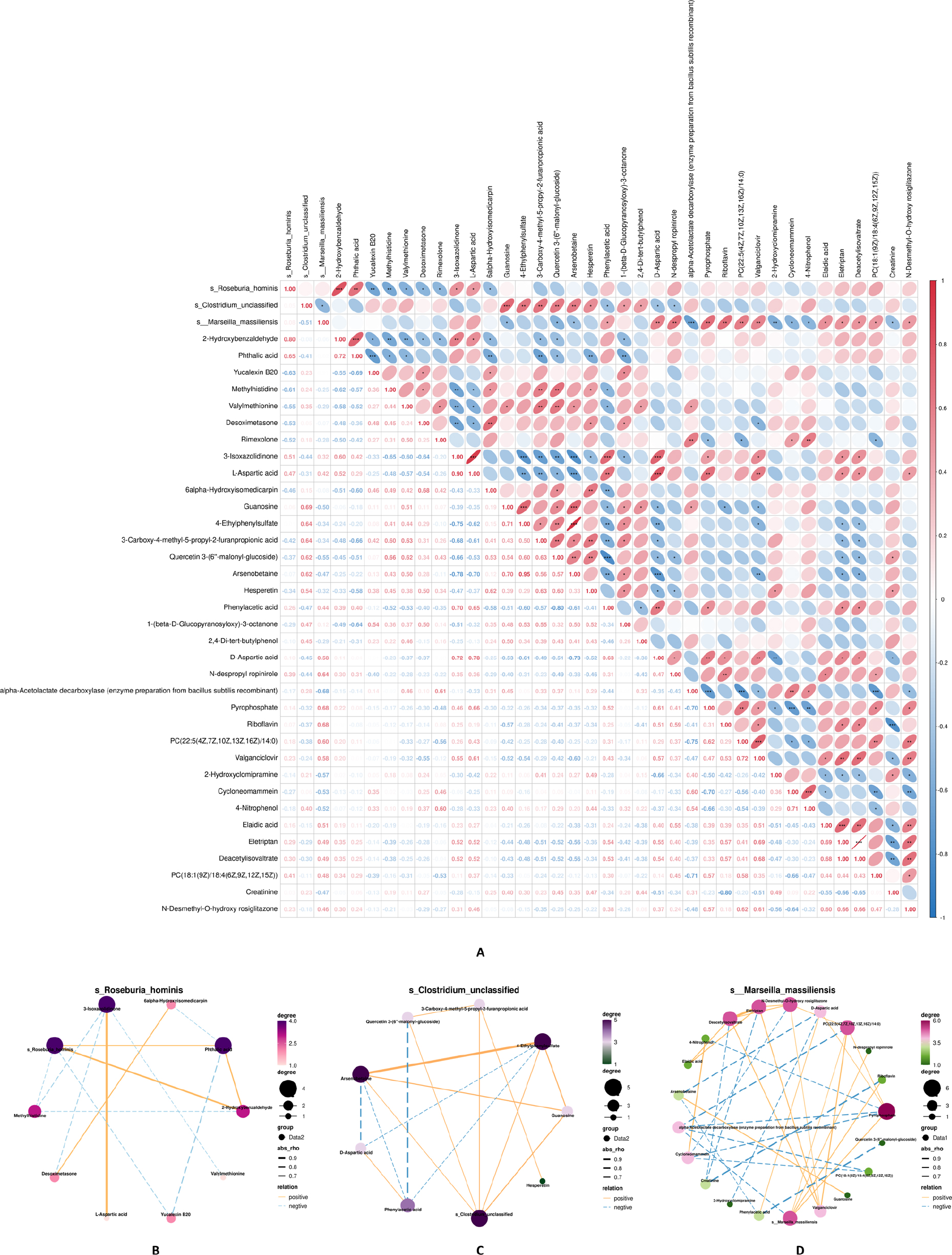
Fig. 8.
Correlation Analysis Between Differential Microbes and Metabolites: (A) Heatmap of Spearman correlation between differential metabolites and microorganisms. Color gradient (red to white to blue) corresponds to correlation coefficients (1 to -1), with warm and cool colors indicating positive and negative correlations, respectively. Numerical labels show exact correlation values, and asterisks denote significance levels: *P < 0.05, **P < 0.01, ***P < 0.001. (B), (C), (D) Network diagrams illustrating correlations between the dif-ferential microorganisms s_Roseburia_hominis, s_Clostridium_unclassified, and s_Marseilla_massiliensis, respectively, and differential metabolites meeting |r| ≥ 0.60 and P < 0.05. Line style and color represent positive (solid/red) or negative (dashed/blue) correlations, line thickness indicates correlation strength, and node size/color intensity reflects the number of correlated entities. The key correlation patterns remained consistent after FDR correction (adjusted P < 0.10), supporting the robustness of these microbe-metabolite associations.
Discussion
In this study, we systematically evaluated the composition and metabolic profiles of the intestinal flora of MSI-H CRC patients treated with immunotherapy and comprehensively depicted the microecological profiles of the different efficacy groups at seven taxonomic levels ranging from kingdom to species. We further carried out differential analyses to explore potential biomarkers closely related to immunological efficacy, which provided a panoramic view for elucidating the complex associations between the intestinal flora and the efficacy of ICIs. Our findings, while derived from an exploratory cohort, suggest that even though the overall microbial Alpha diversity and Beta diversity were not significantly different between the NCB and CBR groups with the current sample size, there were significant differences at the species and metabolite levels, implying that key flora and metabolites may be involved in the immunotherapy response. It is important to acknowledge that fecal metabolomic profiles are inherently susceptible to environmental confounding factors, particularly dietary habits, lifestyle, and non-antibiotic medications. While our inclusion criteria excluded patients with recent antibiotic or probiotic use, we did not systematically collect or control for detailed dietary information (such as fiber intake, protein consumption, or specific food preferences) or other lifestyle parameters (such as physical activity, smoking, or alcohol consumption). Consequently, the differential metabolites identified between the CBR and NCB groups—such as guanosine, pyrophosphate, riboflavin, and specific phospholipids—may partially reflect inter-individual variations in dietary patterns rather than biologically specific signatures of immunotherapy response. For instance, dietary nucleotides from meat or fish consumption could influence fecal guanosine levels; riboflavin abundance may correlate with vitamin B2-rich food intake (e.g., dairy products, eggs, leafy greens); and phospholipid profiles may be modulated by dietary fat composition. Without comprehensive dietary records or nutritional assessments, we cannot definitively disentangle the contributions of host-microbe metabolic interactions from direct dietary inputs. Future studies must incorporate standardized dietary questionnaires, food frequency records, or nutritional biomarkers to control for these confounders and validate the biological specificity of microbiota-metabolite-immunity associations. While our study primarily focuses on the gut microbiome as a systemic regulator of immunotherapy response, emerging evidence highlights the intricate relationship between gut microecology and the intratumoral microbiome within the tumor microenvironment (TME). The TME represents a complex ecosystem where tumor cells, immune cells, stromal components, and microbial communities engage in dynamic crosstalk that fundamentally shapes tumor progression and therapeutic outcomes (Gao Z et al. 2025). Recent studies have demonstrated that intratumoral microbiota exhibit significant spatial heterogeneity and can directly influence tumor cell proliferation, immune evasion, and response to therapy. This “gut-TME” axis suggests that alterations in gut microecology may not only modulate systemic immunity but also parallel or interact with intratumoral microbial communities, creating a holistic-local regulatory network that determines immunotherapy efficacy.
At the microbial level, s_Clostridium_unclassified was significantly enriched in the CBR group, suggesting that members of the genus Clostridium may enhance the efficacy of immunotherapy for colorectal cancer. Although this operational taxonomic unit (OTU) has not yet been fully characterized, it has been demonstrated that butyrate-producing bacteria of the same genus, such as Clostridium butyricum (CBM588), enhance the therapeutic efficacy of immune checkpoint inhibitors (ICIs). In a colorectal cancer (CRC) model, combination treatment with C. butyricum was more effective in increasing the expression of CD4, CD8, and granzyme B than anti-PD-1 monotherapy (Xu H et al. 2023). A retrospective analysis of non-small cell lung cancer (NSCLC) patients treated with ICIs and concomitant CBM588 showed that administration of C. butyricum significantly improved progression-free survival (PFS) and overall survival (OS) compared to those not receiving CBM588 (Tomita et al. 2020). Similarly, in patients with metastatic renal cell carcinoma, the addition of the Clostridium butyricum CBM588 strain to nivolumab-ipilimumab resulted in a significant advantage in PFS and a trend toward improved OS, accompanied by significant increases in chemokines including CCL2 (MCP-1), CCL4 (MIP-1β), CXCL9 (MIG), and CXCL10 (IP-10) (Dizman et al. 2022). Recent studies have shown that Clostridium butyricum activates cytotoxic CD8+ T lymphocytes (CTLs) and inhibits tumor-associated macrophages (TAMs), particularly in combination with anti-PD-1 therapy. The surface protein SecD of Clostridium butyricum binds to the colorectal cancer cell receptor glucose-regulated protein 78 (GRP78), inactivates GRP78, and blocks the PI3K-AKT-NF-κB signaling pathway, thereby reducing the secretion of the immunosuppressive cytokine IL-6 (Xie M et al. 2025). Notably, the PI3K-AKT pathway represents a critical node in the immunoregulatory network, as evidenced by recent findings that pharmacological inhibition of this pathway can simultaneously activate autologous CD8+ T cells and suppress tumor AKT/MAPK signaling, thereby enhancing antitumor immunity (Liao et al. 2020). This suggests that s_Clostridium_unclassified may potentiate immunotherapy efficacy not only through direct immunomodulation but also via modulation of the AKT/MAPK axis, warranting further investigation into whether this unclassified species exerts similar pathway-specific effects. Interestingly, the enrichment of Clostridium species in the gut of responders may parallel their potential presence or functional influence within the TME. Intratumoral microbiota heterogeneity has been shown to modulate local immune infiltration and tumor cell behavior; thus, gut-derived Clostridium or their metabolites might translocate or systemically condition the TME to favor immunogenicity (Gao Z et al. 2025). This “gut-TME parallelism” offers a mechanistic explanation for why specific gut microbiota features can predict treatment response. Therefore, the significant enrichment of s_Clostridium_unclassified in the CBR group suggests that it could represent a class of immunosensitizing strains, providing a new candidate target for hypotheses regarding the association between bacterial flora and therapeutic response. However, due to its unclassified status, its biological function remains unclear, and future studies are needed to resolve the causal relationship between its metabolic profiles and immune regulation using metagenomic binning or single-bacterial isolation techniques. The interplay between gut microbiota and tumor cell death modalities represents an emerging frontier in immuno-oncology. Immunogenic cell death (ICD), characterized by the release of damage-associated molecular patterns (DAMPs) such as calreticulin, HMGB1, and ATP, is essential for initiating antitumor immune responses. Engineered nanomedicines targeting ICD have shown promise in converting “cold” tumors into “hot” tumors by enhancing dendritic cell maturation and cytotoxic T lymphocyte activation (Wang Y et al. 2022). In this context, the enrichment of immune-activating metabolites such as guanosine and quercetin derivatives in the CBR group raises the intriguing possibility that the s_Clostridium_unclassified-associated metabolomic profile may facilitate ICD-like immune activation. Guanosine, for instance, functions as a signaling molecule that can modulate immune cell activity, while flavonoid derivatives like quercetin 3-(6”-malonyl-glucoside) may promote tumor cell apoptosis and enhance immunogenicity. Future studies should investigate whether s_Clostridium_unclassified-derived metabolites can directly induce ICD in colorectal cancer cells or prime the tumor microenvironment for ICD-mediated immune activation.
In contrast, s_Roseburia_hominis and s_Marseilla_massiliensis were significantly enriched in the NCB group. The genus Roseburia comprises five well-characterized species—Roseburia intestinalis, Roseburia hominis, Roseburia inulinivorans, Roseburia faecis, and Roseburia cecicola—all of which produce short-chain fatty acids (SCFAs) such as acetate, propionate, and butyrate (Nie et al. 2021). Roseburia intestinalis and butyrate have been shown to significantly enhance the efficacy of anti-PD-1 therapy in mice bearing microsatellite instability-low (MSI-L) CT26 tumors. The mechanism involves butyrate directly binding to Toll-like receptor 5 (TLR5) on the surface of CD8+ T cells and enhancing its activity through nuclear factor-κB (NF-κB) signaling pathway activation, thereby improving the anti-tumor response (Kang et al. 2023). In a CRC mouse model, treatment with Roseburia intestinalis induced high infiltration of CD8+ T cells within the tumor and demonstrated higher efficacy than a mixture of four Clostridium strains (Montalban-Arques et al. 2021). Additionally, Roseburia intestinalis enhances the sensitivity of CRC to radiotherapy by producing butyrate, which activates the OR51E1/RALB axis, promotes autophagy in CRC cells, inhibits DNA damage repair, and modulates the immune microenvironment, thereby accelerating cancer cell death (Dong et al. 2024). However, the functional heterogeneity within the Roseburia genus extends beyond species-level differences to encompass distinct immunometabolic phenotypes. Ferroptosis, an iron-dependent form of regulated cell death driven by lipid peroxidation, has emerged as a critical determinant of cancer therapy response. Recent evidence indicates that the System Xc⁻/GSH/GPX4 axis, which regulates ferroptosis susceptibility, interacts intimately with the gut microbiome (Gao J et al. 2020). Butyrate and other SCFAs can modulate cellular redox status and lipid metabolism, potentially influencing tumor cell sensitivity to ferroptosis. The paradoxical enrichment of s_Roseburia_hominis in the NCB group, contrary to the reported beneficial effects of Roseburia intestinalis, may reflect species-specific differences in ferroptosis modulation. From a TME perspective, intratumoral microbial heterogeneity can create immunosuppressive niches that facilitate immune evasion. The enrichment of s_Roseburia_hominis in the gut of non-responders may mirror or contribute to a TME characterized by ferroptosis resistance and impaired ICD, where local microbial communities or their metabolites synergize with tumor cells to dampen immune surveillance (Gao Z et al. 2025). Understanding this gut-TME interaction is crucial for explaining why certain microbiota profiles correlate with treatment failure. For instance, distinct Roseburia species may produce varying ratios of SCFAs or differentially regulate host lipid metabolic enzymes such as ACSL4 and LPCAT3, thereby altering the lipid composition of tumor cell membranes and their susceptibility to ferroptotic death. Moreover, the antioxidant metabolites associated with s_Roseburia_hominis (e.g., 2-Hydroxybenzaldehyde) may protect tumor cells from lipid peroxidation, conferring resistance to ferroptosis and subsequently diminishing immunotherapy efficacy. This hypothesis aligns with the observation that ferroptosis induction can enhance antitumor immunity by releasing immunostimulatory signals and promoting dendritic cell maturation (Gao J et al. 2020). Therefore, the “Roseburia paradox” observed in our study may underscore the importance of ferroptosis regulation in microbiome-immunotherapy crosstalk, suggesting that future probiotic interventions should consider species-specific effects on regulated cell death pathways. Notably, this study found significant enrichment of s_Roseburia_hominis in the baseline fecal samples of the NCB group, a finding that contrasts with previous reports indicating that Roseburia intestinalis enhances immunotherapy efficacy in colorectal cancer. This discrepancy highlights the potential for species-or even strain-specific functional diversity within the same genus, where different members may exert distinct or even opposing immunomodulatory effects. This inconsistency may arise from several factors: (1) population heterogeneity and the relatively limited sample size of this study, which may affect statistical validity; (2) functional differences between Roseburia species and even subspecies, which may exhibit unique immunomodulatory properties, including differential modulation of ferroptosis susceptibility and ICD induction; and (3) methodological limitations, such as potential species annotation bias. Future studies should further validate the immunomodulatory mechanisms of specific s_Roseburia_hominis strains through me tag -enome-assembled genomes (MAGs), single-bacterium isolation and culture, and functional experiments. Additionally, no direct association between s_Marseilla_massiliensis and immunotherapy has been reported, warranting further in-depth investigation. Large-sample, multi-center prospective studies are needed to validate these findings and explore the potential biological mechanisms and clinical applications.
At the metabolite level, this study explored the differences in metabolic profiles between the CBR and NCB groups in MSI-H CRC patients receiving immunotherapy through untargeted metabolomics analysis. A total of 78 significantly different metabolites (35 up-regulated and 43 down-regulated) were identified, which may be involved in immune regulation, energy metabolism, and intestinal microenvironment homeostasis, providing a new metabolic perspective for understanding the differences in ICIs efficacy. However, we caution that these metabolic differences should be interpreted with consideration of potential dietary confounders, as discussed above. In the CBR group, s_Clostridium_unclassified was positively correlated with the expression of guanosine, CMPF, Quercetin 3-(6”-malonyl-glucoside), and other molecules associated with immune activation. Guanosine, a purine metabolite, exhibits various biological activities including cell signaling, neuroprotection, immunomodulation, and anti-inflammatory effects. Notably, guanosine can be derived from both endogenous microbial metabolism and dietary nucleotide sources; thus, its enrichment in the CBR group may reflect either enhanced microbial purine biosynthesis or differential dietary intake of nucleotide-rich foods. Guanylate cyclase agonists can enhance the body’s immune response by modulating the activity of immune cells (Bettio et al. 2016). CMPF (3-carboxy-4-methyl-5-propyl-2-furanpropanoic acid) is an endogenous metabolite mainly derived from the metabolism of furan fatty acids (Fu-FAs). Although CMPF may exhibit deleterious effects in certain pathological states, its precursor FuFAs possess antioxidant activity, capable of scavenging reactive oxygen species and mitigating oxidative stress-induced damage (Guo et al. 2021), suggesting that it may influence immunological efficacy through antioxidant responses. Quercetin 3-(6”-malonyl-glucoside), a flavonoid derivative, demonstrates significant antitumor activity. Quercetin and its glycosides are abundant in plant-based foods (e.g., onions, apples, berries); therefore, their detection in fecal samples may partially reflect dietary flavonoid intake rather than solely microbial biotransformation. Molecular dynamics simulation studies have shown that this compound can bind to the anti-apoptotic protein Bcl-2 and inhibit its activity, thereby promoting apoptosis in cancer cells (Kim et al. 2005). The capacity of quercetin derivatives to promote apoptosis intersects with the broader land-scape of regulated cell death in cancer immunotherapy. While apoptosis has traditionally been considered immunologically silent, emerging evidence suggests that certain apoptotic stimuli can trigger immunogenic cell death (ICD) when occurring in the context of endoplasmic reticulum stress and reactive oxygen species production. Nanomedicine-based delivery of ICD inducers, including certain flavonoids and chemotherapeutic agents, has demonstrated enhanced efficacy in preclinical models by promoting dendritic cell maturation and tumor-specific T cell responses (Wang Y et al. 2022). In the present study, the characteristic elevation of quercetin 3-(6”-malonyl-glucoside) in the CBR group suggests that this metabolite could contribute to immunotherapy sensitivity not merely through direct cytotoxicity but potentially by lowering the threshold for ICD in tumor cells or by creating an immunostimulatory microenvironment that facilitates antigen presentation. Furthermore, the combination of guanosine-mediated immune cell activation and quercetin-induced tumor cell stress may represent a synergistic “push-pull” mechanism that enhances the overall antitumor immune response. In the present study, this metabolite was characteristically elevated in the CBR group, suggesting that it could jointly contribute to immunotherapy sensitivity through direct modulation of immune effector cell function and regulation of tumor cell biological behavior. These results further emphasize that the response of CRC patients to immunotherapy depends not only on microbial community structural characteristics but may also be synergistically regulated by microbiota-derived metabolites within the immunometabolic network.
The systemic effects of gut-derived metabolites must be considered in the context of TME heterogeneity. Intratumoral microbiota and their metabolites can create spatially distinct microenvironments within the tumor, leading to regional differences in immune cell infiltration and therapeutic sensitivity (Gao Z et al. 2025). For example, gut-derived guanosine and quercetin metabolites may not only enhance systemic immunity but also directly modulate the TME by promoting M1 macrophage polarization, enhancing dendritic cell function, or inhibiting regulatory T cell recruitment within the tumor bed. This dual action—systemic immune priming and local TME modulation—provides a comprehensive mechanistic basis for the predictive value of gut microbiota features. Future studies employing spatial transcriptomics and metabolomics are warranted to dissect the precise contributions of gut-derived versus intratumoral microbial metabolites in shaping the immunotherapy-responsive TME.
In contrast, the metabolites enriched in the NCB group were more associated with impaired energy metabolism and immunosuppressive states. s_Roseburia_ hominis was positively correlated with 2-Hydroxy-benzaldehyde, an aromatic aldehyde with antioxidant capacity that scavenges free radicals and protects cells from oxidative damage (Xie R et al. 2016), suggesting that s_Roseburia_hominis may reduce immunotherapy efficacy through antioxidant responses. This antioxidant profile may have profound implications for ferroptosis regulation. The System Xc/GSH/GPX4 axis, which represents the primary cellular defense against lipid peroxidation and ferroptosis, relies heavily on glutathione (GSH) and nicotinamide adenine dinucleotide phosphate (NADPH) availability. Metabolites such as riboflavin (positively correlated with s_Marseilla_massiliensis) serve as precursors for FAD-dependent enzymes including glutathione reductase, which regenerates reduced glutathione from its oxidized form. Thus, the enrichment of antioxidant-associated metabolites in the NCB group may reflect a metabolic program that suppresses ferroptosis in tumor cells, thereby limiting the release of damage-associated molecular patterns and attenuating ICD (Gao J et al. 2020). This “ferroptosis-resistant” phenotype, characterized by robust antioxidant defenses and intact lipid repair mechanisms, may render tumor cells less susceptible to immunotherapy-induced cell death and impair the generation of antitumor immunity. The TME in non-responders may thus represent a “double jeopardy” scenario: systemically impaired immune priming due to gut dysbiosis combined with locally immunosuppressive, ferroptosis-resistant tumor niches. Intratumoral microbial communities in such environments may further exacerbate immune evasion by producing immunosuppressive metabolites or recruiting myeloid-derived suppressor cells (Gao Z et al. 2025). Understanding these parallel systemic and local mechanisms is essential for developing combination strategies that simultaneously restore gut homeostasis and reprogram the TME. Targeting these metabolic vulnerabilities-perhaps through the use of System Xc⁻ inhibitors such as sulfasalazine or GPX4 inhibitors such as RSL3z-could represent a promising strategy to overcome immunotherapy resistance in patients harboring the NCB-associated microbiome and metabolome. s_Marseilla_massiliensis showed positive correlations with pyrophosphate (PPi), riboflavin, and PC(22:5(4Z,7Z,10Z,13Z,16Z)/14:0), indicating that this bacterium or its metabolites may promote PPi production or inhibit its hydrolysis. High concentrations of PPi can inhibit ATP synthase, leading to insufficient energy supply, which may trigger HIF-1α-mediated glycolytic reprogramming and ultimately drive T cells toward a terminally exhausted phenotype (Tex) (Wu H et al. 2023). Riboflavin (vitamin B2) serves as a precursor for two coenzymes, flavin mononucleotide (FMN) and flavin adenine dinucleotide (FAD), and is involved in energy metabolism, antioxidant defense, cell signaling, drug metabolism, and growth. It supports a synergistic antioxidant system through FMN/FAD-dependent enzymes such as glutathione reductase (GR), superoxide dismutase (SOD), and catalase (CAT), as well as through direct free radical scavenging and regeneration of vitamins C and E. These antioxidant properties suggest that riboflavin may attenuate antitumor immune responses (Olfat et al. 2022). PC(22:5(4Z,7Z,10Z,13Z,16Z)/14:0) exerts its functions through its unique fatty acid composition, featuring ω-6 docosapentaenoic acid (a polyunsaturated fatty acid) at the sn-2 position and myristic acid (a saturated fatty acid) at the sn-1 position. Although the specific biological activity of PC(22:5/14:0) has not been individually studied, enrichment of polyunsaturated fatty acids has been demonstrated to exhibit antioxidant effects, inhibit lipid peroxidation, and possess anti-atherosclerotic properties (Richard et al. 2008). The enrichment of polyunsaturated fatty acids (PUFAs) in the NCB group presents an intriguing paradox, as PUFAs are typically considered substrates for lipid peroxidation and ferroptosis. However, when coupled with robust antioxidant defenses (e.g., elevated riboflavin and glutathione reductase activity), these lipids may be protected from peroxidation, effectively creating a “ferroptosis-resistant” lipid environment. This metabolic configuration—abundant PUFAs paired with strong antioxidant capacity—may represent an optimal state for tumor cell survival and proliferation, while simultaneously limiting immunogenic cell death. Pharmacological strategies that disrupt this balance, such as inhibitors of glutathione synthesis or lipid peroxidation repair enzymes, could potentially restore ferroptosis sensitivity and enhance ICD, thereby converting the “cold” NCB-associated tumor microenvironment into a “hot”, immunotherapy-responsive state (Wang Y et al. 2022; Gao J et al. 2020).
Beyond the gut microbiome and metabolome, our study identified a significant association between a classic host systemic biomarker and clinical outcome. Serum lactate dehydrogenase (LDH) levels were significantly elevated in the NCB group compared to the CBR group. LDH is a key enzyme in anaerobic glycolysis (the Warburg effect), and its elevated serum level is a well-established biomarker of high tumor burden, aggressive disease, and poor prognosis across various cancer types (Jurisic et al. 2015). This finding aligns with and complements our microbiome data. It suggests that patients who derived less clinical benefit from immunotherapy not only harbor a distinct gut microbial ecosystem but also exhibit a systemic state of heightened tumor metabolic activity. We hypothesize that elevated LDH (reflecting a pro-tumor metabolic microenvironment) and the specific “flora–metabolite” signature observed in non-responders may represent convergent or synergistic biological pathways that collectively foster an immunosuppressive milieu, thereby limiting the efficacy of ICIs. This metabolic-immune crosstalk may intersect with key oncogenic signaling pathways. The AKT/MAPK pathway, which is frequently hyperactivated in colorectal cancer, promotes glycolytic metabolism (the Warburg effect) and concurrently suppresses antitumor immunity by modulating T cell exhaustion and regulatory T cell function. Recent evidence demonstrates that dual targeting of this pathway—through pharmacological inhibition combined with immune modulation—can achieve superior antitumor effects compared to monotherapy (Liao et al. 2020). In our study, the elevated LDH in the NCB group may reflect AKT/MAPK-driven metabolic reprogramming, which could be further amplified by the immunosuppressive microbiome-metabolite profile. Conversely, the CBR group, characterized by s_Clostridium_unclassified enrichment and immune-activating metabolites, may exhibit relative suppression of the AKT/MAPK axis, creating a metabolic environment conducive to effector T cell function and immunotherapy response. Future mechanistic studies should investigate whether s_Clostridium_unclassified -derived metabolites can directly modulate the AKT/MAPK pathway in tumor cells or immune cells, potentially through the SecD-GRP78-PI3K-AKT axis as observed with Clostridium butyricum, or through novel mechanisms involving immunogenic cell death and ferroptosis regulation. The integration of LDH as a metabolic biomarker with gut microbiota profiling offers a promising approach to stratify patients based on both systemic and local TME characteristics. Intratumoral microbial heterogeneity and local lactate metabolism within the TME may further refine this stratification, as regional differences in microbial colonization and metabolic activity can create “hot” and “cold” tumor subregions with differential sensitivity to immunotherapy (Gao Z et al. 2025). Multi-omics approaches combining gut microbiome, circulating metabolites, and intratumoral microbiome profiling will be essential to fully capture the complexity of host-microbe-tumor interactions. While this study does not establish a direct mechanistic link between systemic LDH and gut microbiota, it underscores the potential of an integrated “host–microbe” metabolic axis in determining immunotherapy response. Future investigations should explore the dynamic interplay between circulating metabolic markers like LDH and gut microbiome composition during treatment.
These findings suggest that although certain core species are shared between groups, their differential abundance may drive functional variations in key metabolic pathways, potentially influencing host response to immunotherapy. In summary, the present study demonstrates that MSI-H CRC patients may exhibit two distinct metabolic signature patterns during immunotherapy response. The CBR group was characterized by enrichment of s_Clostridium_unclassified and immune-promoting metabolites such as guanosine and quercetin 3-(6”-malonyl-glucoside), which are proposed to potentially enhance therapeutic efficacy through improving effector T-cell function, promoting apoptotic signaling, and optimizing the immune microenvironment. These mechanisms may converge on the induction of immunogenic cell death (ICD), wherein guanosine-enhanced immune cell activation synergizes with quercetin-mediated tumor cell stress to create a feed-forward loop of antitumor immunity. The potential involvement of the AKT/MAPK pathway in this process-whether through direct microbial modulation or metabolite-mediated signaling—further underscores the complexity of the microbiome-immunity axis (Liao et al. 2020; Wang Y et al. 2022). In contrast, the NCB group showed enrichment of s_Roseburia_ hominis, s_Marseilla_massiliensis, and metabolites associated with antioxidant response and disrupted energy metabolism—such as PPi, riboflavin, and specific phospholipids. These metabolites could contribute to immunosuppression or T-cell exhaustion by impairing energy metabolism and promoting excessive antioxidant activity, potentially leading to a diminished response to immunotherapy. Specifically, the robust antioxidant capacity associated with the NCB signature may confer resistance to ferroptosis, a regulated cell death modality that has been shown to enhance antitumor immunity when appropriately induced. The resulting “ferroptosis-resistant” and “ICD-deficient” tumor microenvironment may represent a formidable barrier to immunotherapy efficacy, suggesting that combination strategies incorporating ferroptosis inducers or ICD promoters could be particularly beneficial for this patient population (Wang Y et al. 2022; Gao J et al. 2020). From a holistic perspective, the efficacy of immunotherapy is determined by the intricate interplay between systemic immune status, shaped by the gut microbiome, and local TME characteristics, influenced by intratumoral microbial heterogeneity (Gao Z et al. 2025). Our findings suggest that gut microbiota features may serve as accessible biomarkers that reflect or predict the immunological state of the TME. However, the precise mechanisms linking gut and intratumoral microbial communities—whether through direct bacterial translocation, metabolite-mediated systemic conditioning, or immune cell trafficking—remain to be elucidated. Future studies should employ multi-site sampling (fecal, blood, and tumor tissue) and advanced spatial omics technologies to dissect these complex interactions and develop integrated biomarker panels that capture both systemic and local determinants of immunotherapy response. These preliminary findings provide new molecular hypotheses regarding the mechanisms underlying both response and resistance to immunotherapy in CRC, and suggest that targeting specific metabolic pathways merits investigation as a promising strategy for improving treatment outcomes. Further validation in large, multi-center cohorts, combined with functional experiments and clinical translational studies, will be essential to develop novel microbiomeor metabolite-based combination immunotherapies.
The integration of our findings with emerging concepts in cancer immunotherapy suggests several promising directions for translational research. First, the identification of s_Clostridium_unclassified as a potential immunosensitizing strain warrants further investigation into its capacity to induce ICD or modulate ferroptosis sensitivity. Engineered delivery systems, such as lipid nanoparticles or bacterial vesicles, could be employed to enhance the bioavailability of s_Clostridium_unclassified-derived metabolites and maximize their immunomodulatory effects (Wang Y et al. 2022). Second, the “Roseburia paradox” highlights the need for strain-specific functional characterization, as different species within the same genus may exert opposing effects on regulated cell death pathways. Precision microbiome modulation—targeting specific strains based on their metabolic output and immunomodulatory phenotype—may represent a more effective strategy than broad-spectrum probiotic administration. Finally, the metabolic vulnerabilities identified in the NCB group, particularly the antioxidant-dependent ferroptosis resistance, suggest rational combination therapies involving System Xc⁻ or GPX4 inhibitors to restore immunogenic cell death and enhance ICI efficacy. Notably, these interventions should be designed with consideration of TME heterogeneity, as intratumoral microbial communities may create spatially variable therapeutic sensitivities. Strategies that combine systemic microbiome modulation with local TME-targeted therapies—such as intratumoral injection of ferroptosis inducers or ICD-promoting agents—may offer synergistic benefits by simultaneously addressing systemic immune priming and local immune evasion (Gao Z et al. 2025). These strategies, grounded in the mechanistic understanding of microbiome-metabolite-immunity interactions, hold promise for overcoming immunotherapy resistance in MSI-H colorectal cancer.
However, this study has several limitations: (1) First and foremost, this exploratory study is limited by a relatively small sample size (n = 20), which was influenced by the strict exclusion criteria and a limited six-month collection period. We acknowledge that this constrains the statistical power and may affect the generalizability of the conclusions. Nevertheless, it is representative of the preliminary, hypothesis-generating nature of this work. Future studies should expand the sample size and conduct multicenter prospective investigations to further validate and generalize the findings regarding intestinal microbiome characteristics in MSI-H CRC patients undergoing immunotherapy. (1a) Importantly, the lack of systematic dietary and lifestyle data collection represents a significant methodological limitation. The fecal metabolome is profoundly influenced by dietary habits (fiber intake, protein and fat consumption, specific food groups), lifestyle factors (physical activity, smoking, alcohol use), and non-antibiotic medications (e.g., proton pump inhibitors, NSAIDs), which we did not assess or control for in this study. Consequently, we cannot exclude the possibility that the observed metabolic differences between CBR and NCB groups partially reflect inter-individual variations in dietary patterns rather than biologically specific signatures of immunotherapy response. Future studies must incorporate comprehensive dietary questionnaires (e.g., food frequency questionnaires, 24-hour dietary recalls), anthropometric measurements (BMI, body composition), and lifestyle assessments to disentangle the effects of environmental confounders from host-microbe metabolic interactions. Additionally, the absence of baseline clinical characteristic comparisons (e.g., BMI, dietary preferences) between response groups precludes our ability to determine whether differential metabolites are specific to immunotherapy response or merely surrogates of lifestyle heterogeneity. (2) The metagenomic and metabolomic data were derived from a cross-sectional design, which prevents the establishment of causal relationships. Subsequent studies should include longitudinal sampling at multiple time points before, during, and after treatment to dynamically track temporal changes in microbial composition and metabolic profiles. (2a) Crucially, future studies should incorporate paired tumor tissue sampling to characterize intratumoral microbiota and their spatial relationship with immune cell infiltration and metabolic activity. This will enable direct comparison of gut and intratumoral microbial communities and elucidation of their respective contributions to TME heterogeneity and immunotherapy response (Gao Z et al. 2025). (3) This study did not incorporate data on intestinal immune cell infiltration or transcriptomic profiles, limiting a comprehensive understanding of the “flora–metabolism–immunity” axis. Future research should specifically investigate the impact of s_Clostridium_unclassified and s_Roseburia_hominis on T cell infiltration, dendritic cell maturation, and the expression of key regulators of ferroptosis (e.g., GPX4, SLC7A11, ACSL4) and ICD (e.g., calreticulin, HMGB1, ATP) within the tumor micro-environment. Single-cell transcriptomics and spatial metabolomics could provide unprecedented resolution of these host-microbe interactions. Future research should integrate single-cell transcriptomics, functional validation experiments, and animal models to elucidate the mechanisms through which key microbial and metabolic features influence treatment response and to evaluate their potential as therapeutic targets. (4) Fecal samples were collected after patients had completed 2–3 cycles of immunotherapy rather than at baseline prior to treatment initiation, owing to the retrospective nature of the study design. Consequently, we cannot exclude the possibility that some observed microbiome and metabolome differences may reflect alterations induced by immunotherapy itself rather than pre-existing features predictive of treatment response. Future prospective studies with pre-treatment sample collection are warranted to validate the predictive value of the identified microbial and metabolic biomarkers. (5) This study focused exclusively on MSI-H patients, and therefore lacks a comparison group of microsatellite-stable (MSS) patients. Consequently, it remains unclear whether the observed microbiome and metabolome signatures are specific to the MSI-H population or represent general features associated with immunotherapy response across different molecular subtypes of colorectal cancer. Moreover, the molecular mechanisms underlying the differential efficacy of ICIs in MSI-H versus MSS colorectal cancer—particularly regarding the involvement of the AKT/MAPK pathway, ferroptosis sensitivity, and ICD induction—remain to be elucidated. Furthermore, the relative importance of gut versus intratumoral microbiota in mediating immunotherapy response may differ between MSI-H and MSS subtypes, given their distinct TME characteristics and baseline immune infiltration patterns (Gao Z et al. 2025). Comparative studies incorporating both molecular subtypes could clarify whether the microbiome-mediated modulation of these pathways is subtype-specific or represents a universal determinant of immunotherapy response. Future studies should include both MSI-H and MSS cohorts to enable comparative analyses and to delineate subtype-specific microbial and metabolic determinants of immunotherapy efficacy. (6) The microbiota-metabolite associations presented in this study are based on Spearman correlation analysis, which does not establish causation. While we have identified significant correlations between specific species (e.g., s_Clostridium_unclassified) and metabolites (e.g., guanosine), we cannot confirm whether these metabolites are directly produced or metabolized by the corresponding bacteria. A more definitive “gene-metabolite” consistency analysis would require metagenome-assembled genomes (MAGs) and targeted metabolic pathway validation, which are beyond the scope of this exploratory study. Future research integrating deep metagenomic sequencing, stable isotope tracing, and in vitro culture experiments is warranted to establish causal relationships.
Conclusions
In this study, the integration of microbiomics and metabolomics revealed a unique “Flora-Metabolites” interaction pattern in MSI-H CRC patients with differential responses to immunotherapy. The results demonstrated that the CBR group was associated with the enrichment of s_Clostridium_unclassified and its putatively immune-enhancing metabolites, such as guanosine, CMPF, and quercetin 3-(6”-malonyl-glucoside), suggesting a potential role in promoting antitumor immune responses. In contrast, the NCB group exhibited associations with s_Roseburia_hominis, s_Marseilla_ massiliensis, and potentially immunosuppression-related metabolites, including PPi, riboflavin, and PC(22:5(4Z,7Z,10Z,13Z,16Z)/14:0), indicating their possible involvement in the development of immunotherapy tolerance. These findings, while exploratory, provide novel molecular insights worthy of further validation into the role of host-microbe co-metabolism in shaping immunotherapy outcomes establish a foundation for future research into interventions targeting specific microbial or metabolic pathways to enhance combination immunotherapy strategies.
Notes
[2] Availability of data and material
The raw metabolomics data generated in this study have been deposited in the MetaboLights repository under accession code MTBLS13265. The raw metagenomic sequencing data have been deposited in the NCBI Sequence Read Archive (SRA) under Bio-Project accession code PRJNA1357844. These datasets are currently under embargo until 2026-11-05 and 2027-11-01, respectively, but are fully accessible to editors and reviewers via the following private link for Metabolights: https://www.ebi.ac.uk/metabolights/reviewer9e943d43-e584-4b33-a98b-0532696fe81e. The data will be made publicly available upon article publication or on the specified dates, whichever is earlier.
[3] Ethical statement
The study was approved by the Ethics Committee of Zhengzhou Central Hospital (Approval No. ZXYY2025033). All eligible patients were thoroughly informed of the study’s rationale and clinical significance. Written informed consent was obtained from each participant prior to enrollment, and both patients and their families were adequately informed before signing the consent forms. All research-specific procedures, including sample collection and data analysis for this study, were conducted only after ethical approval was obtained. Written informed consent for publication of their clinical details and/or clinical images was obtained from all participants involved in the study.
[4] Contributed by Author contributions
Fang Liang and Jing Li: conceptualization, methodology, formal analysis, writing – original draft preparation, writing – review & editing. Yingkun Yue: software, data curation. Jiaxin Pan: visualization, investigation. Chenyu Liu: formal analysis, data curation. Duo Cheng: project administration, funding acquisition. Nan Zhang: methodology, writing – review & editing. Kunkun Li: supervision, writing – review & editing. Huili Wu and Feifei Chu: resources, supervision. All authors have read and approved the final version of the manuscript and agree to be accountable for their own contributions.
[5] Contributed by Conflict of interest
The authors do not report any financial or personal connections with other persons or organizations, which might negatively affect the contents of this publication and/or claim authorship rights to this publication.