Fig. 1.
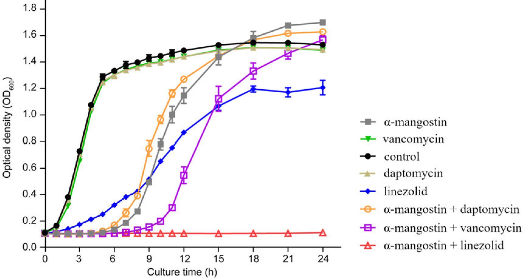
Fig. 2.
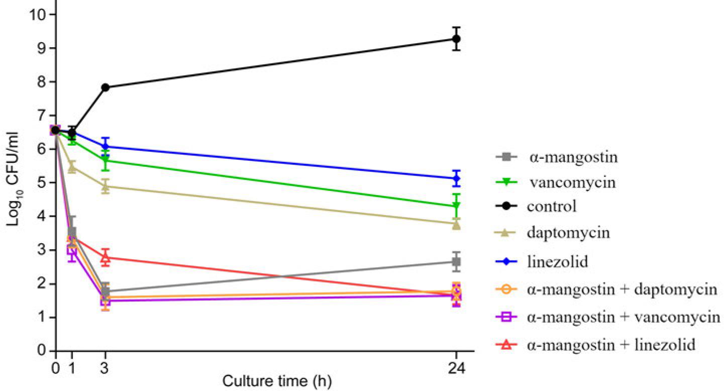
Fig. 3.
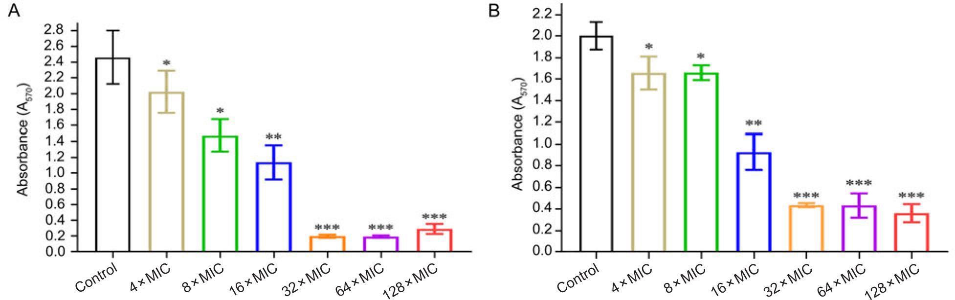
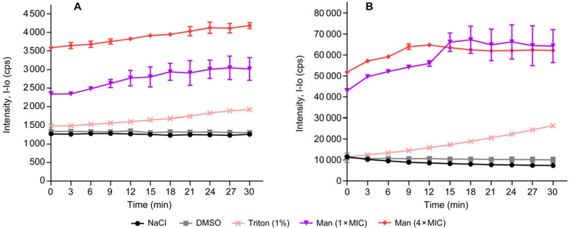
Fig. 4.
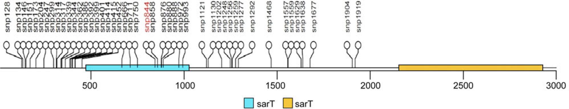
Fig. 5.
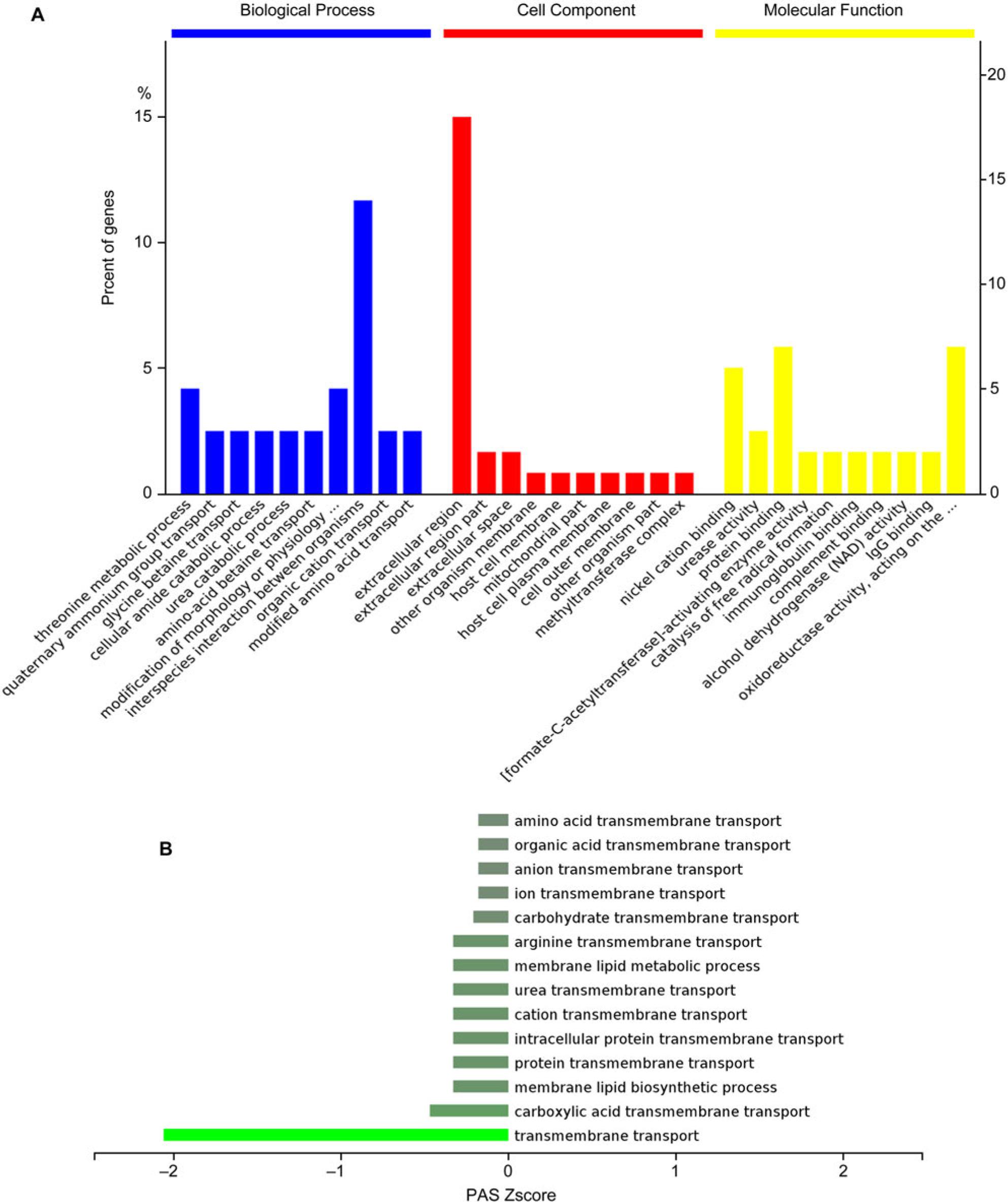
Fig. 6.
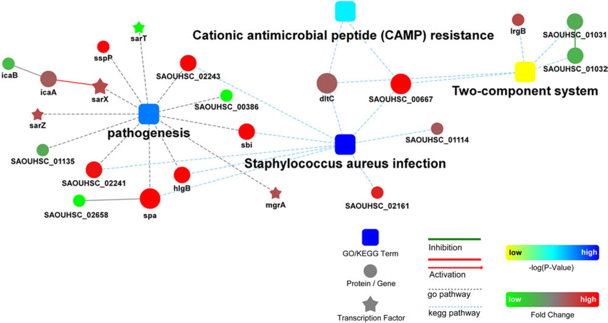
Fig. 7.
Staphylococcus aureus susceptibility to α-mangostin_
| S. aureus | The MICs (μM) of α-mangostin | |||
|---|---|---|---|---|
| 1.56 | 3.13 | 6.25 | MIC50/MIC90 | |
| MSSA (n = 190) | 16 | 163 | 11 | 3.13/3.13 |
| MRSA (n = 138) | 13 | 117 | 8 | 3.13/3.13 |

| S. aureus | The MICs (μM) of α-mangostin | |||
|---|---|---|---|---|
| 1.56 | 3.13 | 6.25 | MIC50/MIC90 | |
| MSSA (n = 190) | 16 | 163 | 11 | 3.13/3.13 |
| MRSA (n = 138) | 13 | 117 | 8 | 3.13/3.13 |
© 2023 Xiangbin Deng, Hongbo Xu, Duoyun Li, Jinlian Chen, Zhijian Yu, Qiwen Deng, Peiyu Li, Jinxin Zheng, Haigang Zhang, published by Polish Society of Microbiologists
This work is licensed under the Creative Commons Attribution-NonCommercial-NoDerivatives 4.0 License.