Introduction
Honeybees can provide humans with various bee products, such as pollen, honey, royal jelly, and propolis (Nainu et al. 2021). Honeybees are also important pollinators worldwide (Khalifa et al. 2021), increasing crop yields and maintaining the stability of ecosystems and the diversity of plant communities (Dai et al. 2018). A previous study has shown that 85% of the main crops directly related to food rely on pollination by insects such as honeybees (Klein et al. 2007). The gut microbial composition of honeybees is simpler than that of humans (Kwong and Moran 2016). The honeybee gut harbors a diverse microbial community, among which lactic acid bacteria (LAB) and bifidobacteria are beneficial in the intestine (Ge et al. 2021). On the contrast, some pathogenic bacteria such as Paenibacillus larvae and Melissococcus pluton have been found to cause diseases in honeybees (Fünfhaus et al. 2018). Both probiotics and prebiotics benefit the intestinal health of animals and humans by improving the gut microbiota balance, epithelial barrier, and immune function of hosts (Sanders et al. 2019). Thus, maintaining gut homeostasis is of great significance in honeybees.
Apis cerana, a native honeybee species in China, has a beekeeping history of more than 1700 years (Wang et al. 2021). In 2017, the number of A. cerana was estimated to be two million in China and play a critical role in crop production (Chen et al. 2017). Antibiotics have been commonly used in bee colonies to treat and control diseases of honeybees caused by bacterial pathogens such as P. larvae (Raymann et al. 2017). However, emerging evidence has reported that antibiotic treatment could reduce gut microbial diversity, weaken nutrient metabolism and immune function, affect physiological and behavioral development, and even increase mortality in honeybees (Raymann et al. 2017; Ortiz-Alvarado et al. 2020; Duan et al. 2021). Therefore, the need for alternatives to antibiotics to maintain honeybee fitness has emerged.
A prebiotic is defined as “a substrate that is selectively utilized by host microorganisms conferring a health benefit” by the International Scientific Association for Probiotics and Prebiotics (ISAPP) (Gibson et al. 2017). Commonly used prebiotics include inulin (INU), fructo-oligosaccharides (FOS), xylo-oligosaccharides (XOS), and isomalto-oligosaccharides (IMO) (Carlson et al. 2017; Poeker et al. 2018; Sorndech et al. 2018; Tandon et al. 2019). Prebiotics can alter the gut microbiota composition and promote host health by accumulating organic acids (Rastall and Gibson 2015). Prebiotics, which mainly favor bifidobacteria and lactobacilli, can be fermented to generate short-chain fatty acids (SCFAs) with colonic bacteria to fight chronic diseases (Tornero-Martínez et al. 2019). A synbiotic is a mixture comprising live microorganisms and substrate(s), selectively utilized by host microorganisms that confer a health benefit on the host (Swanson et al. 2020). Lactic acid bacteria, the most widely used probiotics, antagonize the pathogenic bacteria and influence the gut microbiota of the honeybees (Williams 2010; Audisio et al. 2015; Ramos et al. 2020). These studies highlight the potential of prebiotics and synbiotics modifying the gut microbiota of honeybees and consequently improving host health. However, little is known about the effects of prebiotics and synbiotics on the intestinal microbiota of A. cerana. This study aimed to determine the prebiotic properties of selected commonly used prebiotics using in vitro fermentation of gut microbiota of A. cerana. The most prebiotic potential was observed in IMO. Furthermore, IMO’s in vitro symbiotic effect with lactobacilli, which has shown probiotic properties in our previous study, was determined. The obtained results may provide insight into the potential outcomes of IMO to improve the health of honey bees.
Experimental
Materials and Methods
LAB strains and incubation condition. Strains Limosilactobacillus helveticus KM7 (CGMCC No. 16042) and Lactobacillus reuteri LP4 (CGMCC No. 16043) deposited in the China General Microbiological Culture Collection Center, were isolated from the intestines of adult worker honeybees from A. cerana in our previous study and showed probiotic properties (Wang 2018; Lei et al. 2020). Each strain was incubated with Man Rogosa and Sharpe (MRS) broth at 37°C and grown to stationary phase for 24 h in an anaerobic jar (Anaero-Jar TM 2.5l, Oxoid Ltd., Basingstoke, UK), including a gas-generating package (AnaeroPack, Mitsubishi Gas Chemical Co., Tokyo, Japan) prior to experiments. After centrifuging at 3,944 × g for 10 min at 4°C, the pellets were resuspended in MRS broth.
In vitro fermentation of A. cerana gut microbiota in the presence of prebiotics
Gut homogenate. Thirty-six naturally foraging adult worker bees of A. cerana, aged 18 days, were collected in summer (July) from a single colony, with robust population and without identified diseases, maintained in the apiary of the Yunnan Agricultural University in Kunming, China (longitude 102°45′30.5″ E, latitude 25°8′5.8″ N). Collected bees were placed in 50 ml sterilized centrifuge tubes and then pumped in CO2 until the honeybees were unconscious. The whole bees were washed in 75% (v/v) ethanol. The guts were dissected aseptically using forceps and 1 g of gut samples were diluted with 10 ml of phosphate buffer saline (PBS), followed by cutting into pieces in an anaerobic workstation. The collection of gut samples were processed within 15 min. The experiment was performed in triplicates.
Gut fermentations. FOS (catalog No. S11133), INU (catalog No. S11143), IMO (catalog No. S11134), and XOS (catalog No. S11137) (Shanghai Yuanye Biological Technology Co., Ltd, Shanghai, China) were chosen, and glucose (GLU) was used as a nonselective control. In vitro, static batch culture fermentations were performed with five FOS, INU, IMO, XOS, and GLU treatments in three replicates using the serum bottle, as described by Rycroft et al. 2001) the simple modification. Briefly, the basal medium (peptone 2 g/l, yeast extract 2 g/l, NaCl 0.1 g/l, K2HPO4 0.04 g/l, KH2PO4 0.04 g/l, MgSO4 · 7H2O 0.01 g/l, CaCl2 · 6H2O 0.01 g/l, NaHCO3 2 g/l, L-cysteine 0.5 g/l, bile salt 0.5 g/l, Tween80 2 ml/l, vitamin K1 10 μl, and hemin chloride 0.05 g/l) (100 ml) supplemented with 1% GLU, 1% FOS, 1% INU, 1% IMO, or 1%XOS was placed into the serum bottle and sterilized. According to the literature (Likotrafiti et al. 2014; Henrique-Bana et al. 2020) and our preliminary study, selected prebiotics were added to a final concentration of 1% (m/v). Afterward, the bottles were refluxed with O2-free N2 and covered. Each bottle filled with 100 ml sterilized medium was then inoculated 1 ml of the gut homogenate and maintained at 37°C in an anaerobic jar (AnaeroJar TM 2.5l, Oxoid Ltd., Basingstoke, UK) including a gas-generating package (AnaeroPack, Mitsubishi Gas Chemical Co., Tokyo, Japan). Samples were collected after 0, 6, 12, and 24 h of fermentation to analyze viable microbial cells and pH. In the preliminary study, no total cultivable bacteria, Bifidobacterium, LAB, Enterococcus, and total fungi were detected in the basal medium in in vitro culture of gut microbiota of A. cerana. The explanation may be that the basal medium without supplemented carbohydrates could not support the in vitro growth of honeybees gut microbe (Long et al. 2015). Therefore, in the present study, the effect of the basal medium in the absence of prebiotics or glucose was not investigated.
Enumeration of Bifidobacterium and LAB. The number of Bifidobacterium and LAB was determined using the plate count method described by Abdel-Moneim et al. (2020). Briefly, 1 ml fermentation broth was 10-fold serially diluted in PBS. The viable count was enumerated by plating onto MRS agar (Catalog No.: HB0384-5, Qingdao Haibo Biotechnology Co., Ltd, Qingdao, China) or Bifidobacterium selective medium agar containing 10 g/l of peptone, 5 g/l of liver extract, 3 g/l of beef extract, 5 g/l of yeast extract, 8 g/l of peptone from casein, 0.5 g/l dissolved starch, 1 g/l of NaCl, 1 g/l of K2HPO4, 1 g/l of KH2PO4, 10 g/l of GLU, 0.01 g/l of FeSO4 · 7H2O, 0.005 g/l of MnSO4, 0.5 g/l of L-cysteine, 1 ml of Tween80, and 20.0 g/l of agar (Catalog No.: HB0394, Qingdao Haibo Biotechnology Co., Ltd, Qingdao, China) within 15 min and incubated at 37°C in an anaerobic jar (AnaeroJar TM 2.5L, Oxoid Ltd., Basingstoke, UK) including an anaeroPack (Mitsubishi Gas Chemical Co., Tokyo, Japan) for 48 h. The colonies grown on Bifidobacterium selective medium for further microscopic examination were identified. Enumeration was repeated three times, and the number of microbial colonies (CFU) was recorded.
Measurement of pH. After fermentation at 0, 6, 12, and 24 h, 10 ml of fermentation broth from each culture bottle was taken and transferred to 50 ml sterile centrifuge tube. The pH meter (Shanghai INESA Instrument Co., Ltd, Shanghai, China) was used to measure pH.
In vitro fermentation of A. cerana gut microbiota in the presence of prebiotic IMO combined with L. helveticus KM7 or L. reuteri LP4
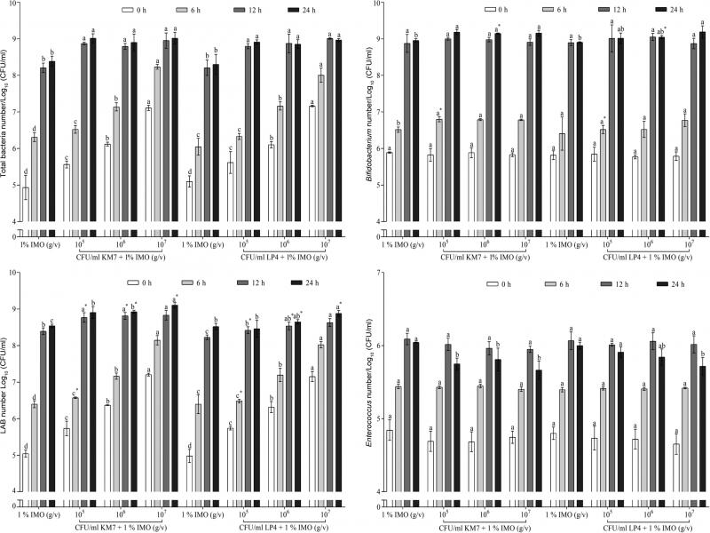
Gut fermentations. Gut samples and basal medium with 1% (g/v) IMO were prepared as described above. One milliliter of gut homogenate and 1 ml of freshly cultured L. helveticus KM7 and L. reuteri LP4 (105, 106, or 107 CFU/ml) was seeded into 100 ml of basal medium supplemented with IMO and maintained at 37°C in an anaerobic jar (AnaeroJar TM 2.5L, Oxoid Ltd., Basingstoke, UK) including an anaeroPack (Mitsubishi Gas Chemical Co., Tokyo, Japan). IMO without the presence of Lactobacillus strains was served as control. Samples were collected after 0, 6, 12, and 24 h of fermentation to measure total bacteria, bacteria, Bifidobacterium, fungi, and Enterococcus, respectively.
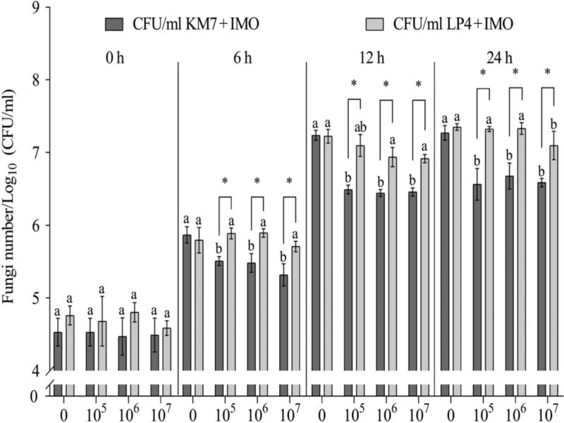
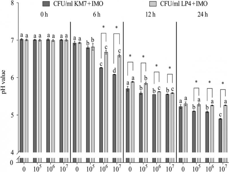
Enumeration of bacteria and fungi and analysis of pH. After fermentation of 0, 6, 12, and 24 h, 1 ml of fermentation broth was collected and mixed with 9 ml of PBS. Enumeration of A. cerana gut microbiota was performed using plate count methods. Serial dilutions were performed as described above and placed onto different selective media to determine the microbial amounts. Agars of MRS and Bifidobacterium selective medium were used as described above to count LAB and Bifidobacterium, respectively. The diluted broth was placed onto Luria Bertani nutrient agar (Catalog No.: HB0129, Qingdao Haibo Biotechnology Co., Ltd, Qingdao, China) and incubated at 30°C for 48 h to count total aerobic bacteria (Abdel-Moneim et al. 2020). The dilution was placed onto Enterococcus selective agar (Bile Aesculin Azide Agar, catalog No.: HB0133, Qingdao Haibo Biotechnology Co., Ltd, Qingdao, China) and incubated at 37°C for 48 h to count Enterococcus sp., which can be recognized by producing colonies with a black halo around (hydrolysis of esculin) (Śliżewska et al. 2019). To enumerate total fungi, the dilutions were placed onto potato dextrose agar (Catalog No.: HB0233, Qingdao Haibo Biotechnology Co., Ltd, Qingdao, China) with 0.01% (w/v) chloramphenicol and incubated at 28°C for 72 h to count total fungi number with the addition of microscopic examination, based on China National Standard Microbiological Examination of Foods (GB4789.15-2016). For pH measurement, fermentation broth samples were collected and subjected to analysis as described above.
Statistical analysis. Experimental data analysis was performed using SPSS18.0. One-way ANOVA was used to compare the differences between the data of multiple different treatment groups. The independent student t-test was performed to compare the data differences between the two treatment groups. p < 0.05 Indicated a significant difference. The results were expressed as mean ± standard deviation (SD).
Results
In vitro fermentation of A. cerana gut microbiota in the presence of prebiotics
Bididobacterium and LAB enumeration. As shown in Table I, Bifidobacterium multiplied over time in each group. Compared to the GLU group, the Bifidobacterium number in the IMO group significantly rose from fermentation for 6 h until 24 h (p < 0.05). At the fermentation of 12 h, the number of Bifidobacterium in INU, IMO, and XOS group was significantly higher than that in the GLU group (p < 0.05), and in the IMO group the number was significantly higher than in INU and XOS groups (p < 0.05). At 24 h, Bifidobacterium amount in the IMO group was significantly more than those in GLU, INU, FOS, and XOS groups (p<0.05). Bifidobacterium multiplication benefited from four kinds of prebiotics: IMO, INU, FOS and XOS. IMO provided the best promotion effect in the intestinal of A. cerana.
Table I
Number of Bifidobacterium and LAB in the prebiotic fermentation broth of A. cerana gut microbiota.
| Number of gut microbiota / Log10 (CFU/ml) | Time (h) | Treatment | ||||
|---|---|---|---|---|---|---|
| GLU | FOS | INU | IMO | XOS | ||
| Bifidobacterium | 0 | 5.85 ± 0.06Ca | 5.82 ± 0.05Ca | 5.83 ± 0.02Ca | 5.82 ± 0.01Ca | 5.80 ± 0.02Ca |
| 6 | 5.63 ± 0.03Db | 5.66 ± 0.16Cab | 5.83 ± 0.13Cab | 5.94 ± 0.19Ca | 5.81 ± 0.15Cab | |
| 12 | 6.99 ± 0.08Bc | 6.46 ± 0.25Bd | 7.79 ± 0.08Bb | 8.31 ± 0.36Ba | 7.43 ± 0.24Bb | |
| 24 | 8.15 ± 0.05Ad | 8.41 ± 0.08Ac | 8.85 ± 0.02Ab | 9.10 ± 0.12Aa | 8.52 ± 0.18Ac | |
| LAB | 0 | 5.01 ± 0.02Da | 4.99 ± 0.03Ba | 5.00 ± 0.02Da | 5.01 ± 0.05Ca | 4.98 ± 0.02Ca |
| 6 | 6.03 ± 0.03Cb | 5.06 ± 0.16Bc | 6.10 ± 0.09Cab | 6.34 ± 0.19Ba | 5.21 ± 0.15Cc | |
| 12 | 7.29 ± 0.08Bc | 5.76 ± 0.25Ad | 8.09 ± 0.08Bab | 8.28 ± 0.33Aa | 7.73 ± 0.24Bb | |
| 24 | 7.76 ± 0.14Ac | 5.67 ± 0.35Ad | 8.36 ± 0.11Aab | 8.61 ± 0.02Aa | 8.19 ± 0.16Ab | |
| Time (h) | pH value | ||||
|---|---|---|---|---|---|
| GLU | FOS | INU | IMO | XOS | |
| 0 | 6.98 ± 0.03Aa | 7.00 ± 0.04Aa | 6.98 ± 0.06Aa | 7.01 ± 0.02Aa | 6.99 ± 0.03Aa |
| 6 | 6.89 ± 0.04Ba | 6.91 ± 0.05Ba | 6.89 ± 0.07Aa | 6.89 ± 0.03Ba | 6.90 ± 0.06Ba |
| 12 | 6.13 ± 0.07Cb | 6.31 ± 0.03Ca | 5.71 ± 0.02Bd | 5.57 ± 0.07Ce | 5.97 ± 0.05Cc |
| 24 | 5.78 ± 0.07Db | 6.22 ± 0.04Da | 5.59 ± 0.04Cc | 5.48 ± 0.03Dd | 5.65 ± 0.03Dc |