
Introduction
For many years, tuberculosis has remained relevant in veterinary and human medicine worldwide and in Ukraine. According to WHO, tuberculosis is one of the top ten causes of death in the world. In 2017, 10,000,000 people had a tuberculosis infection, and 1,600,000 died from the disease. In 2018, according to WHO, the overall incidence of tuberculosis in Ukraine was 36,000 (per 37,000,000 population).
Complexities of overcoming tuberculosis and mycobacteriosis can be interrelated to the variability of the bacterial cells that enable pathogens to survive both in hostile macrophages (Kumar and Sanyal 2012) and outside the organism. Mycobacteria have an extraordinary capacity to adapt to the changing environment (Velayati and Farni 2012). It has been shown that nutrient deficiency, hypoxia, temperature, pH, NaCl, and various exogenous stress can significantly influence the metabolism of mycobacteria and, at the same time, the microorganism’s morphology (Vera and Rettger 1940; Smeulders et al. 1999; Bentrup and Russell 2001; Shleeva et al. 2002; Young et al. 2005; Anuchin et al. 2009; Velayati et al. 2009; 2011; Velayati and Farnia 2012).
It has been established that mycobacteria have a high degree of variability, and changes in the biological properties of Mycobacterium bovis (cultural, tinctorial, and morphology) may be accompanied by changes in their metabolism (Tkachenko et al. 2020b). The biological properties of mycobacteria and M. bovis, in particular, are far from being fully characterized. It leads to the tense situation of tuberculosis in individual countries, both among animals and humans.
Understanding the processes in a bacterial cell in response to the external factors could push the knowledge on the complex mechanism of mycobacterial adaptation and survival in macroorganisms and the environment.
There is no sufficient research on the impact of long-term storage of vaccine strains on the effectiveness of preventive immunization for infectious diseases. The BCG strain is a good example. Long-term storage of a parent strain in various countries led to its variability and the formation of BCG sub-strains: Russia (Moscow), Japan (Tokyo), Moreau (Brazil, Rio), Sweden (Gothenburg), Birkhaug, Danish (Denmark, Copenhagen), Prague (Czech Republic), Glaxo (London), China, Phipps (New York, Park, Philadelphia), Tice (Chicago), Frappier (Montreal), Pasteur (Paris), Bulgaria (Sophia), Connaught (Toronto), etc. BCG sub-strains slightly differ in genetic traits and immunogenicity. Therefore, considering potential changes in bacteria’s biological features after the long-term storage is essential for a comprehensive assessment of vaccines’ safety and efficiency (Ritz et al. 2008; Zhang et al. 2016).
Therefore, there was necessary to investigate the morphology, growth rate, tinctorial properties, biochemical activity, pathogenic, and sensitizing properties of mycobacteria in long-term conditions of hypoxia, nutrient deficiency, and low temperature (3°C).
Experimental
Materials and Methods
Experiments were performed in a laboratory of the Department of Epizootiology and Infectious Diseases of Animals of the Dnipro State Agrarian and Economic University using the museum’s fast-growing virulent strain M. bovis. The strain had been passaged many times (Tkachenko 2004) through solid Lövenstein-Jensen medium at pH 6.5 and 7.1. As a control (the initial mycobacterial strain), a pathogenic (parent) strain of M. bovis was used. The fast-growing strain of M. bovis was characterized by the formation of colonies after 2–3 days at 37°C, the absence of nitrate reduction reaction, hydrolysis of Tween-80, growth on the Lövenstein-Jensen medium containing sodium salicylate, and low catalase, dehydrogenase, and peroxidase activity (Tkachenko 2004; Kovaleva 2005; Glebenjuk and Telizhenko 2015; Tkachenko et al. 2020a).
Our study was approved according to the guidelines and roles of the Animal Researches Committee of the Dnipro State Agrarian and Economic University.
First, long-term storage of cultures (10–13 years at 3°C) in the test tubes was performed. The passages (variants of subculture, first-generation bacteria) No. 54, 115, 135, 135, 171, and 180 at pH 6.5, and passage No. 143 at pH 7.1 were grown in tubes covered with rubber stoppers. Each culture was stored in a nutrient medium in two tubes. It ensured the persistence of mycobacteria in an anoxic environment. Microscopy of smears was carried out, studying the tinctorial properties and morphology of mycobacteria. The M. bovis long-stored strains were also inoculated onto Lövenstein-Jensen supportive nutrient medium (six test tubes), and subcultures were grown at 3°C and 37°C. The pigmentation, colonies, and their consistency were assessed. In the smears prepared from the first generations of mycobacteria (Manchenko et al. 1994), their tinctorial properties (acid resistance) and morphology were studied under the immersion system. The smears were stained by the Ziel-Nielsen method and viewed under a MicroMed XS-3330 microscope using an immersion lens (magnification 100-folds). The image fixation was performed using an eyepiece camera for the MDC-500 microscope and the СyberlinkYouCam program.
The culture purity was verified as follows: The bacterial suspension was divided into two parts, and one of them was heated at 130°C for 5 minutes. Then, both parts were inoculated on Lövenstein-Jensen medium at 37°C and Sabouraud agar at 30°C. Cultures were considered pure in the absence of growth of any foreign microorganisms on Lövenstein-Jensen nutrient medium after inoculation of heat-treated suspensions and the absence of colonies on Sabouraud agar.
At the second step, the pathogenic properties of mycobacteria were investigated. Seven guinea pigs were infected for each of six variants of subculture (a first passage). A control group of pigs was infected with the pathogenic parental strain of M. bovis. The suspension (2 × 10⁵ CFU/ml) was injected parenterally (subcutaneous) from the inner side of the thigh. In total, 49 guinea pigs were infected. After euthanasia of the animals (on day 90th of the experiments), tissue specimens were examined bacteriologically after growth at 3°C and 37°C on the Lövenstein-Jensen nutrient medium. The pre-inoculation material was prepared separately from each guinea pig according to the method of Alikayeva. The inoculation of suspensions into ten tubes containing the Lövenstein-Jensen medium was performed (Manchenko et al. 1994).
During the experiments (within 90 days), animals were weighed, an ulcer formation was studied in the injection site, and the manifestation of allergic reaction to PPD for mammals at a dose of 25 IU in 0.1 cm3 isotonic solution was monitored. The allergic tests were performed at 30, 60, and 90 days after infection of the animals, and the reaction was recorded 24–48 hours after tuberculin injection.
The guinea pigs were then infected for a second time (a second passage) with the mycobacteria recovered from the organs of guinea pigs infected with the bacteria of the first passage. The microorganisms recovered from the animal organs infected with the second passage were used to infect the next group of guinea pigs (a third passage). In the second and third rounds of infections, 84 (42 + 42) guinea pigs were infected (the control group’s guinea pigs were not infected).
At the third step of our studies, M. bovis ability to grow on simple (basal) media (nutrient agar and nutrient broth) at 37°C, in the Lövenstein-Jensen medium with sodium salicylate at 3°C and 37°C was investigated. For this purpose, the suspensions (2 × 10⁵ CFU/ml) were prepared from mycobacteria culture, and three tubes with the nutrient medium were inoculated (Manchenko et al. 1994).
To determine the biochemical activity of M. bovis, the first-generation bacteria were grown for 3–4 weeks. The activity of catalase, peroxidase (following modified Bogen method), and dehydrogenase, as well as nitrate reduction, and Tween-80 hydrolysis (following modified Wayne method) were assayed as described by Zhurylo et al. (2012).
The polymerase chain reaction was used to confirm or refute the mycobacterial changes in genetic material. DNA was isolated from cultures of the original highly virulent fast-growing strain (at the second passage), long-term variants of subculture (passages), as well as Vallee strain (KMIEV-9 modification, provided by the Suny National Agrarian University) and BCG strain (sub-strain of Russia BCG-1, used for vaccine production). These strains were inoculated at a volume of 0,2 cm3 and a concentration of 2 × 10⁵ CFU/ml and grown in a nutrient medium for 30 days.
For RT-PCR, an amplifier iCycler iQ5 (Bio-Rad, USA) and reagent kits for DNA isolation and PCR («MIKO-GEN», producer NGO DNA-Technology, Russian Federation) were used.
Results
Initial cultures on a Lövenstein-Jensen nutrient medium were characterized by a predominantly moderate growth rate in the inoculation line (Fig. 1), the color of ivory. Some strains also grew as individually formed colonies. The colony surface was rough (R) in three subculture variants (passages No. 135, 171, and 180) or smooth (S) – also in three subculture variants (passages No. 54, 115, and 143).
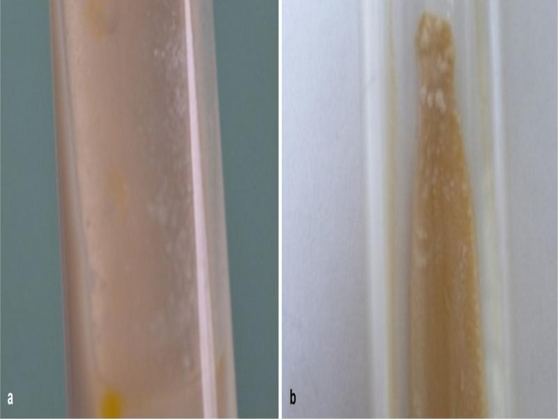
Fig. 1.
Cultures of Mycobacterium bovis: a) passage No. 54, b) passage No. 180.
The mycobacteria from passage 54 appeared as the acid-fast forms (thick rod with rounded ends and single grains) (Fig. 2a). All other subculture variants (passages No. 115, 135, 145, 171, and 180) displayed mixed morphological forms (Fig. 2b), i.e., acid-fast rods, non-acid-fast filamentous and rod-shaped forms, and grains.
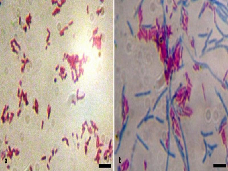
Fig. 2.
Morphology of initial cultures of Mycobacterium bovis: a) passage No. 54, b) passage No. 180 (stained by Ziehl-Neelsen). Bar = 10 µm.
After inoculation of the Lövenstein-Jensen nutrient medium, the mycobacteria were capable of growing at two temperatures (3°C and 37°C) (passages No. 115, 171), at one selected temperature 37°C (passages No. 54, 143, 180), or 3°C – one strain (passage No. 135). The subcultures obtained (Fig. 3) differed from the initial cultures, as shown in Fig. 1.
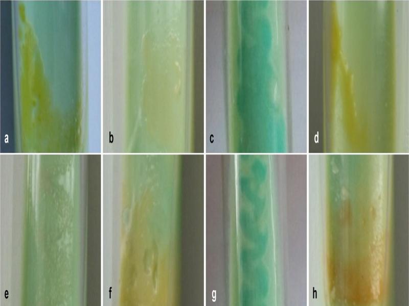
Fig. 3.
Subcultures of Mycobacterium bovis: a) passage No. 54, b) passage No. 115 (at 37°C), c) passage No. 115 (at 3°C), d) passage No. 135, e) passage No. 143, f) passage No. 171 (at 37°C), g) passage No. 171 (at 3°C), h) passage No. 180.
At 37°C, the bacterial growth became visible on the fourth day – passages No. 115, 143, on the fifth day – passages No. 171 and 180, and on the twentieth day – the passage No. 54. The growth rate in all cultures was considerable. In four subculture variants (passages No. 54, 115, 171, and 180), the growth was characterized by an oily coating (fur) of yellow or orange colors (Fig. 3), in one subculture variant (passage No. 143) – small translucent ivory-colored S-colonies were located in clusters.
At 3°С, the growth was noted on the sixth day – passages No. 115 and 171, and on the fiftieth day – passage No. 135. In two cultures (passages No. 115, 171), there was considerable growth in the form of blue-green mucous ropy fur, and in one culture (passage No. 135) – yellow fur.
Microscopy of smears prepared from the isolated cultures revealed the difference between the parental strain of M. bovis (as shown in Fig. 2) and the organisms that were stored for many years (Fig. 4). Thus, in passage No. 54 only blue thick thread-like forms with a barely noticeable red granularity inside could be found. The morphology of mycobacteria was quite diverse: short and long, straight and curved, thin and thick rod-shaped bacteria with rounded ends, coccal and rod-shaped forms, and grains. In passages No. 171 and 180, the L-forms and other rod-shaped mycobacteria, thread-like forms, and grains were noticed. In passage No. 143 only L-forms with a different optical density were recorded. As proof of the affiliation of L-forms to mycobacteria is the destruction of vesicular L-forms and the release of acid fast-negative grains and acid fast-positive shaped bacteria (rods).
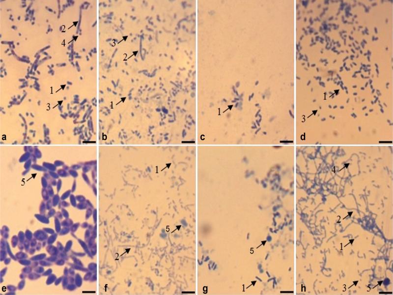
Fig. 4.
Morphology of Mycobacterium bovis subcultures: a) passage No. 54, b) passage No. 115 (at 37°C), c) passage No. 115 (at 3°C), d) passage No. 135, e) passage No. 143, f) passage No. 171 (at 37°C), g) passage No. 171 (at 3°C), h) passage No. 180; 1 – rod-shaped bacteria, 2 – thread-like forms, 3 – grains (elementary bodies), 4 – output of grains from thread-like forms, 5 – L-forms. Bar = 10 µm.
At the end of the animal experiments and euthanasia, an autopsy was performed to reveal any pathological-anatomical changes characteristic for tuberculosis in the experimental animals. The infection of guinea pigs by all studied mycobacterial subcultures (passages) was not accompanied by sensitization of the macroorganism to PPD for mammals, the formation of ulcers at the site of mycobacteria suspension, or bodyweight decrease.
Guinea pigs infected with the parental pathogenic strain of M. bovis responded to tuberculin after 30 and 60 days of the experiment, and their body weight decreased. At the point of inoculation of the mycobacterial suspension, the ulcers were formed after 27 days. The death of one guinea pig was reported on day 69. The other guinea pigs were euthanized after 90 days. The pathological changes characteristic of tuberculosis were noted in infected all guinea pigs (Fig. 5).
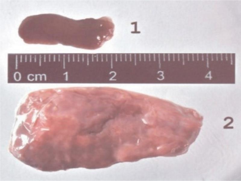
Fig. 5.
The spleen of the guinea pig: 1) normal, 2) with tuberculous foci.
Through bacteriological investigations of specimens from each guinea pig, eight cultures of mycobacteria were isolated: after four days of culture, there were three cultures at 3°C (passages No. 115, 135, and 171) and five cultures at 37°C (passages No. 54, 115, 143, 171, and 180). Non-acid-fast rod-shaped mycobacteria and grains were therefore visible under the immersion in the microscope.
In animals infected with a second passage, no allergic reactions to PPD for mammals, formation of ulcers in the injection site, or tuberculosis characteristic features were observed. However, the bacteria growth from tissue specimens at 37°C was observed on the 6th day (passages No. 54 and 180) and on the 10th day (passages No. 171 and 143). Under microscopy, the smears from cultures revealed acid fast-negative rod-shaped bacteria and grains.
The animals infected with a third passage did not suffer allergic reactions to the PPD for mammals, the formation of ulcers in the site of inoculation of material, decrease in body weight, or and symptoms of tuberculosis. No bacteria were grown from the animal tissues.
These mycobacteria could grow on nutrient media. In the nutrient broth, colony growth was observed for all passages except for passage No. 54. In the Lövenstein-Jensen medium with sodium salicylate at 3°C, colony growth was observed for all passages except for 54, 135, and 171 (Fig. 6).
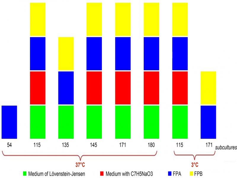
Fig. 6.
Growth of pathogenic Mycobacterium bovis strain and its subcultures (variants) on nutrient media. FPA – nutrient agar, FPB – nutrient broth.
Concerning the biochemical activity of the cultures studied, variations in the activity of mycobacterial enzymes were ascertained depending on the number of passages and the cultivation temperature. The tendency to change the biochemical activity of the microorganisms studied was established: catalase activity increased along with the decrease of the cultivation temperature in passages 115 and 171 (Fig. 7).
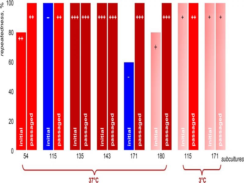
Fig. 7.
Catalase activity of Mycobacterium bovis initial subcultures and the subcultures passaged through the guinea pigs (repeatedness, %): “–” – negative reaction, “±” – doubtful reaction, “+”, “++”, “+++” – the magnitude of the positive reaction.
Peroxidase activity was observed only in the control samples of the highly virulent strain. Dehydrogenase activity in the first 15–30 minutes was not detected in any culture, but after 24 hours was observed in almost all cultures, except those on the 54 passage (Fig. 8).
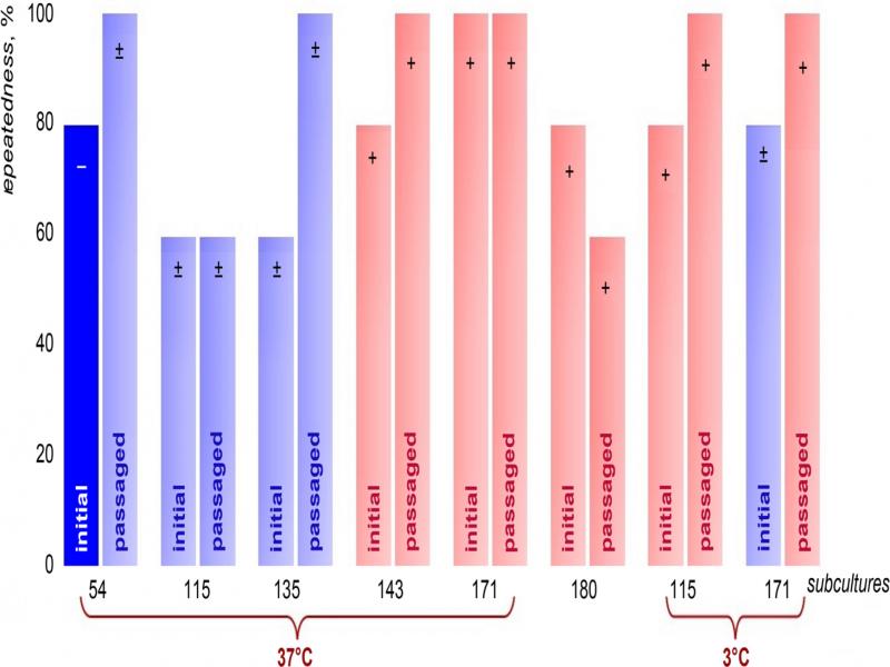
Fig. 8.
Dehydrogenase activity of Mycobacterium bovis initial subcultures and the subcultures passaged through the guinea pigs (repeatedness, %): “–” – negative reaction, “±” – doubtful reaction, “+”, “++”, “+++” – the magnitude of the positive reaction after 24 hours.
Nitrate reduction was recorded in all cultures except for passages No. 143 and 171 grown at 37°C (Fig. 9).
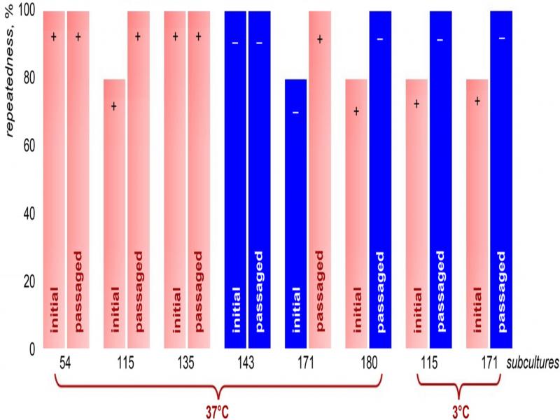
Fig. 9.
Reduction of nitrates by Mycobacterium bovis initial subcultures and the subcultures passaged through the guinea pigs (repeatedness, %): “–” – negative reaction, “±” – doubtful reaction, “+”, “++”, “+++” – the magnitude of the positive reaction.
Hydrolysis of Tween-80 in the first 4 hours was not found in all subcultures (passages) of M. bovis. Passages No. 115 and 171, which were grown at a low temperature, had a higher ability to hydrolyze Tween-80 on the fifth day.
When the biochemical activity of mycobacteria at subsequent passages in guinea pig was compared with the initial strain, the following phenomena become apparent: an increase in the activity of the enzyme dehydrogenase – passages No. 54, 135 (grown at 37°C) and 171 (grown at 3°C); loss of ability to reduce nitrates, especially in cultures grown at low temperature (3°C) – passages No. 115, 171, and at 37°C – for the passage No 195; increase of catalase activity – passages No. 115 and 171 at both cultivation temperatures (3°C and 37°C); the ability to hydrolyze Tween-80 in the vast majority of cultures was decreased for all passages except passage No. 54 (Fig. 10).
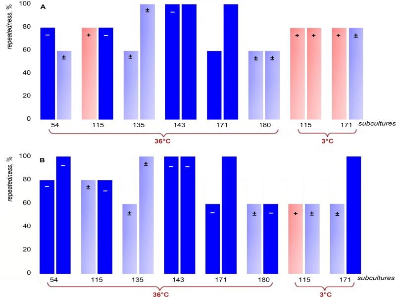
Fig. 10.
Tween-80 hydrolysis by Mycobacterium bovis initial subcultures and the subcultures passaged through the guinea pigs: A) after five days, B) after ten days.
It should also be noted that the biochemical activity of one subculture strain No. 143 after passage through the body of guinea pigs remains unchanged.
Using PCR, the amplicons were detected in the initial pathogenic M. bovis fast-growing strain and both Vallee and BCG strains (Table I). At the same time, the amplicons were not observed after amplification of DNA derived from the stored for long-time M. bovis fast-growing strain.
Table I
The results of DNA amplification of mycobacteria
| Type of mycobacteria | Strain of mycobacteria | Result of detection of amplification products |
|---|---|---|
| Mycobacterium bovis | fast-growing (original) | + |
| after the long-term storage and reinoculation of cultures on the nutrient medium), passages 54, 115, 135, 135, 171, and 180 | – | |
| Vallee strain | + | |
| BCG strain | + |