
Introduction
Mugwort (Artemisia argyi) is found in Europe (Artemisia vulgaris), Africa (Artemisia vulgaris), India (Artemisia vulgaris), Asia (Artemisia argyi), and America (Artemisia douglasiana). Early humans may have transported this plant throughout the world for its medicinal and food value (Adams et al. 2012). Its leaves are rich in essential oils, flavonoids, sugars, and other major components with pharmacological properties, such as bacteriostatic, insect-resistant, anti-inflammatory, antitussive, expectorant, soothing, antiallergic, antioxidant, and antitumor compounds, etc. Jiang et al. 2019a, 2019b). They are widely used in traditional Chinese medicine as well as in pharmaceutical products, animal feed, disinfectants, and other everyday products. With the increasing use of mugwort products, research on the quality and yield of mugwort cultivation to meet the growing global demand has increased.
Recent studies have shown that endophytic fungi, as a natural constituent of the plant micro-ecosystem, live in the tissues of healthy plants without causing any disease symptoms. These fungi have the functions of promoting plant growth, increasing plant disease resistance, inhibiting pathogenic bacteria, and even affecting the yield and quality of plants (Chu et al. 2020). Furthermore, endophytic fungi in plant leaves can not only directly or indirectly affect the synthesis of major therapeutic constituents of medicinal plants (Jiang et al. 2008). However, they can also have a significant influence on the internal microenvironment of roots, stems, and leaves, as well as affect the quality of these plants (Zhang 2017).
In a previous study, Zhang et al. (2011) isolated 19 strains of endophytic Actinomycetes from the leaves of A. argyi using plate culture screening method combined with crude extract of fermentation broth. Among these isolates, 11 strains presented extracellular amylase activity and exhibited protease activity, and eight strains showed cellulase activity. Besides, two strains had antagonistic activity against pathogenic bacteria, and three strains presented antagonistic activity against penicillin-resistant Staphylococcus aureus. Furthermore, Shi et al. (2014) isolated and screened ten strains of Actinomycetes from the collective site of A. argyi rhizome by combining the primary screening method of confrontation culture with the rescreening method of fermentation broth, and noted that 70% of the isolates presented different degrees of antibacterial activity. Liu et al. (2019) used the plate culture method to isolate 13 strains of endophytic fungi belonging to three genera from the stems and leaves of North A. argyi grown in Tangyin, China and detected six species and three genera of endophytic fungi in the leaves. However, all these studies had employed traditional culture techniques, which cannot support the growth of unculturable microorganisms in the stems and leaves of A. argyi. To date, studies on the diversity of the endophytic fungal community in A. argyi based on high-throughput sequencing have not yet been reported. Therefore, to evaluate the influence of endophytic fungi on the growth and quality of A. argyi, the present study employed high-throughput amplicon sequencing technology to compare and analyze the diversity and composition of endophytic fungal community structure in the leaves of A. argyi cultivated in different regions in China. Besides, the degree of health and biocontrol application potential of A. argyi was also examined. The results obtained can provide a scientific basis and guidance for large-scale cultivation of A. argyi and its application for the biocontrol of plant diseases and pests.
Experimental
Materials and Methods
Overview of the test site. The experimental site was located in the A. argyi cultivation base of Anyang Institute of Technology in Henan Province, China (N36°0′, E114°35′). The region has a warm, temperate, continental monsoon climate with mild weather conditions and four distinct seasons. The annual average temperature is 14.9°C, the annual sunshine duration is 2,500 h, and the annual average precipitation is 538.4 mm, with 206 days of frost-free period and sandy loam soil.
Sample collection.A. argyi, commonly known as Chinese mugwort, is widely distributed in Henan, Hebei, Hubei, Anhui, and Zhejiang in China. Different ecological species have different biological flora. The leaves of 2-year-old A. argyi without diseases and pests on its surface were collected in June 2020 at the A. argyi cultivation base in Anyang Institute of Technology. Five kinds of A. argyi leaves were collected from Tangy in Beiai in Henan (BA), Qichun Qiai in Hubei (QA), Wanai in Nanyang in Henan (WA), Haiai in Ningbo in Zhejiang (HA), and Anguo Qiai in Anguo in Hebei (AQA). The experimental field was weeded manually without the use of herbicides. Fertilizers were not applied, and two crops were cultivated in a year.
During sampling, the third leaf of A. argyi was selected, and 15 samples (three biological replicates per A. argyi variety and five A. argyi varieties in total) were put into sterile plastic bags with labels (indicating the sample number, name, place, date, and collector), transported to the laboratory, and stored in a –80°C refrigerator for the subsequent isolation of DNA.
Extraction and electrophoresis of endophytic fungi total DNA fromA. argyileaves. Firstly, the samples were surface-washed with 70% ethanol solution three times, then washed with 1 × PBS solution three times, dried, and then extracte with liquid nitrogen grinding or tissue disrupter. Then, the endophytic fungi total DNA was extracted with the OMEGA kit, qualitatively detected using agarose gel electrophoresis, quantified by nucleic acid quantitative spectrophotometer (Nanodrop, USA), and stored in a refrigerator at –20°C for the subsequent analysis.
PCR amplification and sequencing of ITS1-ITS2 region of 18S rRNA. The extracted total DNA was used as a template, and the internal transcribed spacer (ITS) region of fungi (ITS1-ITS2 region) was amplified using specific PCR primers (ITS1F: 5’-CTTGGTCATTTAGAGGAAGTAA-3’; ITS2: 5’-GCTGCGTTCATCGATGC-3’). The reaction system for PCR comprised the following: Phusion Master Mix (2×), 15 μl; primer (2 μmol/l), 3 μl; DNA (1 ng/μl), 10 μl; and ddH2O, 2 μl. The PCR conditions were as follows: pre-denaturation at 95°C for 3 min, followed by 25 cycles of denaturation at 94°C for 30 s, annealing at 55°C for 30 s, and extension at 72°C for 30 s, and a final extension at 72°C for 7 min. The PCR products were qualitatively detected by electrophoresis using 2% agarose gel in 1 × TAE solution, purified, quantified, and a test library was constructed using Qubit 3.0. Subsequently, the test DNA library was subjected to high-throughput amplicon sequencing performed by Sangon Bioengineering Co., Ltd. (Shanghai) using the Illumina MiSeq sequencing platform (Liao et al. 2020). The raw data of high-throughput amplicon sequencing was uploaded to the NCBI SRA database, and could be downloaded from website (https://www.ncbi.nlm.nih.gov/bioproject/PRJNA714493) and the clean data was obtained by quality control and filtering from the raw data.
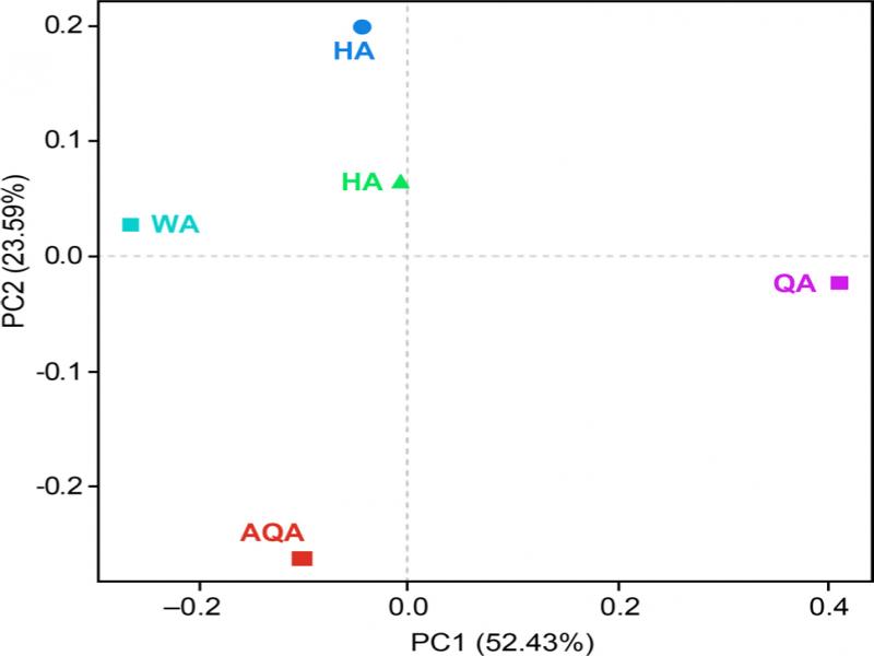
Bioinformatics analysis. UPARSE was used for operational taxonomic unit (OTU) classification of the representative sequences at 97% similarity level, and the fungal community composition in each sample was determined at different classification levels. Mothur software was used to analyze the dilution curves, and Shannon index, Simpson index, species richness index (ACE), and Chao1 index were employed to determine the microbial ecological diversity. The sequences were compared with the functional genes in the NCBI NT database using BLAST. The optimized sequences were identified at the phylum, class order, family, and genus levels according to the reference sequences in the database, and the community composition, abundance, and diversity were compared and analyzed. Ramette’s method was used for principal component analysis (PCA), Excel (2007) was utilized to construct a histogram, and SPSS (19.0) software was employed to statistically analyze the class group test data of endophytic fungi in A. argyi.
FUNGuild functional analysis. The fungi classification and functional analysis was completed by FUN-Guild (Fungi Functional Guild) software. It is a tool for classification and analysis of fungal communities through microecological guild depending on the currently published literature or authoritative website data.
Results
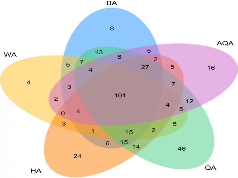
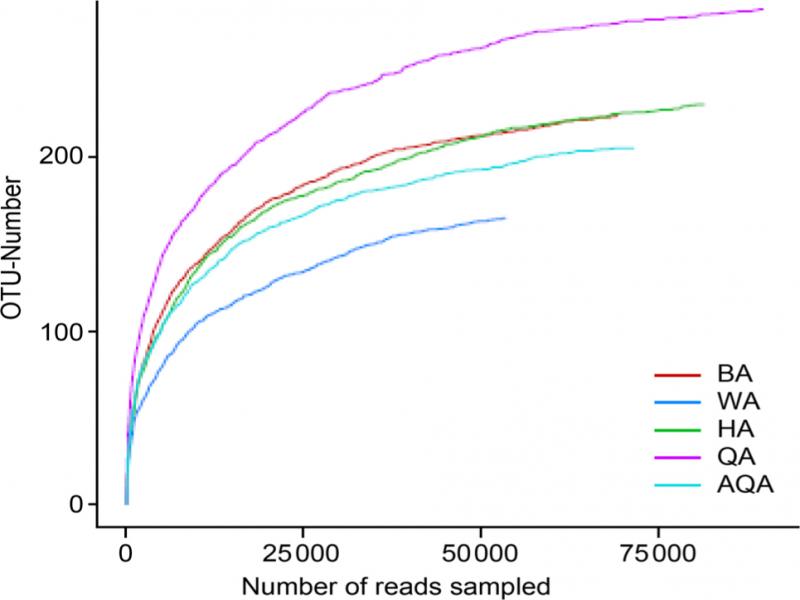
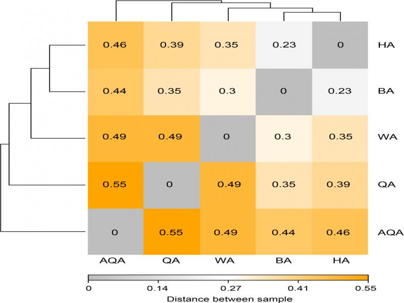
Qualitative analysis of endophytic fungi inA. argyileaves. The optimal sequence and information about the genus or species number (OTUs) of the endophytic fungal community in A. argyi leaves were obtained using high-throughput amplicon sequencing (Table I). After merging and filtering of double-ended reads, the clean tags for endophytic fungi in WA, AQA, HA, BA, and QA samples were 53,504.0, 69,364.0, 71,661.0, 81,561.0, and 89,829.0, respectively. Species classification based on similarity level ≥ 97% revealed 709.0 OTUs, and the number of OTUs in WA, AQA, HA, BA, and QA samples was 165.0, 205.0, 230.0, 224.0, and 285.0, respectively (Fig. 1). QA presented the highest sequence number and species classification, which was mostly consistent with the values of alpha-diversity index. The dilution curve for the endophytic fungi in the five samples is shown in Fig. 2. When the sequencing data reached 50,000, the number of OTUs in the five samples remained flat, indicating that the sequencing depth of the samples was essentially reasonable and that the obtained data could reflect the composition of the fungal community structure in the samples under natural conditions more tangibly and comprehensively.
Table I
Richness and diversity of endophytic fungi in A. argyi leaves.
| Treatments | No. of reads | Shannon index | Chao1 index | ACE index | Simpson index |
|---|---|---|---|---|---|
| WA | 53504.0 | 2.09 ± 0.01a | 180.53 ± 15.03a | 193.54 ± 8.34a | 0.25 ± 0.000a |
| AQA | 69364.0 | 2.19 ± 0.01a | 223.91 ± 9.32a | 226.58 ± 11.53a | 0.24 ± 0.000a |
| HA | 71661.0 | 2.57 ± 0.02a | 239.38 ± 10.02a | 242.84 ± 10.02a | 0.14 ± 0.000a |
| BA | 81561.0 | 2.62 ± 0.04a | 255.54 ± 8.39a | 253.33 ± 9.14a | 0.08 ± 0.000a |
| QA | 89829.0 | 2.99 ± 0.03a | 291.73 ± 15.12a | 297.24 ± 12.02a | 0.12 ± 0.001a |
| Generic | Type | Generic | Type |
|---|---|---|---|
| Aspergillus_intermedius | biocontrol fungi-saprotroph | Phanerochaete | pathotroph |
| Aspergillus_sydowii | biocontrol fungi-saprotroph | Phlebia | pathotroph-saprotroph-symbiotroph |
| Penicillium_catenatum | biocontrol fungi-saprotroph | Phoma | saprotroph-symbiotroph |
| Penicillium_oxalicum | biocontrol fungi-saprotroph | Podosphaera | pathotroph-saprotroph-symbiotroph |
| Botryosphaeria | pathotroph-saprotroph-symbiotroph | Ramichloridium | pathotroph-saprotroph |
| Botrytis | pathotroph-saprotroph | Rhodotorula | saprotroph |
| Cladosporium | pathotroph-saprotroph-symbiotroph | Sarocladium | pathotroph-saprotroph |
| Clonostachys | pathotroph-saprotroph | Schizophyllum | pathotroph-symbiotroph |
| Curvularia | saprotroph | Sphaerulina | saprotroph |
| Cyphellophora | pathotroph-saprotroph-symbiotroph | Sporisorium | saprotroph |
| Cystobasidium | saprotroph | Sporobolomyces | pathotroph-symbiotroph |
| Diaporthe | pathotroph-saprotroph-symbiotroph | Stagonospora | saprotroph |
| Dothidea | saprotroph | Stemphylium | saprotroph |
| Edenia | pathotroph | Tilletiopsis | saprotroph |
| Entocybe | pathotroph | Trametes | pathotroph-saprotroph |
| Entodesmium | pathotroph | Tricharina | pathotroph-saprotroph-symbiotroph |
| Erythrobasidium | pathotroph | Trichomeriaceae | pathotroph |
| Filobasidium | pathotroph-saprotroph-symbiotroph | Trichosporon | pathotroph-saprotroph-symbiotroph |
| Fusarium | pathotroph-saprotroph | Verticillium | pathotroph |
| Gibellulopsis | pathotroph-saprotroph | Alternaria | pathotroph-saprotroph |
| Herpotrichiellaceae | pathotroph-saprotroph-symbiotroph | Amphisphaeriaceae | pathotroph-saprotroph-symbiotroph |
| Knufia | pathotroph | Amphobotrys | pathotroph-saprotroph-symbiotroph |
| Leptosphaeria | pathotroph | Anthracocystis | pathotroph |
| Limonomyces | saprotroph | Apiotrichum | pathotroph |
| Microdochium | pathotroph-saprotroph-symbiotroph | Articulospora | pathotroph |
| Mycosphaerella | pathotroph-symbiotroph | Ascochyta | pathotroph |
| Myrmecridium | saprotroph | Aurantiporus | pathotroph |
| Myrothecium | symbiotroph | Aureobasidium | pathotroph |
| Neosetophoma | pathotroph-saprotroph-symbiotroph | unclassified | other |
| Occultifur | saprotroph | unclassified_Ascomycota | other |
| Paraconiothyrium | pathotroph-saprotroph | unclassified_Fungi | other |
| Paraphoma | pathotroph-saprotroph | unclassified_Phaeosphaeriaceae | other |
| Phaeosphaeria | pathotroph-saprotroph | Others | other |