Have a personal or library account? Click to login
Improved Biosurfactant Production by Enterobacter cloacae B14, Stability Studies, and its Antimicrobial Activity Cover

Improved Biosurfactant Production by Enterobacter cloacae B14, Stability Studies, and its Antimicrobial Activity

Open Access
|Aug 2020

Figures & Tables

Fig. 1.

Growth of E. cloacae B14 using different carbon sources (solid lines with filled square markers) – (a) glucose; (b) sucrose; (c) maltose; (d) lactose and (e) glycerol. Emulsification activity (%E24) of cell-free supernatant obtained from those cultures are shown as dashed lines with filled circle markers. Error bars indicate standard deviations of triplicate data.

Fig. 2.

Growth of E. cloacae B14 on maltose using different nitrogen sources (solid lines with filled square markers) – (a) yeast extract; (b) urea; (c) NH4NO3; (d) NH4Cl and (e) (NH4)2SO4. Emulsification activity (%E24) of cell-free supernatant obtained from those cultures are shown as dashed lines with filled circle markers. Error bars indicate standard deviations of triplicate data.

Fig. 3.

Biosurfactant yielded from E. cloacae B14 grown on maltose media with different nitrogen sources. Error bars indicate standard deviations of triplicate data.

Fig. 4.

Stability of biosurfactant under a range of pH. Error bars indicate standard deviations of triplicate data.

Fig. 5.

Stability of biosurfactant under a range of temperatures. Error bars indicate standard deviations of triplicate data.

Fig. 6.

TLC analysis of the purified B14 BS obtained from an optimized culture. Samples on TLC plates were sprayed using ninhydrin solution (1); phenol-H2SO4 solution (2) and iodine vapor (3).

Fig. 7.

FTIR spectrum of the purified B14 biosurfactant.

Fig. 8.

Antimicrobial activity of B14 supernatant compared to tetracycline (positive control) and non-inoculated medium (negative control) – a) the activity against S. marcescens and b) the activity against B. subtilis.

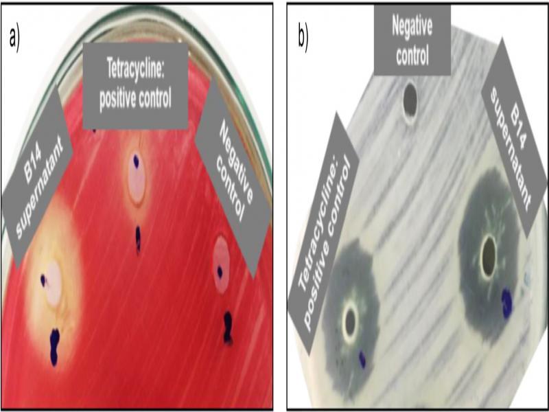
Antimicrobial activity of cell-free supernatant of E_ cloacae B14 against Gram-positive and Gram-negative pathogens using 30 µg/ml tetracycline as a control_ Standard deviations were calculated from the data obtained from triplicate experiments_

Pathogenic bacteriaInhibition zone diameter (mm) ± standard deviation
Cell-free supernatant of B14 culturesTetracycline 30 μg/ml (control)
Gram-negative bacteria
Escherichia coli12.3 ± 1.121.1 ± 0.5
Pseudomonas aeruginosa17.0 ± 1.420.0 ± 0.7
Serratia marcescens  9.7 ± 1.5  0.0 ± 0.0
Gram-positive bacteria
Bacillus cereus20.7 ± 2.030.0 ± 2.0
Bacillus subtilis22.0 ± 1.820.0 ± 1.1
Staphylococcus aureus26.7 ± 2.130.0 ± 1.8
DOI: https://doi.org/10.33073/pjm-2020-030 | Journal eISSN: 2544-4646 | Journal ISSN: 1733-1331
Language: English
Page range: 273 - 282
Submitted on: Mar 9, 2020
|
Accepted on: Jun 19, 2020
|
Published on: Aug 14, 2020
In partnership with: Paradigm Publishing Services
Publication frequency: 4 issues per year

© 2020 JINDARAT EKPRASERT, SASIWIMON KANAKAI, SULADDA YOSPRASONG, published by Polish Society of Microbiologists
This work is licensed under the Creative Commons Attribution-NonCommercial-NoDerivatives 4.0 License.