
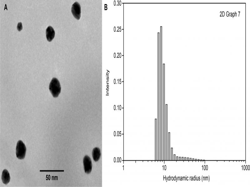
Fig. 1.
Nanoparticles’ shape (A) and dimensions (B).
Table I
AgNP inhibition of the filamentous fungi growth.
| Fungal species | AgNPs in the medium [mg/l] | |||
|---|---|---|---|---|
| 0 | 2.14 | 4.28 | 21.4 | |
| Growth [%] | ||||
| Aspergillus brasiliensis | 100 | 100 | 83.1 | 3.74 |
| Trichoderma virens | 100 | 100 | 33.9 | 9.72 |
| Paecilomyces variotii | 100 | 100 | 0 | 0 |
| Penicillium pinophilum | 100 | 57.3 | 44.8 | 0 |
| Chaetomium globosum | 100 | 63.9 | 61.2 | 0 |
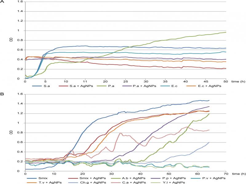
Fig. 2.
Microorganism growth in BioscreenC with and without AgNPs: (A) Bacteria: S.a, Staphylococcus aureus; P.a, Pseudomonas aeruginosa; E.c, Escherichia coli; (B) yeasts Y.l, Yarrowia lipolytica, and fungi: Smix, the mixture of spores of five species of filamentous fungi; A.b, Aspergillus brasiliensis; P.p, Penicillium pinophilum; P.v, Paecilomyces variotii; T.v, Trichoderma virens, and Ch.g, Chaetomium globosum.
Table II
Lag phase duration and final OD after 96 hours of filamentous fungal and yeast growth in BioscreenC without or with AgNPs.
| Filamentous fungi and yeast species | Lag phase [h] | OD | ||
|---|---|---|---|---|
| at 96 h | compared to | |||
| SM [%] | initial [× folds] | |||
| SM: spores mix (control) | 10 | 1.592 | 100 | 36 |
| SM: spores mix +AgNPs | 20 | 1.308 | 82 | 5.8 |
| A. brasiliensis +AgNPs | 40 | 1.600 | 100 | 8.1 |
| P. pinophilum +AgNPs | 30 | 1.517 | 95 | 9.2 |
| P. variotii +AgNPs | ~ 68 | 0.633 | 40 | 3.7 |
| T. virens +AgNPs | 18 | 1.306 | 82 | 6.5 |
| C. globosum +AgNPs | 48 | 1.063 | 67 | 5.2 |
| C. albicans +AgNPs | 15 | 0.953 | – | 3.8 |
| Y. lipolytica +AgNPs | > 96 | 0.194 | – | 0 |
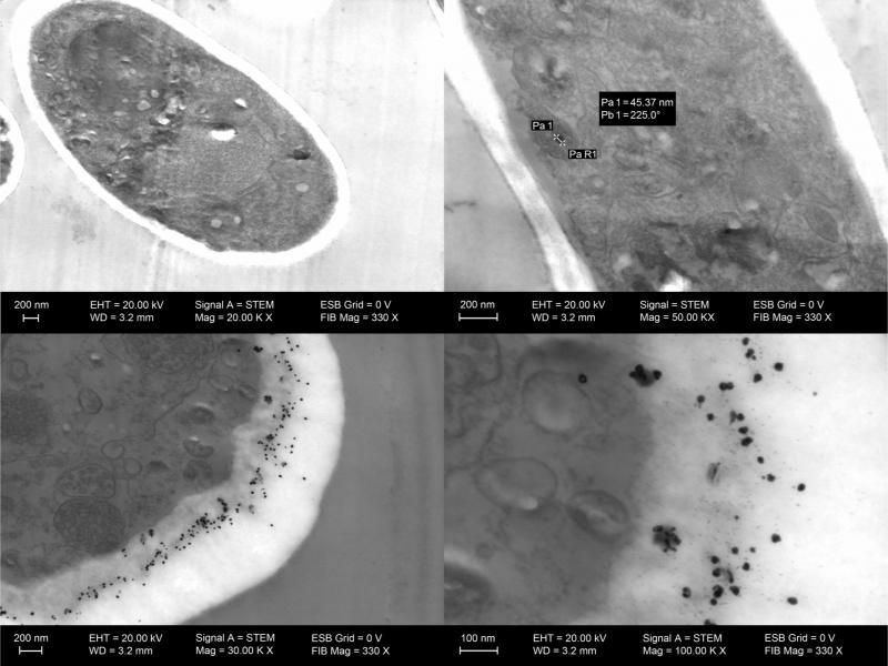
Fig. 3.
TEM images of Aspergillus brasiliensis after the growth in the BioscreenC: (a) without AgNPs; (b) with AgNPs visible inside the cell (black points); (c, d) accumulated within the cell wall.
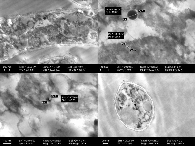
Fig. 4.
TEM images of filamentous fungi after the growth with AgNPs in the Bioscreen C: (a, b) Paecilomyces variotii; (c, d) Penicillium pinophilum; AgNPs, the black structures observed inside the cell and hypha.
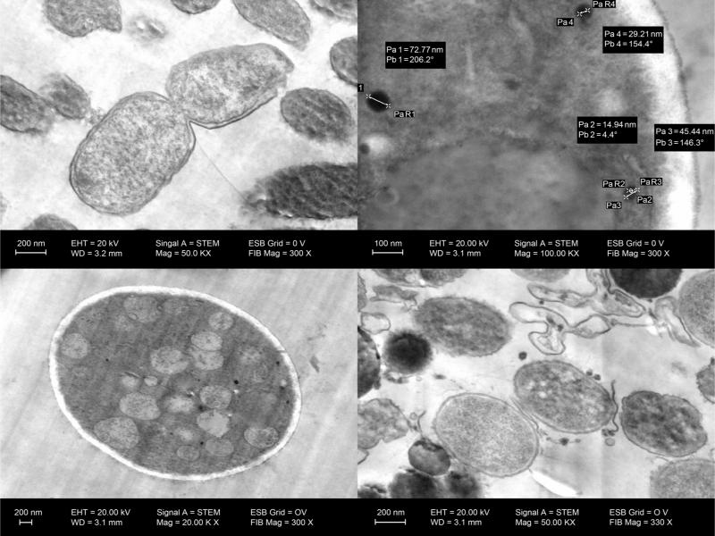
Fig. 5.
TEM images of the bacterium and yeasts after the growth in the BioscreenC: (a) Pseudomonas aeruginosa without AgNPs; (b) P. aeruginosa with AgNPs; (c, d) Candida albicans with AgNPs, the black structures visible inside and outside of cells.
Table III
The size and distribution (inside, outside, and within the cell wall) of AgNPs after their interaction with microorganisms.
| AgNPs Size [nm] | [%] | Cell distribution according o the size [%] | |||
|---|---|---|---|---|---|
| Inside | Outside | In the cell wall | Total | ||
| 21.45 ± 5.9 | 79.1 | 37.4 | 51.6 | 11 | 100 |
| 54.49 ± 9.1 | 13.9 | 93.8 | 6.25 | 0 | 100 |
| 94.62 ± 18.16 | 7.0 | 85.7 | 14.3 | 0 | 100 |
| Total | 100 | 48.7 | 42.6 | 8.7 | 100 |