
Table 1
Demographics of the patients and pancreatic tumor details in the current lot
| Parameters | Total number of patients (n=401) |
|---|---|
| Mean age ±SD, years | 63.99±10.91 |
| Gender, no. (%) | |
| Male | 211 (52.6%) |
| Female | 190 (47.4%) |
| Lesion diameter, mean±SD, mm | 36.59±16.23 |
| Tumor localization, no. (%) | |
| Head/uncinate | 301 (75.1%) |
| Body | 75 (18.7%) |
| Tail | 25 (6.2%) |
| Presence of biliary/pancreatic stent, no. (%) | |
| Yes | 118 (29.4%) |
| No | 283 (70.6%) |
| Number of EUS procedures per pacient, no. (%) | |
| One procedure | 379 (94.5%) |
| Two procedures | 19 (4.8%) |
| Three procedures | 3 (0.7%) |
| Final malignant diagnosis, no. (%) | |
| EUS-FNA/FNB | 365 (91.02%) |
| Surgery | 36 (8.97%) |
Table 2
Positive and false-negative results of the 401 patients included in the study
| Patients with pancreatic neoplasia detected through EUS-FNA/FNB | 365 (91.02%) |
| Positive results after first EUS, n (%) | 348(95.34%) |
| Positive results after second EUS, n (%) | 16 (4.38%) |
| Positive results after third EUS, n (%) | 1 (0.27%) |
| Patients with pancreatic neoplasia false-negative at EUS examination, confirmed by surgery | 36 (8.97%) |
| Negative results after first EUS, n (%) | 31 (86.11%) |
| Negative results after second EUS, n (%) | 3 (8.33%) |
| Negative results after third EUS, n (%) | 2 (5.55%) |
Table 3
Technical aspects and outcomes of FNA and FNB
| Total number of procedures (n=426) | p | ||
|---|---|---|---|
| FNA (n=334, 78.4%) | FNB (n=92, 21.6%) | ||
| Technical EUS success, no. (%) | 334 (100%) | 92 (100%) | |
| Diagnostic EUS yield, no. (%) | |||
| Positive | 281 (84.1%) | 84 (91.3%) | 0.048 |
| False-negative | 53 (15.9%) | 8 (8.7%) | |
| Number of passes, mean±SD | 1.99±0.52 | 1.63±0.60 | <0.001 |
| Lesion diameter, mean±SD, mm | 36.11±15.68 | 37.74±17.08 | 0.388 |
| Tumor localization, no. (%) | |||
| Head/uncinate | 258(77.2%) | 64(69.6%) | |
| Body | 58 (17.4%) | 21 (22.8%) | 0.312 |
| Tail | 18 (5.4%) | 7 (7.6%) | |
| Presence of biliary/pancreatic stent, no. (%) | |||
| Yes | 90 (26.9%) | 36 (39.1%) | 0.017 |
| No | 244 (73.1%) | 56 (60.9%) | |
| Major complications, no. (%) | 0 (0) | 0 (0) | |
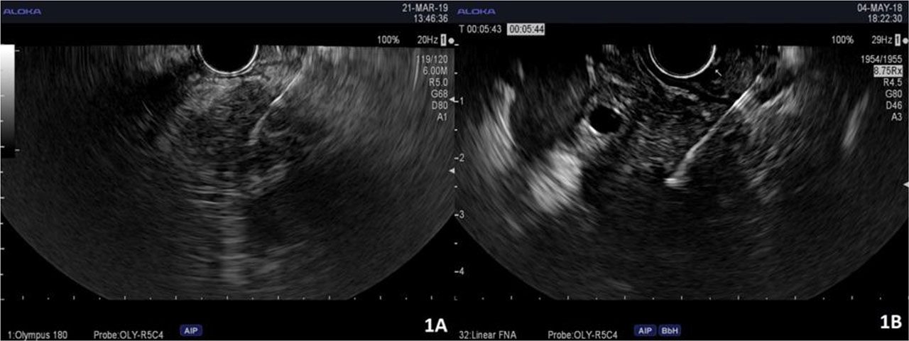
Figure 1.
FNA (1A) and FNB (1B) EUS-guided biopsies from pancreatic tumors.
Table 4
Histopathological results of EUS-FNA, FNB and surgery
| Histopathological diagnosis | FNA positive | FNB positive | FNA/FNB false-negative (surgery positive) |
|---|---|---|---|
| Pancreatic adenocarcinoma (n=332) | 232 | 65 | 35 |
| Pancreatic cystadenocarcinoma (n=18) | 15 | 3 | 0 |
| Pancreatic neuroendocrine tumor (n=26) | 16 | 9 | 1 |
| Non-Hodgkin lymphoma/leukemia (n=7) | 5 | 2 | 0 |
| Neoplastic gastric GIST (n=3) | 1 | 2 | 0 |
| Neoplastic IPMN (n=3) | 2 | 1 | 0 |
| Breast carcinoma metastasis (n=3) | 2 | 1 | 0 |
| Clear-cell renal carcinoma metastasis (n=3) | 2 | 1 | 0 |
| Colonic carcinoma metastasis (n=2) | 2 | 0 | 0 |
| Pulmonary neuroendocrine metastasis (n=1) | 1 | 0 | 0 |
| Hepatocellular carcinoma metastasis (n=1) | 1 | 0 | 0 |
| Pancreatic acinary carcinoma (n=1) | 1 | 0 | 0 |
| Solid pseudopapillary neoplasia (n=1) | 1 | 0 | 0 |
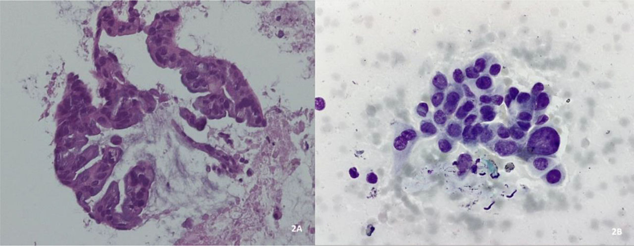
Figure 2.
Histological evaluation of with hematoxylin and eosin staining of pancreatic ductal adenocarcinoma.
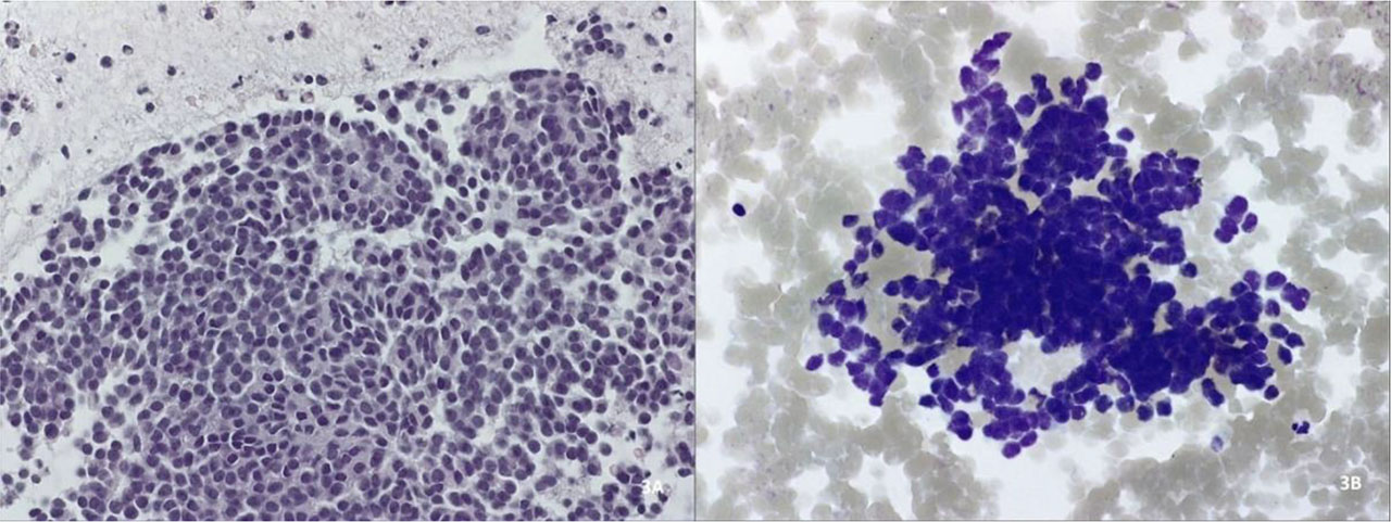
Figure 3.
Cytological examination with hematoxylin-eosin (3A) and Giemsa (3B) staining of a pancreatic neuroendocrine tumor.