
Highlights
- Dressler syndrome (DS) should be considered post-myocardial infarction (MI) with effusion and inflammation.
- Progressive effusion in DS may signal tamponade and needs urgent evaluation.
- Reassess anticoagulation in DS; discontinuation may prevent hemorrhagic effusion.
Introduction
Dressler syndrome (DS), included under the umbrella term of post-cardiac injury syndromes, is a form of secondary pericarditis that typically occurs 1–8 weeks after an acute myocardial infarction (MI). It is considered to be immune-mediated, and although its incidence was estimated at 3%–4% in Dressler's original description [1], with the advent of modern reperfusion strategies, the incidence of the syndrome has dramatically declined [2].
Pericardial effusion (PE) is a common feature of DS, typically mild and responsive to standard anti-inflammatory therapy. However, in rare cases, it may become significant and progress to cardiac tamponade, requiring prompt recognition and intervention [3]. The management is further complicated when patients are on concomitant anticoagulant (AC) therapy, as there are concerns of an increased risk of haemorrhagic PE.
We report the uncommon presentation of a patient who developed DS following an extensive anterior wall MI, complicated by haemorrhagic cardiac tamponade requiring percutaneous drainage. The clinical course was made more challenging by the need for combined AC and dual antiplatelet therapy due to concomitant atrial fibrillation and percutaneous coronary intervention.
Case presentation
A male patient in his 60s with no prior medical history was admitted to the hospital with complaints of a prolonged episode of severe chest pain 10 days earlier, followed by progressive exertional dyspnoea. Physical examination was unremarkable.
Initial laboratory investigations showed elevated cardiac troponins and natriuretic peptides, along with a significant inflammatory syndrome. The 12-lead electrocardiogram demonstrated sinus rhythm with pathological Q waves and ST-segment elevation in the anterolateral leads (Figure 1A). Transthoracic echocardiography (TTE) confirmed the diagnosis of extensive anterior wall myocardial infarction, revealing a small apical aneurysm, severely reduced left ventricular ejection fraction (~30%), elevated filling pressures, grade II mitral regurgitation, a trivial PE and small bilateral pleural effusions. Cardiac telemetry recorded several sustained episodes of atrial fibrillation.
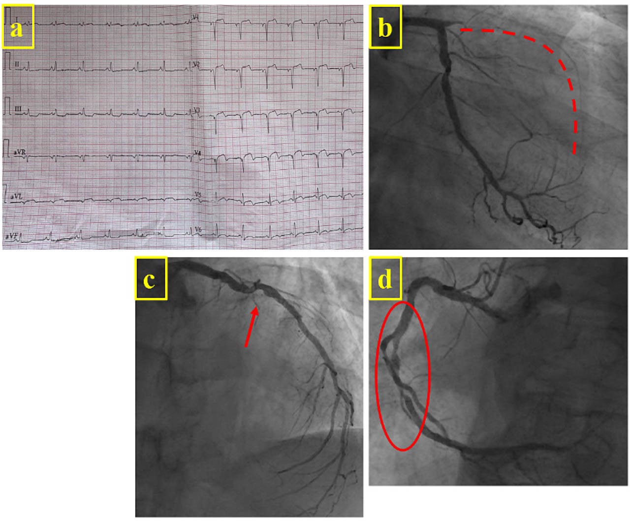
Figure 1-
(A) ECG at admission showing sinus rhythm with pathological Q waves and ST-segment elevation in the anterolateral leads. Coronary angiography showing (B) total occlusion of the left anterior descending artery with TIMI 0 flow (dotted line), (C) a moderate left circumflex lesion (arrow) and (D) two significant mid-right coronary artery lesions (circle).
The patient received standard initial therapy consisting of dual antiplatelets, a direct oral AC (Apixaban 5 mg), high-dose statin, class III antiarrhythmic, SGLT-2 inhibitor, aldosterone antagonist and loop diuretic. ACE inhibitor/ARNI therapy could not be initiated because of persistent hypotension. Subsequent coronary angiography demonstrated three-vessel disease: total occlusion of the left anterior descending artery with TIMI 0 flow and poor retrograde filling via left-to-left collaterals, a moderate left circumflex lesion and two significant mid-right coronary artery lesions (Figure 1B–D). Percutaneous coronary intervention of the right coronary artery was performed with implantation of two 3.5 mm × 23 mm drug-eluting stents, while the decision to revascularize the culprit lesion was deferred pending a future viability assessment.
There were no periprocedural complications. However, the patient developed deterioration in general condition with progressive PE. A diagnosis of DS was made, and treatment with high-dose aspirin and colchicine was initiated, while the AC was discontinued. Despite this, the patient's condition continued deteriorating, with development of orthopnoea, hypotension and multiorgan failure. Repeat TTE revealed a large PE with echocardiographic signs of cardiac tamponade (Figure 2, Supplementary material online, Video 1, Video 2), prompting emergency pericardiocentesis with evacuation of 600 mL of haemorrhagic fluid. Fluid analysis was consistent with an exudate, with elevated LDH and protein concentrations and a cellular composition comparable to peripheral blood. The post-procedural course was uneventful, and the patient was subsequently discharged.
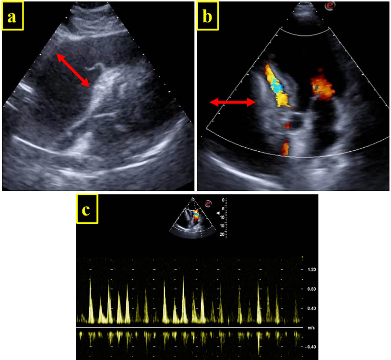
Figure 2-
2D TTE. Large PE (arrow) with signs of cardiac tamponade. Diastolic collapse of the right heart chambers on subcostal long-axis view (A) and apical 4C view with colour Doppler (B). (C) Significant respiratory variation in mitral inflow velocities. PE, pericardial effusion; TTE, transthoracic echocardiography.
At 3-month follow-up, contrast-enhanced cardiac magnetic resonance imaging revealed an ejection fraction of 28%, a mid-apical anteroseptal, inferoseptal and anterior wall aneurysm, and absence of viability on late gadolinium enhancement (Figure 3A–C). A trivial PE without signs of inflammation was also noted (Figure 3D). Based on these findings, a decision was made not to attempt recanalisation of the left anterior descending artery occlusion. The patient was scheduled for cardioverter-defibrillator implantation for primary prevention because left ventricular ejection fraction remained below 35% despite optimal medical therapy. At this point, given his CHA2DS2-VA score of 2, oral AC was reintroduced while Clopidogrel was continued for an additional 3 months according to the recommended post-procedural antithrombotic regimen.
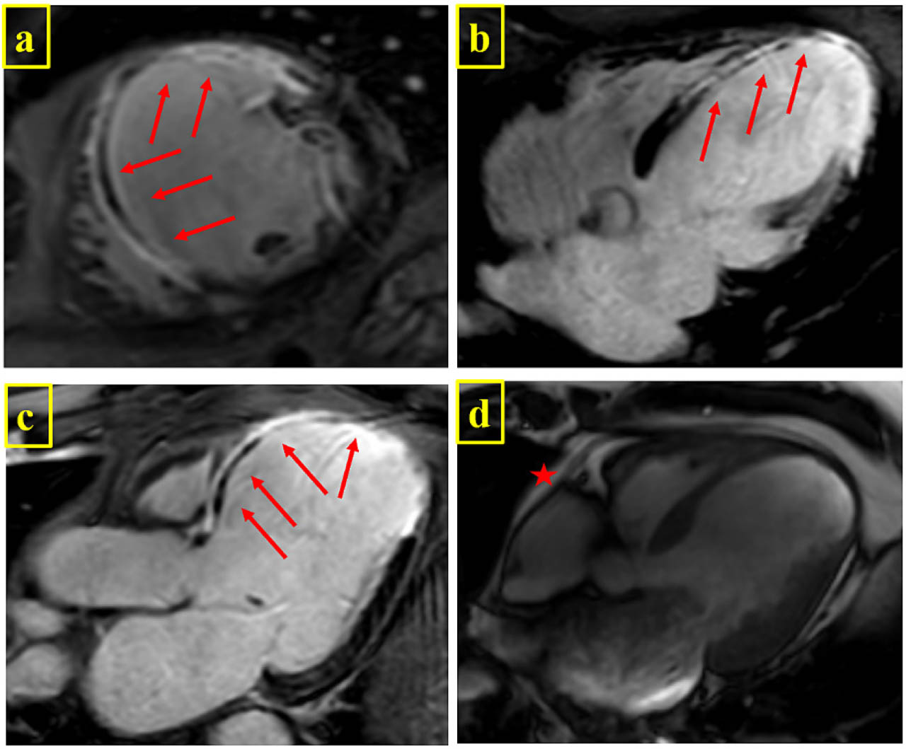
Figure 3-
Contrast-enhanced cardiac magnetic resonance imaging (A) short axis, (B) four-chamber and (C) three-chamber long-axis views showing transmural late gadolinium enhancement at the level of the anterior wall and mid-apical anteroseptal and inferoseptal wall (arrow). (D) A small PE is present (star), with no contrast enhancement. PE, pericardial effusion.
Discussion
This case is remarkable not only for the rarity of DS in current practice but also for its complication with haemorrhagic cardiac tamponade and the controversial role of AC in the setting of pericarditis.
Owing to advances in contemporary cardiovascular care, DS has nowadays become an uncommon diagnosis [2]. The pathogenesis of DS is now thought to involve an autoimmune reaction directed against myocardial necrosis antigens, leading to pericardial injury [3]. A previously proposed concept was that AC might lead to blood leakage through the damaged, highly permeable pericardium, resulting in inflammation and fluid accumulation. Nevertheless, DS was not observed in patients treated with thrombolytics who achieved early reperfusion, all of whom were receiving standard AC regimens [2].
In our case, the diagnosis of DS was supported by the presence of typical post-MI timing, systemic inflammatory response, progressive PE and exclusion of alternative causes of pericarditis. An oozing-type left ventricular free-wall rupture was considered but deemed unlikely based on the absence of recurrent chest pain, the gradual progression of PE over several days, the exudative characteristics of the pericardial fluid and the absence of ventricular rupture or pseudoaneurysm on serial cardiac imaging.
The clinical course of DS is usually benign, associated with mild PE and a favourable response to anti-inflammatory medication. However, in a very small number of cases, it can progress to a large PE and even cardiac tamponade.
Caution is generally advised regarding the use of AC in the setting of acute pericarditis, primarily due to concerns about haemorrhagic transformation. However, no large-scale studies have demonstrated a causal relationship between DS and haemopericardium or tamponade. Only a handful of cases of DS complicated by cardiac tamponade have been reported in the contemporary literature, highlighting the rarity of this presentation. Haemorrhagic PE occurred irrespective of AC status [6,7], while in the case by Tew et al. [8], no such complication developed despite AC. In his original study [1], Dressler recommended caution with AC, although haemopericardium was also observed in patients who had not received such treatment. Moreover, in cases of acute pericarditis unrelated to MI, AC has not been identified as a risk factor for developing haemorrhagic PE [4,5]. Whether the addition of dual antiplatelet therapy to oral AC further increases the risk of haemorrhagic PE in DS remains unknown. Nevertheless, the combined antithrombotic regimen may have contributed to the haemorrhagic nature of the effusion in our patient.
In patients with concomitant atrial fibrillation and coronary artery disease, a therapeutic dilemma arises in balancing the risks of bleeding and thrombosis. In our patient, atrial fibrillation occurred in the subacute phase following MI, and oral AC was initiated according to the patient's CHA2DS2-VA score and current guideline recommendations. However, temporary discontinuation may be justified in the presence of life-threatening haemorrhagic complications. Accordingly, the decision to discontinue AC following the diagnosis of DS was justified by the presence of haemodynamic compromise and the high bleeding risk associated with an enlarging PE. Colchicine and high-dose aspirin were initiated in accordance with current pericarditis management guidelines [9], and pericardiocentesis was successfully performed. Despite the severity of presentation, the patient had a favourable outcome, underscoring the importance of timely recognition and intervention. Oral AC was resumed after the acute phase, following an individualised reassessment of thromboembolic and bleeding risks.
Conclusion
Although rare in the reperfusion era, DS can still lead to life-threatening complications such as cardiac tamponade. This case highlights the need for vigilance in post-MI patients with PE and the importance of rapid intervention and individualised management in the context of combined antithrombotic therapy, where therapeutic decisions remain particularly challenging.
Video captions
Supplementary Video 1. 2D TTE subcostal long-axis view, with large PE and signs of cardiac tamponade-diastolic collapse of the right heart chambers.
Supplementary Video 2. 2D TTE apical 4C view, colour Doppler, with large PE and signs of cardiac tamponade-diastolic collapse of the right heart chambers.
Abbreviations
- AC
anticoagulant therapy
- DS
Dressler syndrome
- MI
myocardial infarction
- PE
pericardial effusion
- TTE
transthoracic echocardiography
Notes
[1] Ethics Approval
The study was approved by an institutional medical research ethics committee and conducted in accordance with the Declaration of Helsinki.