
Introduction
Acute pulmonary thromboembolism (PTE) is a life-threatening cardiovascular emergency with a wide spectrum of clinical, haemodynamic and electrocardiographic manifestations. Electrocardiography (ECG) plays an important adjunctive role in early risk stratification, although findings are often non-specific. According to the 2019 European Society of Cardiology (ESC) Guidelines for the diagnosis and management of acute pulmonary embolism, the most frequent ECG abnormalities include sinus tachycardia, right bundle branch block, right axis deviation and right ventricular (RV) strain patterns, while ST-segment elevation is not considered a typical feature [1].
Nevertheless, rare cases of acute PTE presenting with ST-segment elevation mimicking acute ST-elevation myocardial infarction (STEMI), particularly inferior wall myocardial infarction (IWMI), have been reported [2,3,4,5]. Such presentations pose a major diagnostic challenge, as misinterpretation may lead to inappropriate activation of primary percutaneous coronary intervention pathways and delay in definitive treatment for PTE. This case illustrates a rare but clinically significant ECG manifestation of acute PTE.
We report a case of high-risk acute PTE presenting with inferior and lateral ST-segment elevation, initially referred to as IWMI and discuss the underlying mechanisms with reference to current ESC guidelines and published case literature.
Case presentation
A 44-year-old man presented to the emergency department at approximately 9:30 p.m. with acute-onset chest tightness and dyspnoea of 3 hr duration. He was a known case of Type 2 diabetes mellitus on oral hypoglycaemic agents. There was no prior history of hypertension, thyroid disease, coronary artery disease or venous thromboembolism.
On presentation, he was in distress. His vital parameters were notable for a pulse rate of 150 beats/min (regular), blood pressure of 90/50 mm Hg and oxygen saturation of 76% on room air. Respiratory system examination was unremarkable, with clear lung fields on auscultation. There were no clinical signs of deep vein thrombosis.
1. ECG
The 12-lead ECG demonstrated a regular tachycardia at approximately 150 beats/min with a frontal axis of +60°. ST-segment elevation was noted in leads II, III, aVF, V5 and V6 without clear reciprocal ST-segment depression. An S wave was present in lead I, and the R/S ratio in V2 was approximately 1, suggesting RV pressure overload. Based on these findings, the patient was initially referred with a provisional diagnosis of IWMI.
2. Laboratory investigations
Initial laboratory evaluation revealed:
Troponin T: 0.082 ng/mL (mildly elevated)
D-dimer: 1026 ng/mL
Haemoglobin: 18.5 g/dL
Renal function tests: Blood urea 24 mg/dL, serum creatinine 0.6 mg/dL
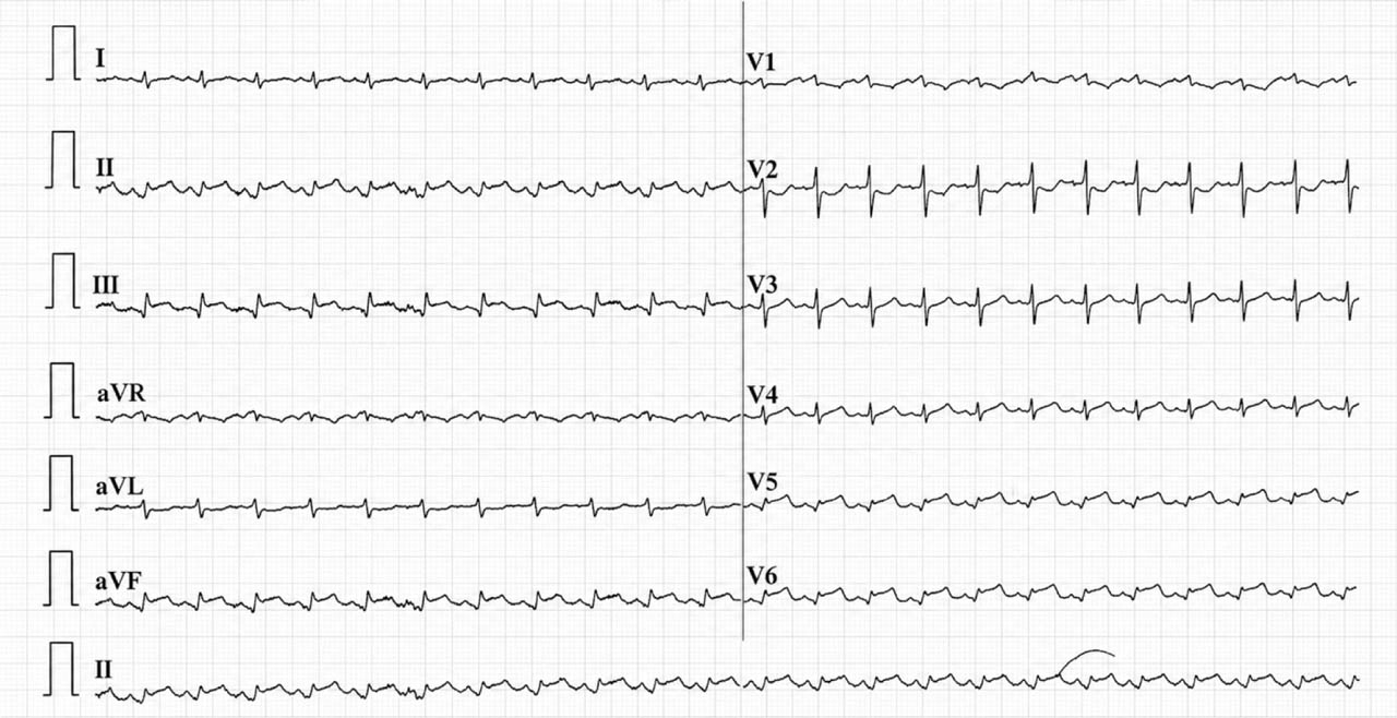
Figure 1-
Admission 12-lead electrocardiogram showing ST-segment elevation in inferior (II, III, aVF) and lateral (V5–V6) leads, with and S wave in lead I and R/S = 1 in V2.
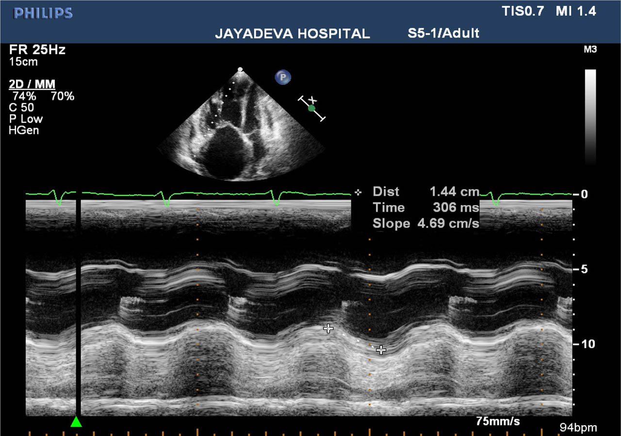
Figure 2-
Transthoracic echocardiography demonstrating dilated right-sided chambers, reduced TAPSE and elevated tricuspid regurgitation gradient suggestive of acute RV pressure overload. RV, right ventricular; TAPSE, tricuspid annular plane systolic excursion.
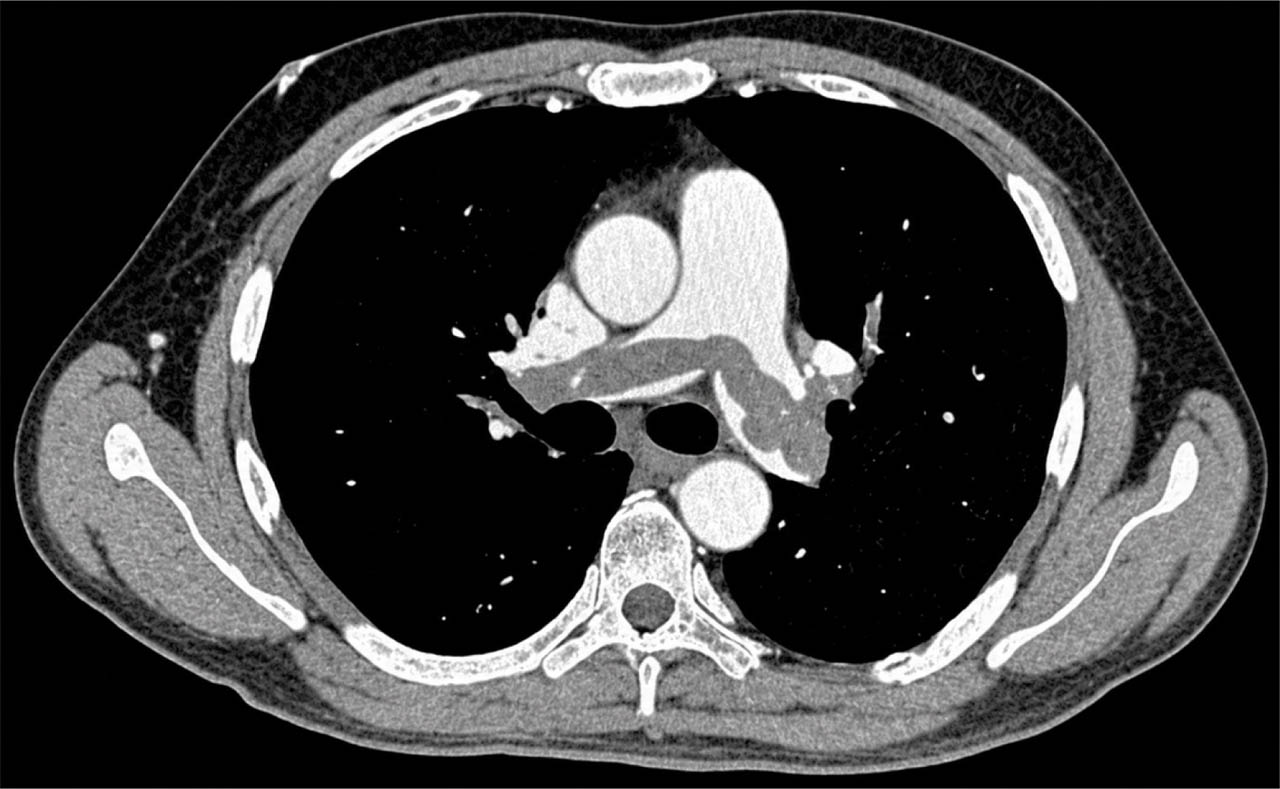
Figure 3-
CTPA showing large filling defects within the main pulmonary artery and right pulmonary artery consistent with acute pulmonary embolism. CTPA, computed tomography pulmonary angiography.
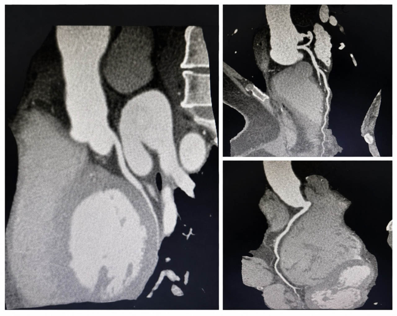
Figure 4-
CT-CAG demonstrating normal coronary arteries without obstructive lesions. CT-CAG, computed tomography coronary angiography.
3. Transthoracic echocardiography
Urgent bedside transthoracic echocardiography showed no regional wall motion abnormalities of the left ventricle. However, there was dilation of right-sided cardiac chambers with evidence of severe pulmonary hypertension. The tricuspid regurgitation jet gradient was estimated at 44 mm Hg. The inferior vena cava measured 2.2 cm with absent inspiratory collapse. Tricuspid annular plane systolic excursion was reduced to 14 mm, consistent with RV systolic dysfunction.
In view of the clinical presentation, severe hypoxaemia, shock and echocardiographic findings suggestive of acute RV pressure overload, a diagnosis of high-risk acute PTE was considered.
4. Imaging and management
Computed tomography pulmonary angiography demonstrated large thrombi within the left pulmonary artery and right pulmonary artery, confirming the diagnosis of acute PTE.
The patient underwent systemic thrombolysis with Tenecteplase 40 mg s/c stat along with UFH 5000 IU qid and subsequently transitioned to oral anticoagulation. Following thrombolytic therapy, he showed marked clinical improvement with stabilisation of haemodynamics, resolution of hypoxaemia and symptomatic relief.
Subsequently, coronary evaluation with computed tomography coronary angiography (CT-CAG) revealed normal coronary arteries, effectively excluding obstructive coronary artery disease.
Discussion
ST-segment elevation in acute PTE is distinctly uncommon, and its true incidence remains unknown. Most large observational studies and guideline documents do not list ST elevation as a characteristic ECG finding in PTE [1]. However, several isolated case reports and small series have described this phenomenon, most often involving the inferior leads (II, III and aVF), occasionally with lateral or anterior extension [2,3,4,5].
The 2026 ACC/AHA guidelines emphasise that ECG changes in acute PTE primarily reflect acute RV pressure overload and myocardial strain rather than primary coronary artery occlusion [1]. Sinus tachycardia remains the most frequent abnormality, while RV strain patterns (T-wave inversion in V1–V4 and inferior leads) are associated with worse prognosis and higher pulmonary artery pressures. ST-segment elevation, although rare, appears to represent an extreme manifestation of acute cardiopulmonary stress.
Several pathophysiological mechanisms have been proposed to explain ST-segment elevation in PTE. Severe hypoxaemia due to sudden obstruction of the pulmonary vasculature can trigger a catecholamine surge, resulting in increased myocardial oxygen demand and transient supply–demand mismatch [3]. This mechanism is supported by reports demonstrating reversible ST elevation with resolution following thrombolysis or anticoagulation, without evidence of obstructive coronary artery disease.
Acute RV pressure overload is another plausible explanation. Sudden elevation in pulmonary artery pressure leads to RV dilatation, increased wall tension and subendocardial ischaemia. Given the anatomical proximity and shared blood supply between the RV and the inferior wall of the left ventricle, RV ischaemia may manifest as ST-segment elevation in inferior leads [4]. In our patient, ST-segment elevation extended beyond the inferior leads to involve V5 and V6. Such changes may represent an extreme manifestation of acute RV pressure overload and myocardial ischaemia secondary to severe pulmonary vascular obstruction. Kukla et al. [3] demonstrated that ischaemic ECG patterns, including ST-segment abnormalities involving inferior and lateral leads, identify intermediate-risk pulmonary embolism patients with more severe RV dysfunction and worse prognosis. The extensive ECG abnormalities observed in our patient are, therefore, consistent with severe haemodynamic compromise and high-risk pulmonary embolism requiring urgent reperfusion therapy.
Paradoxical coronary embolism through a patent foramen ovale or atrial septal defect has also been described as a potential cause of true myocardial infarction in patients with venous thromboembolism [5]. However, normal coronary anatomy on CT-CAG in this case effectively excludes this possibility. The mild elevation in cardiac troponin is consistent with myocardial injury related to acute RV strain, a phenomenon well recognised in high-risk PTE and incorporated into ESC risk stratification algorithms [1].
This case reinforces the importance of integrating ECG findings with clinical context. Profound hypoxaemia, shock and echocardiographic evidence of RV dysfunction should prompt consideration of acute PTE, even in the presence of apparent STEMI patterns on ECG. Bedside echocardiography, as recommended by ESC guidelines for haemodynamically unstable patients, plays a pivotal role in avoiding misdiagnosis and guiding appropriate therapy.
Conclusion
Acute PTE can rarely present with ST-segment elevation mimicking IWMI. Awareness of this atypical presentation is crucial, particularly in patients with severe hypoxaemia, haemodynamic instability and echocardiographic evidence of RV dysfunction. Early recognition and appropriate imaging can prevent misdiagnosis and ensure timely initiation of life-saving therapy.
Learning points
Acute PTE can rarely present with ST-segment elevation mimicking IWMI.
Profound hypoxaemia, shock and RV dysfunction should prompt consideration of pulmonary embolism despite apparent STEMI patterns on ECG.
Bedside echocardiography is critical in haemodynamically unstable patients to differentiate pulmonary embolism from acute coronary syndromes.
Recognition of atypical ECG presentations of pulmonary embolism can prevent diagnostic delay and inappropriate management.
Acknowledgment
None.
Notes
[1] Ethics statement
The study was approved by an institutional medical research ethics committee and conducted in accordance with the Declaration of Helsinki. Written informed consent was obtained from the patient, in accordance with the Declaration of Helsinki.
[2] Patient consent
The participant was provided an informed written consent to participate in the study in accordance with the code of ethics of the World Medical Association (Declaration of Helsinki) for experiments on humans.