
Figure 1
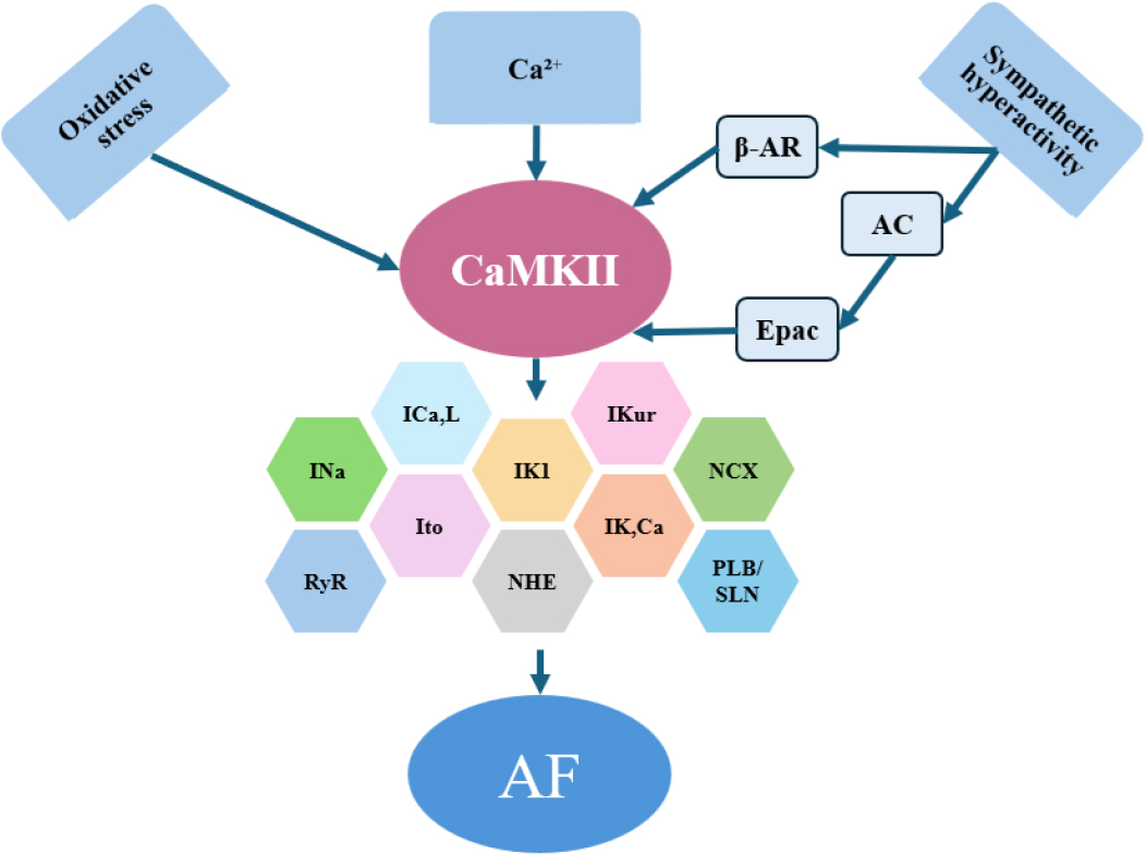
Schematic representation of Ca2+/calmodulin-dependent protein kinase II (CaMKII) pathway in atrial fibrillation (AF).
Schematic representation of CaMKII activation and its downstream effects on atrial electrical remodeling. Increased intracellular Ca2+, sympathetic stimulation, and oxidative stress promote CaMKII activation, leading to altered phosphorylation of ion channels and Ca2+-handling proteins, including RyR2 and phospholamban/sarcolipin. These changes favor sarcoplasmic reticulum Ca2+ leak, triggered activity, and shortening of atrial refractoriness, thereby contributing to the initiation and maintenance of AF.
AF – atrial fibrillation; ICa,L – L-type Ca2+ current; IK1 – inward rectifier K+ current; IK,Ca – Ca2+-activated K+ current; IKur – ultra-rapid delayed rectifier K+ current; INa – Na+ current; Ito – transient outward K+ current; NCX – Na+– Ca2+ exchanger; NHE – Na+-H+ exchanger; PLB/SLN – phospholamban/sarcolipin; RyR – ryanodine receptor
Table 1
Therapeutic strategies targeting signaling pathways involved in atrial fibrillation.
| Signaling pathway | Drug class / examples | Main molecular effects | Effects on atrial remodeling | Clinical AF outcomes | References |
|---|---|---|---|---|---|
| Ca2+/CaMKII | β–blockers | ↓ sympathetic tone; indirect ↓ CaMKII activation | Improved Ca2+ handling; ↓ triggered activity | Reduced AF burden; limited efficacy in persistent AF | [36] |
| Ca2+ channel blockers | ↓ ICa,L; ↓ Ca2+ influx | Electrical stabilization; minimal structural effects | Rate control; no prevention of AF progression | [36] | |
| MAPK | ACE inhibitors / ARBs | ↓ ERK, p38 activation; ↓ Ang II signaling | ↓ atrial fibrosis; ↓ fibroblast activation | Reduced new–onset AF; modest effect in established AF | [35,1] |
| MAPK/TGF–β | Mineralocorticoid receptor antagonists (eplerenone) | ↓ ERK, ↓ TGF–β signaling | Reduced atrial fibrosis; improved conduction homogeneity | Reduced AF recurrence in selected populations | [34,37] |
| PI3K/AKT /mTOR | Statins | Anti-inflammatory; ↓ oxidative stress; indirect AKT modulation | Attenuation of structural remodeling | Inconsistent AF prevention; benefit mainly in postoperative AF | [38,39] |
| mTOR inhibitors (rapamycin) | ↓ mTORC1 activity; ↑ autophagy | Reduced atrial fibrosis (experimental) | No established clinical role | [40,41] | |
| NF–κB | Anti–inflammatory agents | ↓ NF–κB activation; ↓ cytokine expression | Reduced inflammatory atrial remodeling | No consistent benefit in AF prevention | [42,43] |
| TGF–β/Smad | ARBs, MRAs | ↓ Smad phosphorylation | ↓ profibrotic gene transcription | Reduced AF susceptibility; limited reversal of advanced fibrosis | [44,45] |
| Multiple | Statins, RAAS blockers | ↓ oxidative stress; ↓ inflammation | Substrate modification rather than rhythm control | Explains delayed and modest clinical effects | (39,44–46) |
1 AF – atrial fibrillation; AKT – protein kinase B; Ang II – Angiotensin II; ARBs – Angiotensin II receptor blockers; CaMKII – Calcium/Calmodulin– dependent protein kinase II; MAPK – mitogen–activated protein kinase; MRAs – Mineralocorticoid receptor antagonists; mTOR – mammalian target of rapamycin; NF-κB – nuclear factor kappa-light-chain-enhancer of activated B cells; p38 – p38 mitogen–activated protein kinase; PI3K – phosphatidylinositol 3-kinase; RAAS – Renin-angiotensin-aldosterone system; RyR2 – ryanodine receptor 2; TGF-β1 – transforming growth factor beta
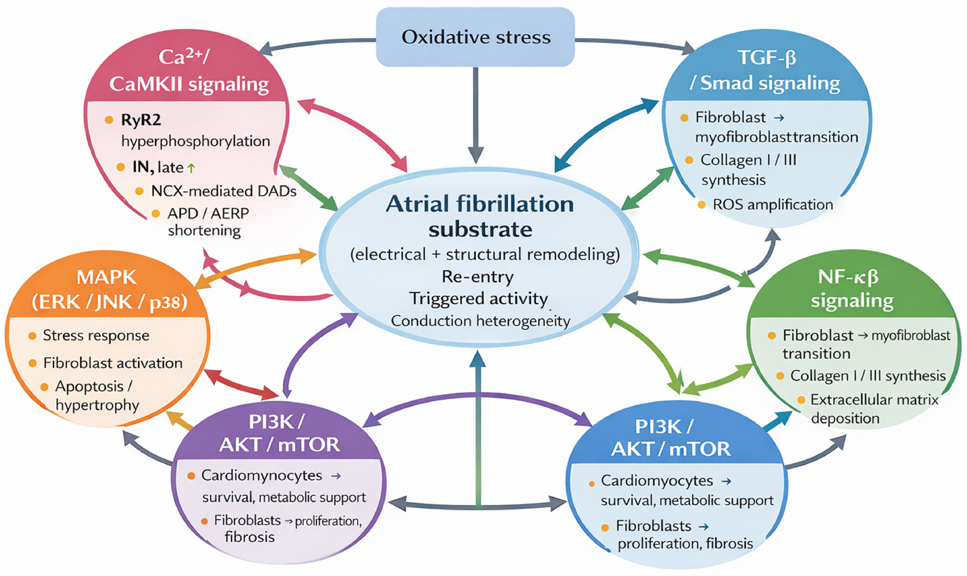
Figure 2
Integrated intracellular signaling network underlying atrial remodeling in atrial fibrillation.
Atrial fibrillation (AF) results from the convergence of multiple intracellular signaling pathways that collectively generate an arrhythmogenic electrical and structural substrate. Ca2+/calmodulin-dependent protein kinase II (CaMKII) plays a central role in electrical remodeling by promoting Ca2+-handling abnormalities and triggered activity. Structural remodeling is driven predominantly by profibrotic signaling through the TGF-β/Smad pathway, with amplification by mitogen-activated protein kinases (MAPK) and inflammatory NF-κB signaling. The PI3K/AKT/mTOR pathway exerts context-dependent effects, supporting cardiomyocyte survival while promoting fibroblast proliferation under pathological conditions. Oxidative stress and inflammatory mediators act as common upstream drivers and reinforce pathway crosstalk. The integrated activity of these signaling networks promotes atrial fibrosis, conduction heterogeneity, and sustained AF.
AKT – protein kinase B; CaMKII – calcium/calmodulin-dependent protein kinase II; ERK – extracellular signal-regulated kinase; IN, late – late sodium channel current; JNK – c-Jun N-terminal kinase; MAPK – mitogen-activated protein kinase; mTOR – mammalian target of rapamycin; NF-κB – nuclear factor kappa-light-chain-enhancer of activated B cells; PI3K – phosphatidylinositol 3-kinase; ROS – reactive oxygen species; RyR – ryanodine receptor; Smad – suppressor of mothers against decapentaplegic; TGF-β1 – transforming growth factor beta 1
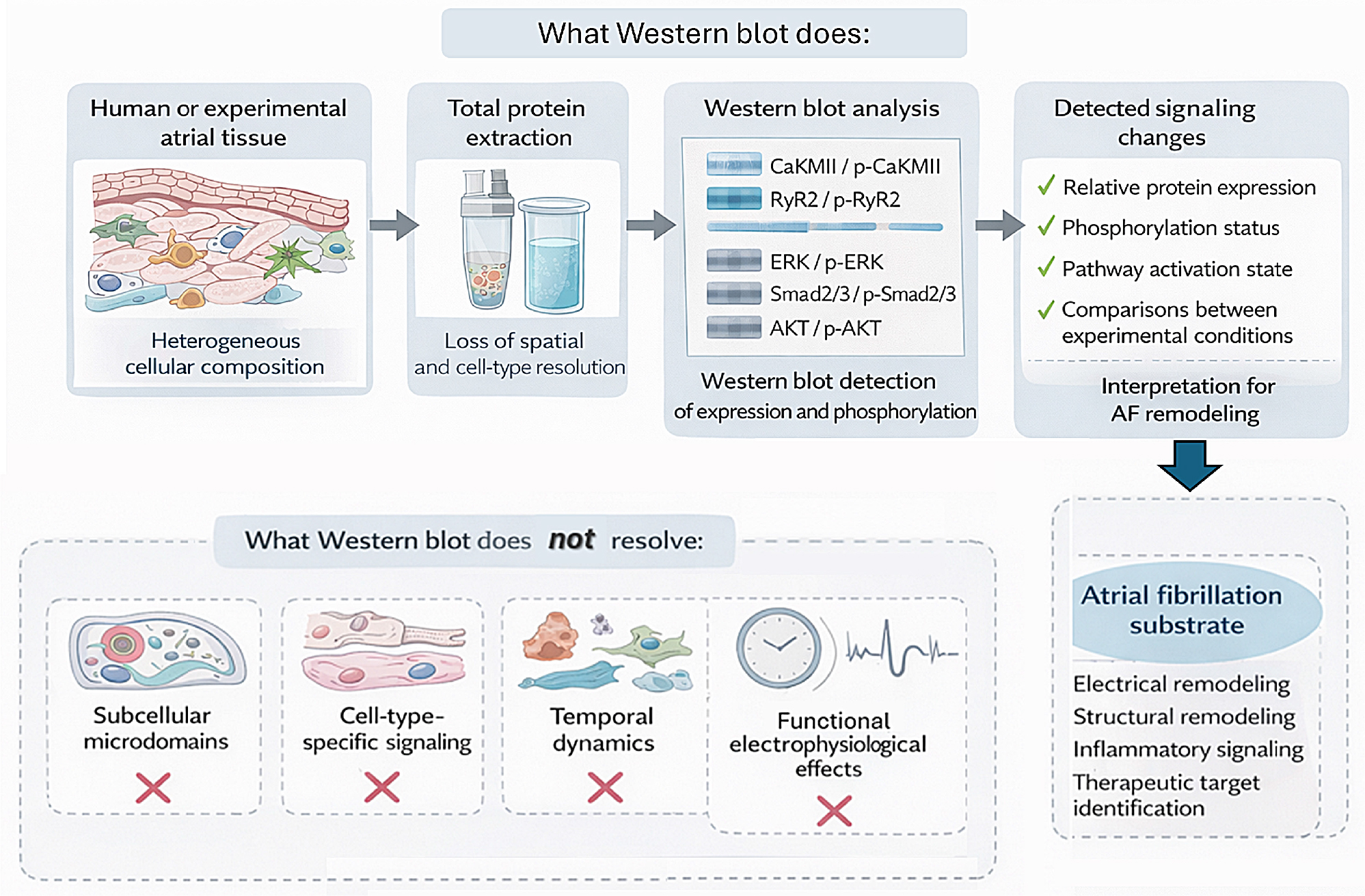
Figure 3
Western blot as a molecular window into atrial fibrillation signaling.
Western blot analysis enables the detection of protein expression and post-translational modifications in atrial tissue samples obtained from experimental models or patients with atrial fibrillation (AF). Following total protein extraction from heterogeneous atrial tissue, Western blot allows the relative quantification of signaling proteins and their phosphorylated forms, providing insights into the activation status of key intracellular pathways involved in electrical and structural remodeling. However, this technique does not preserve spatial resolution, cell-type specificity, or temporal dynamics of signaling events. Therefore, Western blot findings should be interpreted as reflecting global pathway activation and are best integrated with functional, imagingbased, and electrophysiological approaches to fully characterize the AF substrate.
AF – atrial fibrillation; AKT – protein kinase B; CaMKII – calcium/calmodulin-dependent protein kinase II; ERK – extracellular signal-regulated kinase; RyR2 – ryanodine receptor 2; Smad – suppressor of mothers against decapentaplegic