
Table 1
Baseline clinical characteristics of the study cohort, both overall and according to the occurrence of major adverse events.
| Variable | All (n = 215) | No events (n = 175) | MAE (n= 40) | P value |
|---|---|---|---|---|
| Age, years | 61 ± 10 | 59 ± 10 | 59 ± 13 | 0.791 |
| Man, n (%) | 168 (78.1) | 133 (76) | 35 (87.5) | 0.122 |
| Heart rate, bpm | 80 ± 16 | 79 ± 15 | 78 ± 16 | 0.714 |
| SBP, mmHg | 139 ± 22 | 143 ± 23 | 140 ± 23 | 0.451 |
| DBP, mmHg | 86 ± 14 | 87 ± 15 | 87 ± 16 | 0.801 |
| BMI, kg/m2 | 27.10 (24.22–30.86) | 27.68 (24.5–31.8) | 27.23 (24.0–31.0) | 0.877 |
| BSA, m2 | 1.91 (1.79–2.13) | 1.97 (1.82–2.16) | 1.96 (1.74–2.11) | 0.855 |
| Peak CK-MB, U/L | 289.60 ± 211 | 244.14 ± 189.66 | 353.28 ± 291.45 | 0.004 |
| Peak Troponin I, ng/L | 3,351 (314–21,506) | 1,866 (218–18,773) | 13,404 (1434–38,967) | 0.057 |
| eGFR, ml/min/1.73 m2 | 74.08 ± 21.22 | 79.89 ± 20.23 | 80.28 ± 24.47 | 0.917 |
| Total Cholesterol, mg/dL | 192 ±49 | 192 ± 43 | 190 ± 52 | 0.811 |
| LDLc, mg/dL | 121 ± 49 | 120 ± 32 | 117 ± 39 | 0.632 |
| Triglycerides, mg/dL | 108 (86–156) | 121 (93–168) | 123 (85–146) | 0.806 |
| Hemoglobin, g/dL | 14.80 ± 1.64 | 14.69 ± 1.47 | 14.58 ± 1.53 | 0.656 |
| Creatinine, mg/dL | 1.08 (0.92–1.30) | 1.09 (0.96–1.24) | 1.16 (0.85–1.28) | 0.669 |
| Glycemia, mg/dL | 132 (113–176) | 132 (113–167) | 128 (110–160) | 0.186 |
| Leucocytes, 103 / μL | 12.03 (10.24–14.95) | 12.03 (9.93–14.41) | 11.97 (9.78–15.94) | 0.448 |
| Neutrophiles, 103 / μL | 9.56 (7.40–11.28) | 8.75 (7.11–11.28) | 9.19 (6.99–13.16) | 0.436 |
| ESR, mm/h | 10 (6-20) | 10 (6-22) | 12 (10-29) | 0.667 |
| Smoking, n (%) | 157 (73) | 129 (73.7) | 28 (70.0) | 0.633 |
| Dyslipidemia, n (%) | 209 (97.2) | 171 (97.7) | 38 (95) | 0.347 |
| Previous CAD, n (%) | 14 (6.5) | 12 (6.9) | 2 (5.0) | 1.000 |
| Cardiac inheritance, n (%) | 14 (6.5) | 13 (7.4) | 1 (2.5) | 0.476 |
| Diabetes, n (%) | 47 (21.9) | 39 (22.3) | 8 (20.0) | 0.752 |
| Hypertension, n (%) | 154 (71.6) | 130 (74.3) | 24 (60.0) | 0.071 |
| Killip Class I, n (%) | 177 (82.3) | 149 (85.1) | 28 (70) | 0.024 |
| Killip Class II, n (%) | 30 (14) | 21 (12) | 9 (22.5) | 0.084 |
| Killip Class III, n (%) | 3 (1.4) | 1 (0.6) | 2 (0.5) | 0.090 |
| Killip Class IV, n (%) | 5 (2.3) | 4 (2.3) | 1 (2.5) | 1.000 |
| CKD, n (%) | 22 (10.2) | 16 (9.1) | 6 (15.0) | 0.259 |
| Thrombolysis, n (%) | 56 (26.0) | 45 (25.7) | 11 (27.5) | 0.816 |
| Single-vessel disease, n (%) | 90 (41.9) | 81 (46.3) | 9 (22.5) | 0.006 |
| Two-vessel disease, n (%) | 74 (34.4) | 59 (33.7) | 15 (37.5) | 0.649 |
| Three-vessel disease, n (%) | 51 (23.7) | 35 (20.0) | 16 (40.0) | 0.007 |
| Anterior STEMI, n (%) | 91 (42.3) | 75 (42.9) | 16 (40.0) | 0.741 |
| Treatment at discharge | ||||
| Aspirin, n (%) | 212 (98.6) | 173 (98.9) | 39 (97.5) | 0.462 |
| P2Y12i, n (%) | 215 (100) | |||
| Betablocker, n (%) | 169 (78.6) | 140 (80.0) | 29 (72.5) | 0.297 |
| Nitrate, n (%) | 92 (42.8) | 69 (39.4) | 23 (57.5) | 0.037 |
| ACE inhibitor, n (%) | 125 (58.1) | 109 (62.3) | 16 (40.0) | 0.010 |
| ARBs, n (%) | 1 (0.5) | 1 (0.6) | 0 (0.0) | 1.000 |
| MRAs, n (%) | 170 (79.1) | 136 (77.7) | 34 (85.0) | 0.307 |
| Furosemide, n (%) | 159 (74.0) | 123 (70.3) | 36 (90.0) | 0.010 |
| SGLT2i, n (%) | 44 (20.5) | 37 (21.1) | 7 (17.5) | 0.606 |
| ARNI, n (%) | 7 (3.3) | 5 (2.9) | 2 (5.0) | 0.617 |
| Statins, n (%) | 215 (100) |
1 Values are expressed as mean ± standard deviation or median (interquartile range). MAE, major adverse events; SBP, systolic blood pressure; DBP, diastolic blood pressure; BMI, body mass index; BSA, body surface area; CK-MB, creatine-kinase isoform MB; eGFR, estimated glomerular filtration rate; LDLc, low-density lipoprotein cholesterol; ESR, erythrocyte sedimentation rate; CAD, coronary artery disease; CKD, chronic kidney disease; ACE, angiotensin-converting enzyme; ARBs, angiotensin II receptor blockers; MRAs, mineral-corticoid receptor antagonists; SGLT2i, sodium-glucose co-transporter 2 inhibitor; ARNI, angiotensin receptor-neprilysin inhibitors.
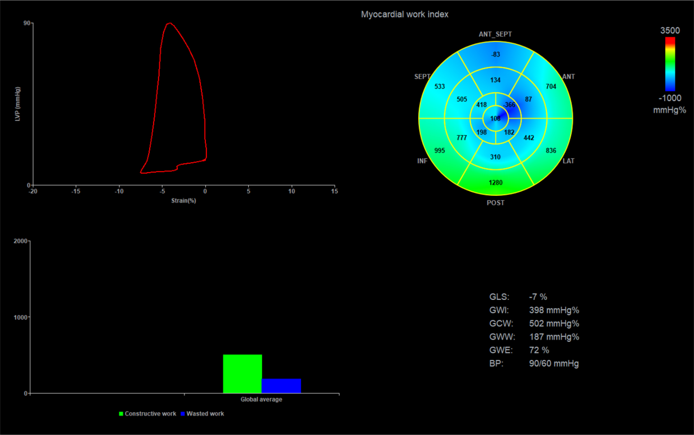
Figure 1
Myocardial work analysis in a patient with acute anterior myocardial infarction, showing reduced myocardial work indices (GWI, GCW, GWE), elevated GWW and impairment of GLS. Abbreviations: GLS, global longitudinal strain; GWI, global work index; GCW, global constructive work; GWE, global work efficiency; BP, blood pressure.
Table 2
Transthoracic echocardiography results.
| Echocardiographic parameters | All (n = 215) | No events (n = 175) | MAE (n = 40) | P value |
|---|---|---|---|---|
| LVEDV, ml | 107 ± 26 | 108 ± 27 | 113 ± 30 | 0.276 |
| LVESV, ml | 65 (45–75) | 62 (52–73) | 60 (45–74) | 0.568 |
| LA diameter, mm | 40 ± 5.5 | 39 ± 4.9 | 40 ± 4.7 | 0.827 |
| LAVi, mL/m2 | 55.78 ± 19.86 | 29.55 ± 9.89 | 29.94 ± 10.10 | 0.785 |
| RV diameter, mm | 26 ± 3.0 | 25 ± 3.4 | 26 ± 3.9 | 0.212 |
| PASP, mmHg | 22 (21–31) | 21 (21–28) | 23 (18–30) | 0.324 |
| TAPSE, mm | 20 ± 2.8 | 26 ± 1.7 | 20 ± 3.8 | 0.466 |
| IVST, cm | 1.17 ± 0.15 | 1.20 ± 0.19 | 1.13 ± 0.15 | 0.370 |
| PWT, cm | 1.12 ± 0.12 | 1.14 ± 0.15 | 1.11 ± 0.17 | 0.279 |
| AV Pmax, m/s | 4.8 (4.0–7.0) | 5.2 (4.0–7.0) | 5.2 (4.0–6.1) | 0.236 |
| E wave, m/s | 0.64 ± 0.20 | 0.63 ± 0.19 | 0.64 ± 0.18 | 0.742 |
| A wave, m/s | 0.78 ± 0.18 | 0.76 ± 0.17 | 0.73 ± 0.19 | 0.467 |
| Mean E’, m/s | 0.06 ± 0.01 | 0.06 ± 0.01 | 0.06 ± 0.01 | 0.786 |
| E/E’ ratio | 10.57 (8.30–12.57) | 9.33 (7.69–12.15) | 10.61 (8.31–15.30) | 0.633 |
| Mean S’, m/s | 0.07 ± 0.01 | 0.07 ± 0.01 | 0.07 ± 0.01 | 0.125 |
| LVEF, % | 41 ± 6 | 43 ± 6 | 42 ± 6 | 0.708 |
| GLS, % | -12.21 ± 3.51 | -12.77 ± 3.3 | -11.75 ± 3.44 | 0.081 |
| MD, ms | 67.60 (54.40–83.33) | 67.80 (54.06–83.33) | 66.95 (51.25–83.57) | 0.123 |
| GWI, mmHg% | 1105 ±479 | 1210 ± 433 | 1077 ± 474 | 0.086 |
| GCW, mmHg% | 1293±493 | 1392 ± 437 | 1258 ± 448 | 0.084 |
| GWW, mmHg% | 139 (95–187) | 147 (90–187) | 126 (96–200) | 0.355 |
| GWE, % | 86 ± 8 | 86 ± 7 | 83 ± 8 | 0.022 |
1 Values are expressed as mean ± standard deviation or median (interquartile range). LVEDV, left ventricular end-diastolic volume; LVESV, left ventricular end-systolic volume; LA, left atrium; LAVi, left atrial volume index; RV, right ventricle; PASP, estimated pulmonary artery systolic pressure; TAPSE, tricuspid annular plane systolic excursion; IVST, end-diastolic interventricular septal thickness; PWT, end-diastolic posterior wall thickness; AV Pmax, aortic valve peak pressure gradient; E, peak early diastolic mitral flow velocity; A, late transmitral flow velocity; E’, mean peak early diastolic mitral annulus velocity; S’, mean peak systolic mitral annulus velocity; LVEF, left ventricular ejection fraction; GLS, global longitudinal strain; MD, mechanical dispersion; GWI, global work index; GCW, global constructive work; GWW, global wasted work; GWE, global work efficiency
Table 3
Cox regression analysis to evaluate the risk of major adverse events during follow-up.
| Univariable | Multivariable | ||||||
|---|---|---|---|---|---|---|---|
| Parameter | Unit interpretation | HR1 | 95% CI2 | P | HR | 95% CI | P |
| Age | per 1-year increase | 1.010 | 0.982–1.038 | 0.486 | 1.025 | 0.995–1.057 | 0.101 |
| Sex | Male/female | 0.493 | 0.193–1.258 | 0.139 | |||
| Peak CK-MB3 | per 100 U/L increase | 1.159 | 1.039–1.293 | 0.008 | 1.126 | 0.998–1.270 | 0.054 |
| Creatinine | per 1 mg/dL increase | 1.409 | 0.640–3.106 | 0.395 | |||
| Hypertension | Yes/No | 0.499 | 0.265–0.941 | 0.032 | 0.426 | 0.216–0.842 | 0.014 |
| LVEF4 | per 1% increase | 0.985 | 0.940–1.033 | 0.536 | |||
| GLS5 | per 1% increase | 1.096 | 0.996–1.207 | 0.060 | |||
| MD6 | per 10 ms increase | 1.115 | 1.000–1.242 | 0.050 | |||
| GWI7 | per 100 mmHg% increase | 0.922 | 0.854–0.996 | 0.040 | |||
| GCW8 | per 100 mmHg% increase | 0.922 | 0.854–0.996 | 0.039 | |||
| GWW9 | per 10 mmHg% increase | 1.018 | 0.992–1.045 | 0.177 | |||
| GWE10 | per 5% increase | 0.811 | 0.688–0.955 | 0.012 | 0.835 | 0.693–0.985 | 0.033 |
| LVEDV11 | per 1 mL increase | 1.007 | 0.996–1.018 | 0.188 | |||
| LAVi12 | per 1 mL/m2 increase | 1.004 | 0.974–1.035 | 0.792 | |||
| E/E’13 | per 1-unit increase | 1.024 | 0.959–1.092 | 0.480 | |||
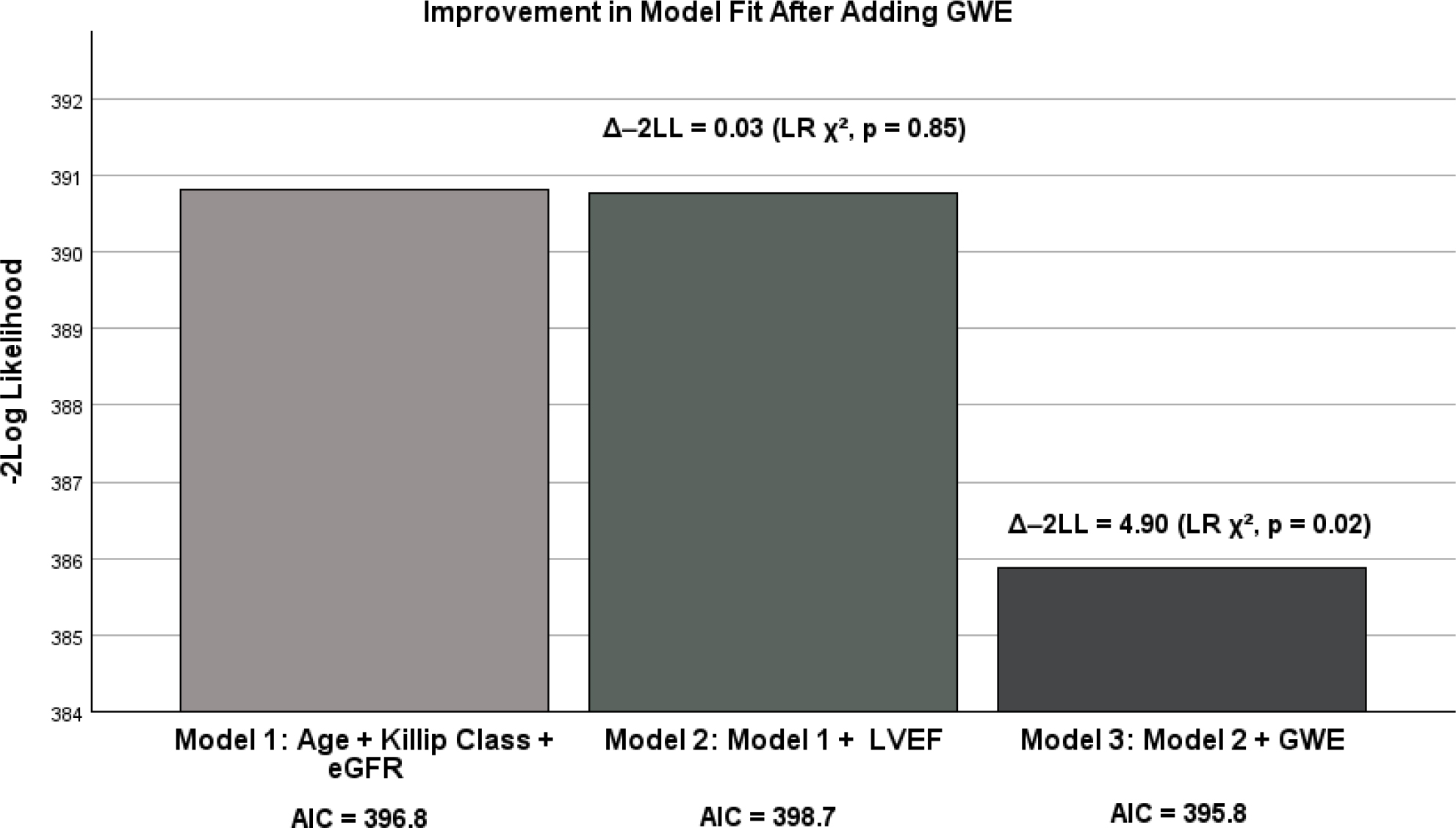
Figure 2
Incremental prognostic value of GWE for MAE. The bar chart shows –2 log likelihood for three sequential Cox models: the baseline clinical model (age, Killip class and eGFR), the model adding LVEF, and the model further including GWE. Data labels indicate the Akaike information criterion for each model. Addition of LVEF resulted in a minimal and non-signif¡cant improvement in model fit (likelihood ratio X2= 0.03, df =1, p = 0.85). Further inclusion of GWE produced a substantial reduction in –2LL, significantly improving model fit (likelihood ratio X2 = 4.90, df = 1, p = 0.02). GWE, global work efficiency; -2LL, -2 log likelihood; LR, likelihood ratio; eGFR, estimated glomerullar filtration rate; LVEF, left ventricular ejection fraction; AIC, akaike information criterion.