
Table 1
Distribution of demographic and cardiovascular risk factors among the studied groups.
| HFpEF Group (N = 39) | Control Group (N = 20) | P | ||
|---|---|---|---|---|
| Age (years) | 52.7 ± 8.6 | 48.5 ± 10.6 | 0.106 | |
| Sex | Male | 10 (25.6%) | 4 (20%) | 0.63 |
| Female | 29 (74.4%) | 16 (80%) | ||
| BMI (kg/m2) | 23.27 ± 1.5 | 22.13 ± 1.4 | 0.006* | |
| Total Cholesterol (mg/dL) | 182.56 ± 16.4 | 167.5 ± 11.5 | 0.001* | |
| Comorbidities | Smoking | 6 (15.4%) | 0 (0%) | 0.064 |
| Hypertension | 5 (12.8%) | 0 (0%) | 0.094 | |
| Diabetes Mellitus | 4 (10.2%) | 0 (0%) | 0.138 | |
Table 2
Heart rhythm and conventional echocardiographic parameters in the studied groups.
| HFpEF Group (N = 39) | Control Group (N = 20) | P | ||
|---|---|---|---|---|
| ECG | AF | 15 (38.5%) | 0 (0%) | 0.001* |
| Sinus Rhythm | 24 (61.5%) | 20 (100%) | ||
| IVSd (cm) | 1.3 ± 0.24 | 0.82 ± 0.12 | < 0.001* | |
| LVIDd (cm) | 4.7± 0.3 | 5.2 ± 0.3 | < 0.001* | |
| LVIDs (cm) | 3.01 ± 0.3 | 3.1 ± 0.2 | 0.367 | |
| LVPWd (cm) | 1.2 ± 0.2 | 0.8 ± 0.1 | < 0.001* | |
| EDV (ml) | 102.2 ± 31.4 | 114.6 ± 25.6 | 0.133 | |
| EF (%) | 66.17 ± 5.89 | 66.4 ± 4.48 | 0.884 | |
| SV (ml) | 80 ± 8.7 | 75.15±15.3 | 0.12 | |
| Conventional echocardiography | FS (%) | 36.48 ± 4.7 | 36.5 ± 2.9 | 0.991 |
| LA diameter (mm) | 39.41± 8.8 | 30.66 ± 8.2 | < 0.001* | |
| Ao (mm) | 30.4 ± 7.1 | 27.05 ± 6.09 | 0.07 | |
| e’ lateral (cm/s) | 6.5 ± 0.6 | 14 ± 3 | < 0.001* | |
| E (cm/s) | 82 ± 15 | 95 ± 13 | < 0.001 | |
| E/e’ | 12.91 ± 2.02 | 6.7 ± 1.79 | < 0.001* | |
| MAPSE (cm) | 1.56 ± 0.15 | 2.25 ± 0.21 | < 0.001* | |
| TAPSE (cm) | 1.72 ± 0.23 | 2.23 ± 0.21 | < 0.001* | |
| PASP (mmHg) | 43.57± 11.03 | 22± 5 | < 0.001 | |
1 Data is presented as mean ± SD or frequency (%). P value < 0.05 is statistically significant. AF: Atrial fibrillation; Ao: Aortic diameter; EDV: End-diastolic volume; E/e’: Ratio of early diastolic velocity to early diastolic filling; EF: ejection fraction; FS: Fractional shortening; HFpEF: Heart failure with preserved ejection fraction; IVSd: Interventricular septal thickness in diastole; LA: Left atrium; LVIDd: Left ventricular internal diameter in diastole; LVIDs: Left ventricular internal diameter in systole; LVPWd: Left ventricular posterior wall thickness in diastole; MAPSE: Mitral annular plane systolic excursion; PASP: Pulmonary artery systolic pressure; SV: Stroke volume; TAPSE: Tricuspid annular plane systolic excursion; e’: Early diastolic velocity.
Table 3
Right ventricular and right atrial diameters and strain parameters, together with N-terminal pro-brain natriuretic peptide (NT-proBNP) level, among the studied groups.
| HFpEF Group (N=39) | Control Group (N=20) | P> | |
|---|---|---|---|
| RV basal diameter (cm) | 3.13 ± 0.54 | 3.26 ± 0.63 | 0.43 |
| RV mid diameter (cm) | 2.56 ± 0.53 | 2.78 ± 0.58 | 0.15 |
| RV longitudinal diameter (cm) | 5.59 ± 0.76 | 5.34 ± 0.69 | 0.21 |
| RA septal-lateral diameter (cm) | 3.7 ± 0.5 | 3.4 ± 0.2 | 0.001* |
| RA apico-basal diameter (cm) | 4.6 ± 0.6 | 4.2 ± 0.4 | 0.003* |
| Right ventricle longitudinal strain | |||
| RV free wall % | 19.6 ± 7.33 | 28 ± 4.31 | < 0.001* |
| RV septal wall % | 15.2 ± 3.49 | 32.5 ± 5.93 | < 0.001* |
| RV GLS % | 16.79 ± 3.96 | 25.35 ± 1.89 | < 0.001* |
| Right atrial strain % | 20.94 ± 8.78 | 39.95 ± 2.03 | < 0.001* |
| NT-proBNP pg./ml | 610 ±15.6 | 103 ±7 | < 0.001* |
Table 4
Correlations between NT-proBNP and RV longitudinal and RA strain in the studied group.
| NT-proBNP pg./ml | ||
|---|---|---|
| r | P | |
| RA strain % | -0.739 | < 0.001* |
| RV longitudinal strain % | ||
| RV free-wall % | -0.505 | < 0.001* |
| RV septal-wall % | -0.849 | < 0.001* |
| RV GLS % | -0.737 | < 0.001* |
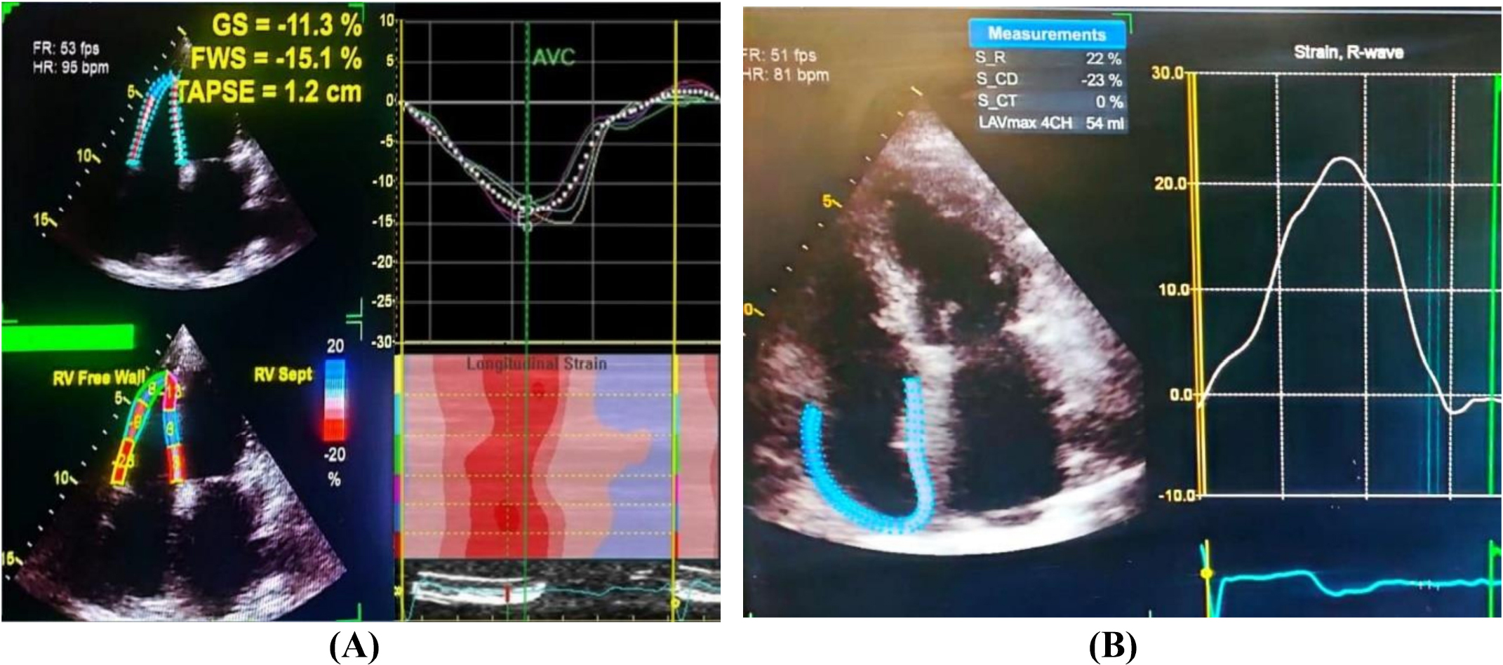
Figure 1
(A) Right ventricular strain analysis: GS: 11.3%, FWS: 15.1%, TAPSE: 1.2 cm (all below normal), HR: 95 bpm, FR: 53 fps. Right panel shows longitudinal strain curve and color-coded strain map (red = higher strain, blue = lower strain). (B) Right atrial strain analysis: HR: 81 bpm, FR: 51 fps, peak strain: 22%, S_CD: 23%, S_CT: 0%, RAVmax: 54 ml. Right panel displays strain rate curve with peak at 22%. Abbreviations: GS = global strain; FWS = free-wall strain; TAPSE = tricuspid annular plane systolic excursion; FR = frame rate; HR = heart rate; S_CD = strain at end-diastole; S_CT = strain at end-systole; RAVmax = maximum RA volume in 4-chamber view.