
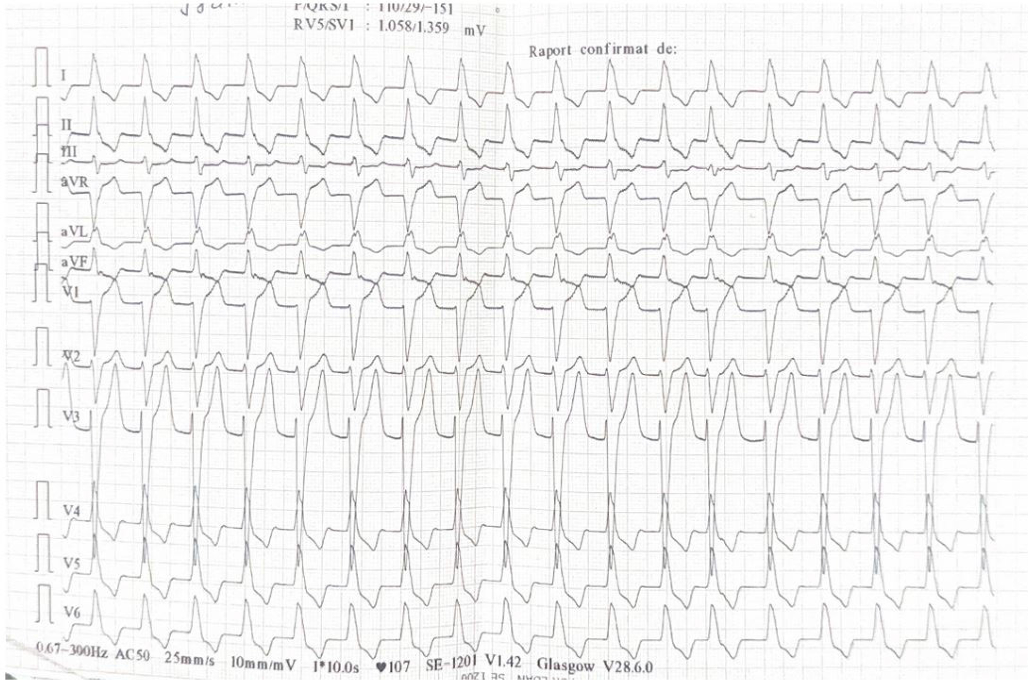
Figure 1
Atrial fibrillation, left bundle branch block.
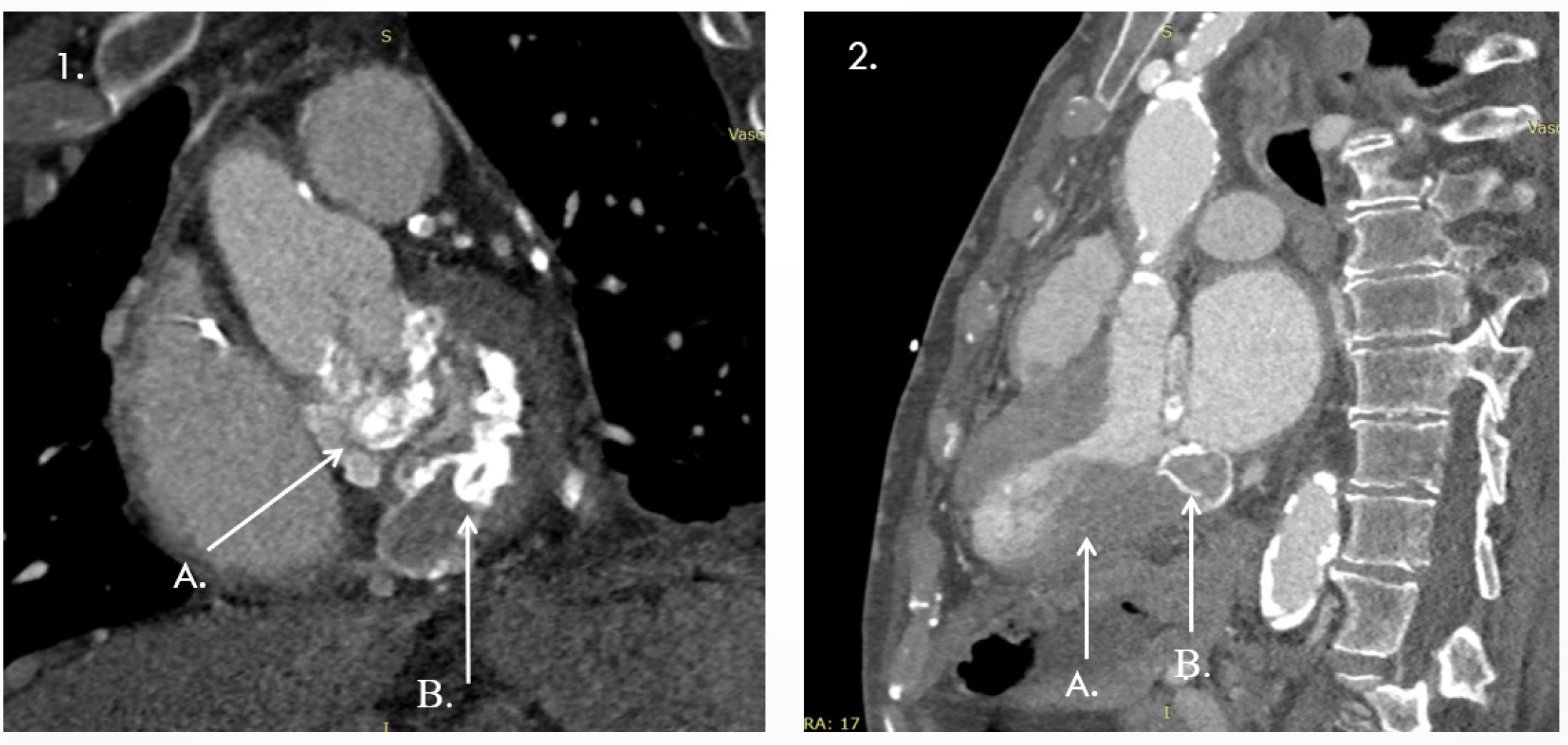
Figure 2
1 A, B Annular calcification extending to the mitral-aortic continuity and aortic root; 2 A-Important left ventricular hypertrophy; B-Toothpaste tumour.
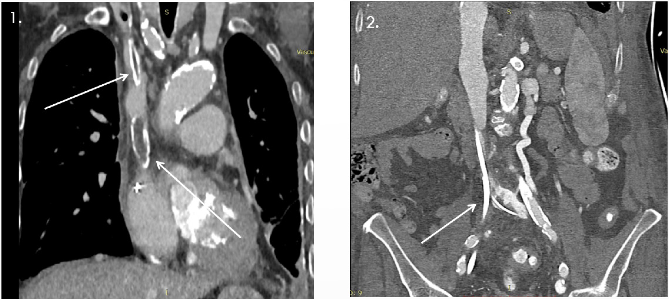
Figure 3
1- Oclussion VCS due to ‘forgotten* dialysis catheter; 2 - Stenosis and oclussion of inferior vena cava and collateral circulation between the left common iliac vein and the left renal vein.
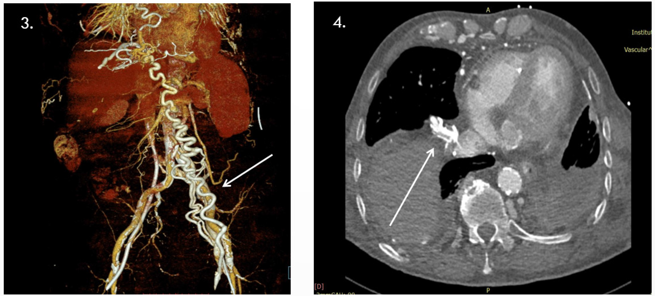
Figure 4
3- Important collateral circulation thoraco-abdominal; 4- Important collateral circulation thoraco-abdominal.
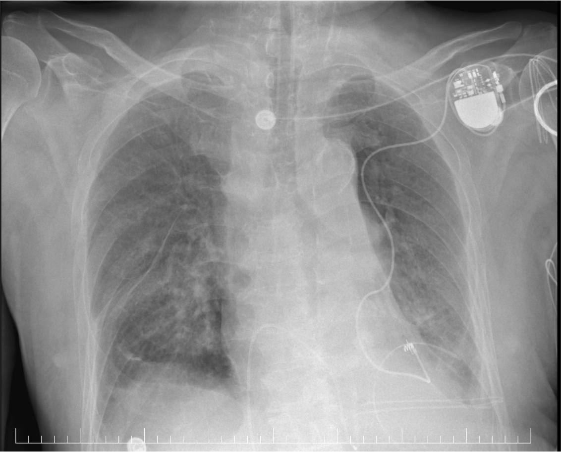
Figure 5
X ray-pacemaker and epicardial lead placement.