
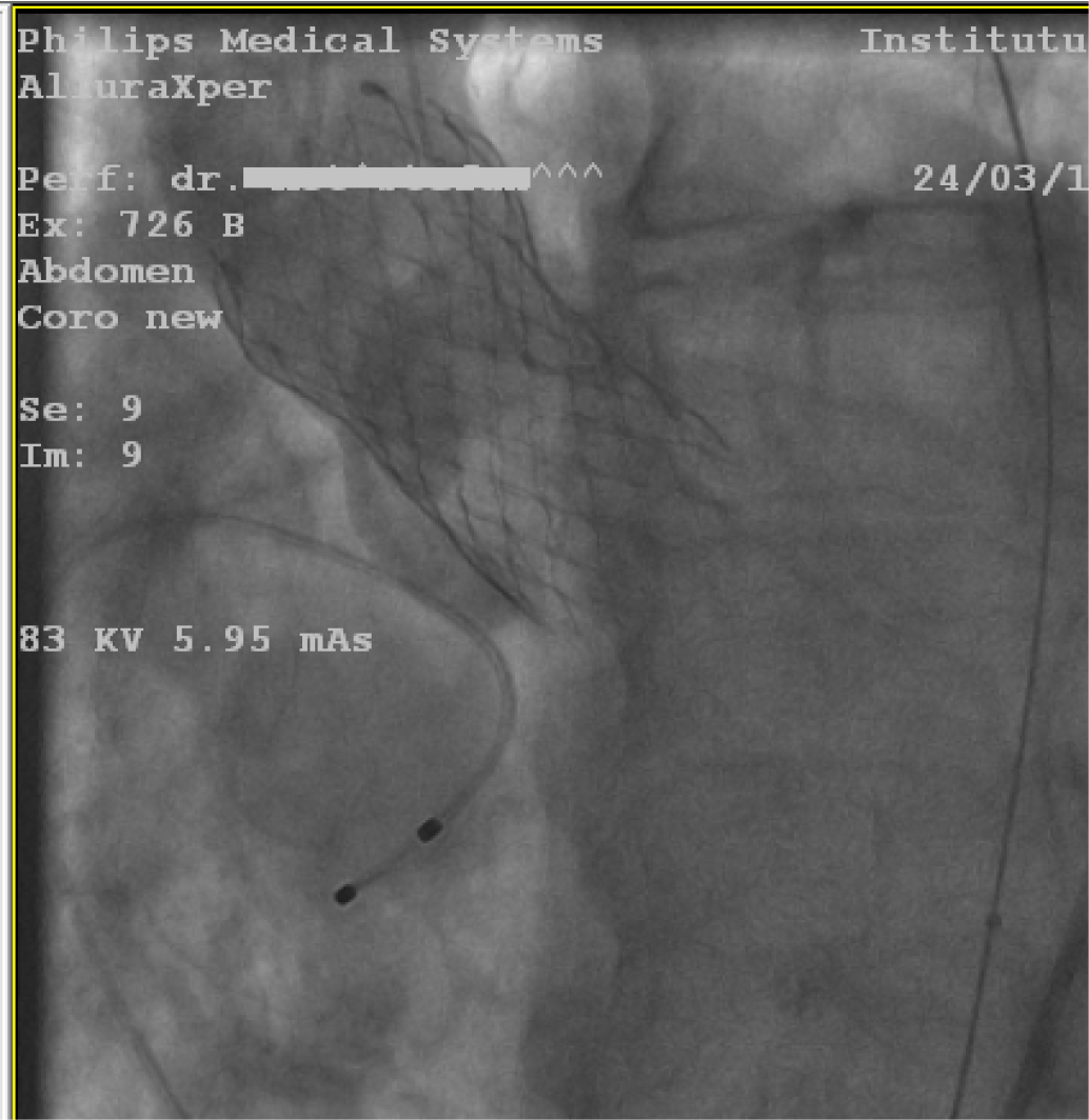
Figure 1
Fluoroscopic image: deployment of the Medtronic valve.
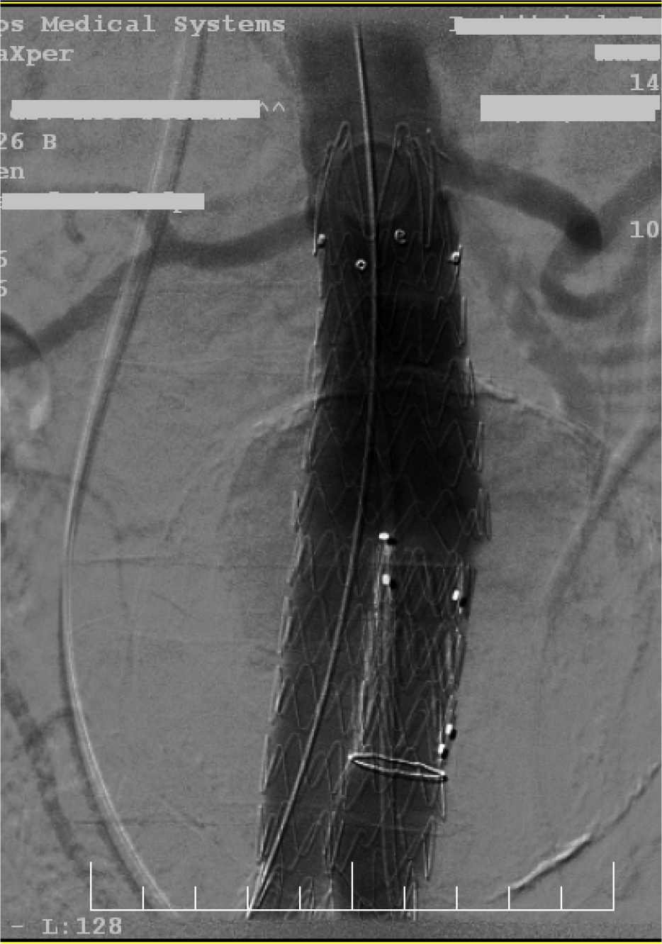
Figure 2
Fluoroscopy: deployment of the aortic endograft.
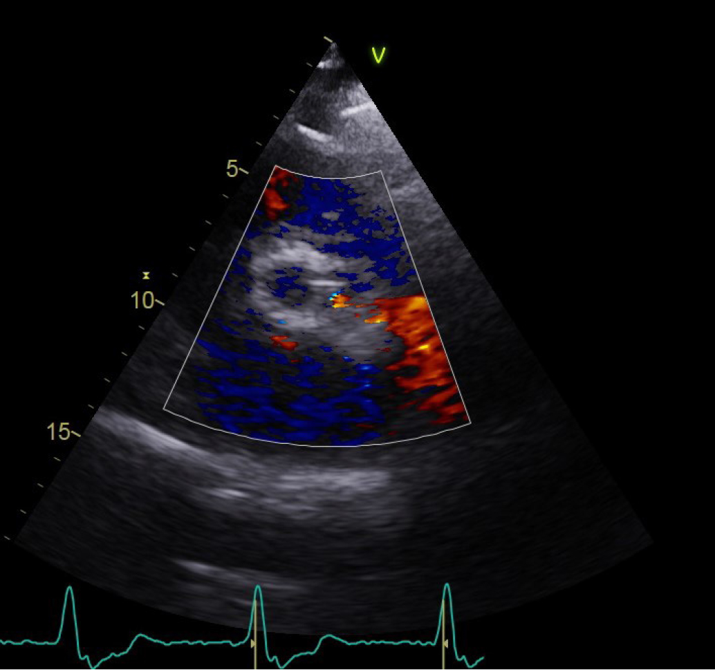
Figure 3
Transthoracic echocardiogram, parasternal short axis view of the paravalvular leak.
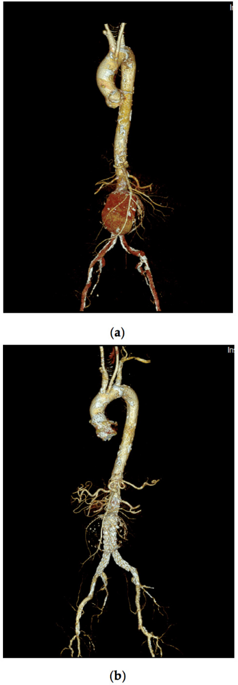
Figure 4
Computed Tomography Angiography – 3D reconstruction of the aorta (a) before, and (b) after aortic endo-graft implant.
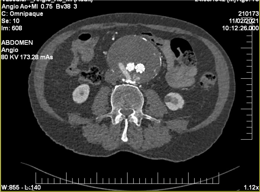
Figure 5
Computed Tomography Angiography at 6 months follow-up.
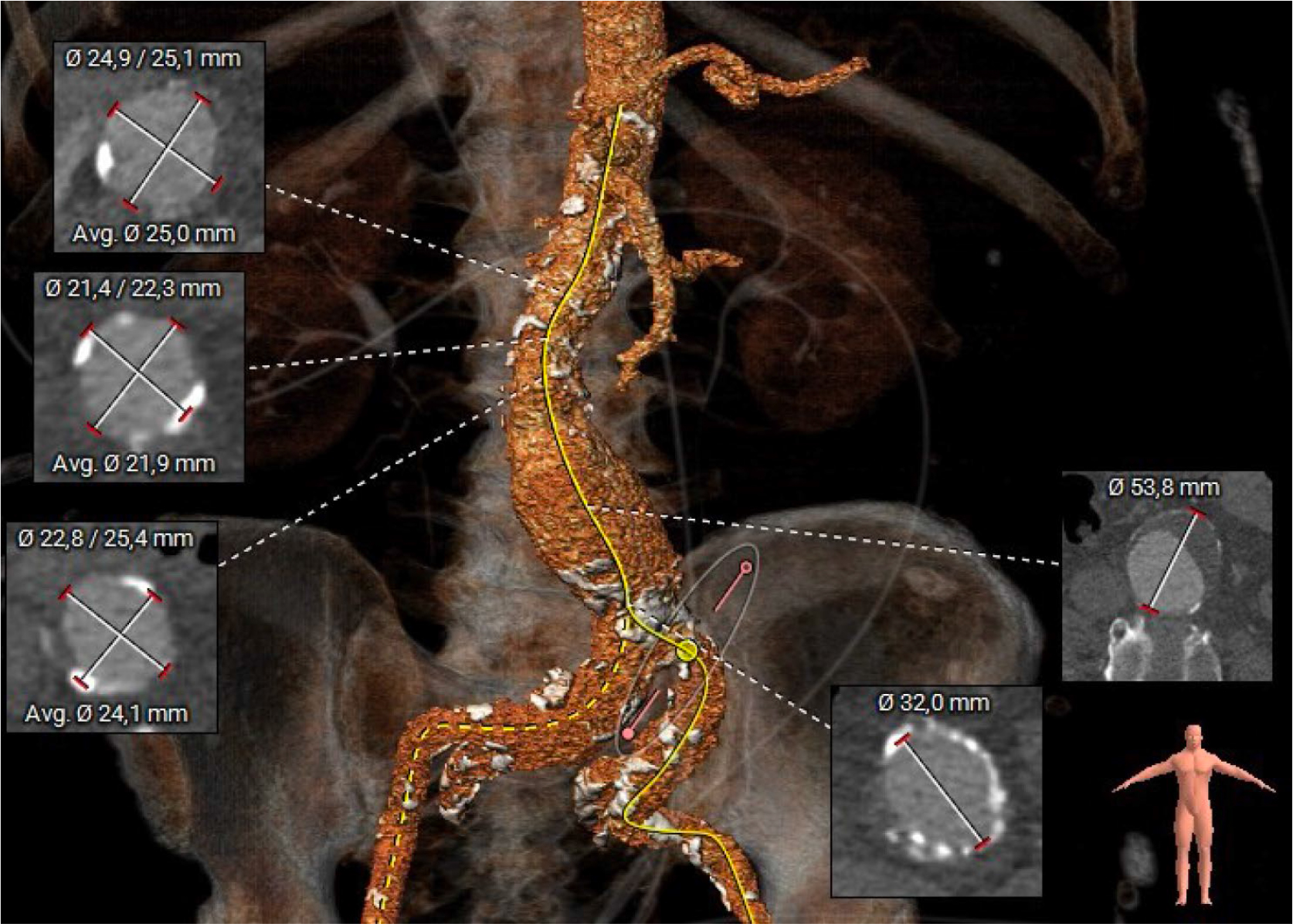
Figure 6
Computed Tomography Angiography showing the AAA and the RCIA aneurysm. An accessory right renal artery is also described.
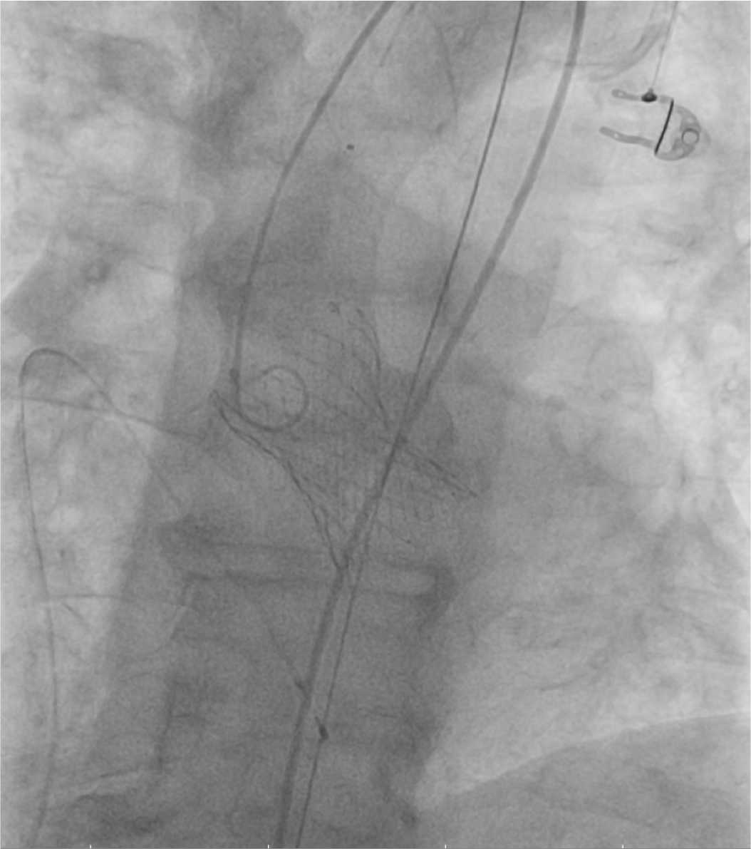
Figure 7
Fluoroscopic image: deployment of the pre-dilated Medtronic valve.
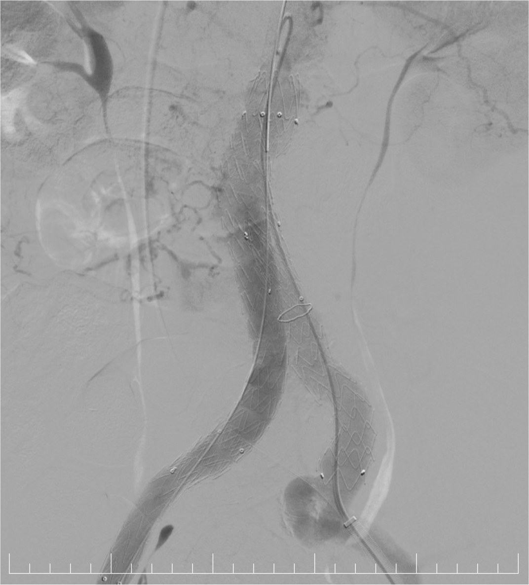
Figure 8
Fluoroscopic image: EVAR successfully performed.