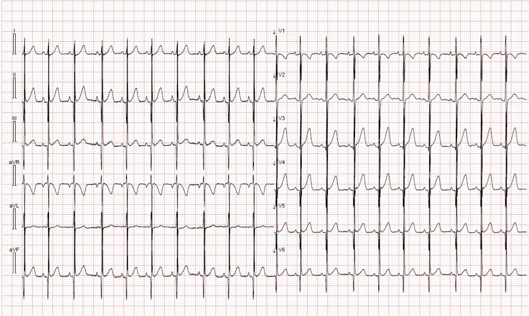
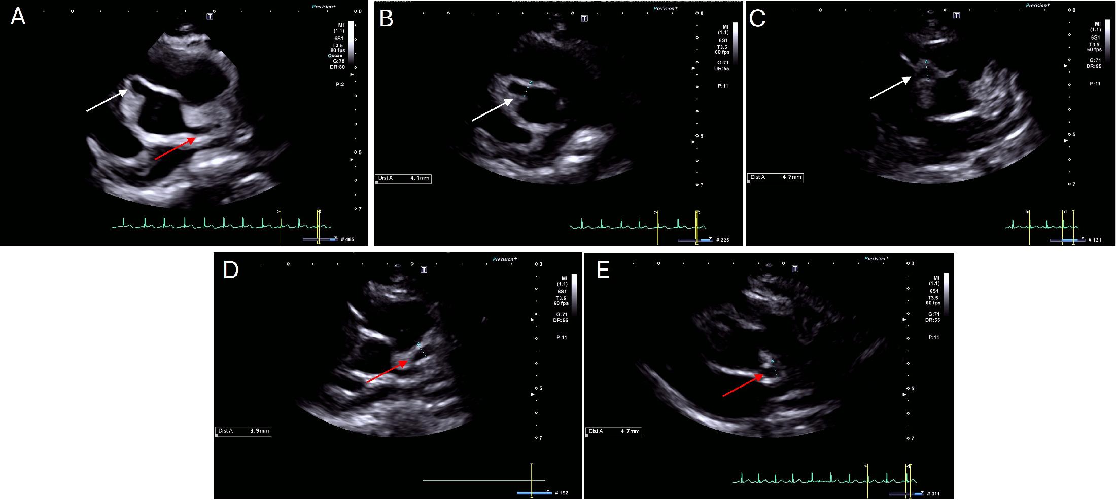
Have a personal or library account? Click to login
A case of severe Kawasaki disease: from multiple coronary aneurysms to interleukin-1 inhibition Cover

A case of severe Kawasaki disease: from multiple coronary aneurysms to interleukin-1 inhibition

Open Access
|Sep 2025

Figures & Tables

Figure 1

Surface 12-lead ECG depicting sinus tachycardia, with a normal repolarization pattern.

Figure 2

Transthoracic echocardiography in the parasternal short-axis view showing a right coronary artery aneurysm (Panel A, white arrow) and a left main coronary artery aneurysm (Panel A, red arrow). The proximal portion of the right coronary artery (Panel B, white arrow) has a diameter of 4.1 mm (Z-score +7.4), while the distal portion of the right coronary artery (Panel C, white arrow) has a diameter of 4.7 mm (Z-score +7.8). The left main coronary artery bifurcation (Panel D, red arrow) measured 3.9 mm (Z-score +4.9), while the proximal left anterior descending artery (Panel E, red arrow) measured 4.7 mm (Z-score +8.1).

Figure 3

Cardiac computed tomography showing axial plane after multiplanar reconstruction of the left main coronary artery (Panel A) with a maximum diameter of 4.6 mm (Z-score +5.3); the left anterior descending artery (Panel B) with a diameter of 4.6 mm (Z-score +10.0); the circumflex artery with a diameter of 3.5 mm; The right coronary artery in axial reconstruction (Panel D) depicts a proximal diameter of 5 mm (Z-score +9.0).

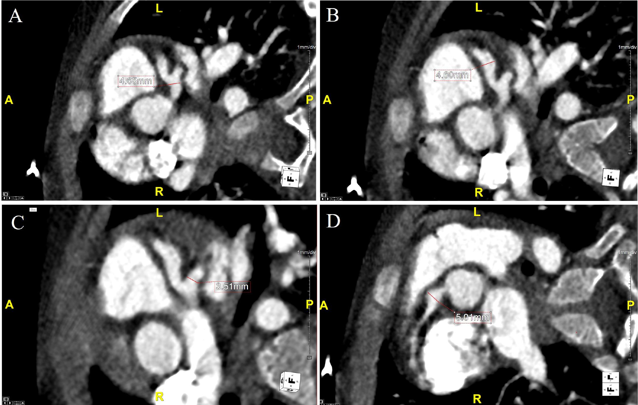
Overview of the clinical course and ongoing management of Kawasaki disease in a young patient_

SymptomsLeft main coronary arteryLeft anterior descending arteryRight coronary arteryTreatment
DiagnosisFever, maculopapular rash1.8 mm (Z-score -0.23)-1.5 mm (Z-score -0.49)IVIG, aspirin, corticosteroids
Follow-up 1 (15 days)Asymptomatic3 mm (Z-score +1.6)4 mm (Z-score +9.0)5 mm (Z-score +7.8)Aspirin, clopidogrel, heparin, warfarin, anakinra
Follow-up 2 (64 days)Asymptomatic3.8 mm (Z-score +2.1)4.1 mm (Z-score +9.0)4.4 mm (Z-score +7.2)Aspirin, clopidogrel, warfarin, anakinra
Follow-up 3 (145 days)Asymptomatic3.4 mm (Z-score +2.4)2.6 mm (Z-score +2.5)2.7 mm (Z-score +2.3)Aspirin, clopidogrel
Follow-up 4 (163 days)Asymptomatic2.8 mm (Z-score +0.9)2.1 mm (Z-score +0.67)2.1 mm (Z-score +0.56)Aspirin
Follow-up 5 (462 days)Asymptomatic2.4 mm (Z-score -0.11)2.2 mm (Z-score +0.55)2.0 mm (Z-score -0.11)Stop aspirin
DOI: https://doi.org/10.2478/rjc-2025-0015 | Journal eISSN: 2734-6382 | Journal ISSN: 1220-658X
Language: English
Page range: 213 - 219
Published on: Sep 30, 2025
Published by: Romanian Society of Cardiology
In partnership with: Paradigm Publishing Services
Publication frequency: 4 issues per year

© 2025 Dan-Alexandru Cozac, Diana Di Paolantonio, Giovanni Di Salvo, Alina Scridon, published by Romanian Society of Cardiology
This work is licensed under the Creative Commons Attribution-NonCommercial-NoDerivatives 4.0 License.