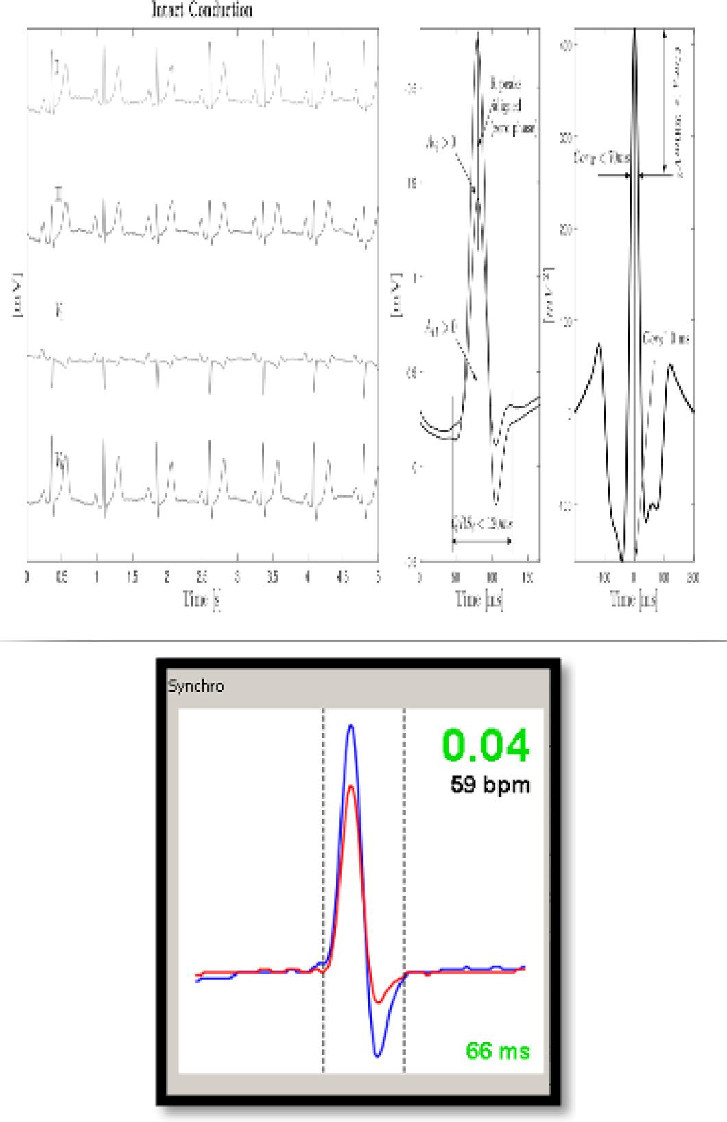
Have a personal or library account? Click to login
The Electrocardiogram and New Methods for Measuring Electrical Synchrony in Cardiac Resynchronization Therapy and Conduction System Stimulation Cover

The Electrocardiogram and New Methods for Measuring Electrical Synchrony in Cardiac Resynchronization Therapy and Conduction System Stimulation

Open Access
|Mar 2025

Figures & Tables

Figure 1

a and b. a. Left panel: Chart of electrical synchrony evaluation by Synchromax® method. Curves are divided according to the synchrony index value: between 0 and 0.4 are synchronous; between 0.41 and 0.7 are intermediate; and between 0.71 and 1 are dyssynchronous. Note the different curves in each column. b. Right panel: Screen delivered by the device for electrical synchrony evaluation

Figure 2

Online electrical synchrony evaluation by Synchromax ® method in patients with CRT. Post-implant optimization. Different synchrony curves and indexes are evident after V-V interval programming

Figure 3

Synchromax curves in true left bundle branch block (LBBB) (index 1) and pseudo LBBB (index 0.4)

Figure 4

a and b. a. Cross correlation time and amplitude between DII and V6. Parameters evaluated:

-Maximum QRS amplitude correlation
-QRS width correlation 70% amplitude
-Correlation of changes to the peak of the signal.
-QRS duration from spike to last QRS
-Area under QRS V6 with cross-correlation between II and V6 b. Synchrony index.
a and b. a. Cross correlation time and amplitude between DII and V6. Parameters evaluated: -Maximum QRS amplitude correlation -QRS width correlation 70% amplitude -Correlation of changes to the peak of the signal. -QRS duration from spike to last QRS -Area under QRS V6 with cross-correlation between II and V6 b. Synchrony index.

His selective and no selective criteria_ CHUNG et al_, 2023 HRS, APHRS, LAHRS, guideline on cardiac physiologic pacing

BaselineNormal QRS durationHis-Purkinje conduction disease
With correctionWithout correction
Selective HBP
  • S-QRS = H-QRS with isoelectric interval

  • Discrete Local ventricular electrogram in HBP lead with S-V = H-V

  • Paced QRS = native QRS

  • Single capture threshold (His bundle)

  • S-QRS ≤ H-QRS with isoelectric interval

  • Discrete local ventricular electrogram in HBP lead

  • Paced QRS < native QRS

  • 2 distinct capture thresholds (HBP with BBB correction, HBP without BBB correction)

  • S-QRS ≤ or > H-QRS with isoelectric interval

  • Discrete local ventricular electrogram in HBP lead

  • Paced QRS = native QRS

  • Single capture threshold (HBP with BBB)

Nonselective HBP
  • S-QRS < H-QRS (usually 0, S-QRSend = H-QRSend) with or without isoelectric interval (pseudodelta wave +/–)

  • Direct capture of local ventricular electrogram in HBP lead by stimulus artifact (local myocardial capture)

  • Paced QRS > native QRS with normalization of precordial and limb lead axes with respect to rapid dV/dt components of the QRS

  • 2 distinct capture thresholds (His bundle capture, RV capture)

  • No QRS slur/notch in leads I, V1, or V4–V6, and V6 R-wave peak time ≤ 100 ms

  • Change in V6 RWPT > 12 ms between stimulus and His to V6 RWPT confirms lack of His capture (99.1% sensitivity and 100% specificity)

  • S-QRS < H-QRS (usually 0, S-QRSend < H-QRSend) with or without isoelectric interval (pseudodelta wave +/–)

  • Direct capture of local ventricular electrogramin HBP lead by stimulus artifact

  • Paced QRS ≤ native QRS

  • 3 distinct capture thresholds (HBP with BBB correction, HBP without BBB correction, RV capture)

  • S-QRS < H-QRS (usually 0) with or without isoelectric interval (pseudodelta wave +/–)

  • Direct capture of Local ventricular electrogram in HBP Lead by stimulus artifact

  • Paced QRS > native QRS

  • 2 distinct capture thresholds (HBP with BBB, RV capture)

j_rjc-2025-0010_utab_001

Pacing typeCriteria
Left ventricular septal pacing
  • Deep septalplacement of the pacing Lead (confirmed by fulcrum sign, contrast, echocardiogram, or CT) and

  • Right bundle branch conduction delay pattern in lead V1 (rare exceptions)

Left bundle branch area pacing
  • Evidence for LV septal pacing in addition to any one of the following LBB capture criteria:

LBB capture criteria
  • Nonselective to selective LBBP or nonselective to septal capture transition during threshold testing

  • Abrupt shortening of RWPT or LVAT in V6≥10 ms at high output during deep septal position with subsequent short and constant LVAT at low output with further advancement of the Lead

  • V6 RWPT <74 ms in non-LBBB and <80 ms in LBBB

  • V6–V1 interpeak interval >44 ms

  • Physiology-based criteria

    • QRS onset to RWPT ≤ native RWPT (+10 ms)

    • Stimulus to RWPT ≤ LBB potential to V6 RWPT (+10 ms)

    • Stimulus to V6 RWPT + 10 ms < (intrinsicoid deflection time - transseptal conduction time) in LBBB

  • Programmed deep septal stimulation demonstrating differential capture

  • Change in V6 RWPT between (corrective) HBP and LBBP>8 ms in LBBB

  • Demonstration of LBB potential with injury current

  • Demonstration of stimulus to retrograde His <35 ms or anterograde left conduction system potential preceding ventricular electrogram during LBBP

Recommendations of CRT in sinus rhythm

RECOMMENDATIONSClassLevel
CRT is recommended in patients in sinus rhythm with symptomatic HF, LVEF less than 35%, R-wave width longer than 150 ms and LBBB morphology, despite optimal medical treatment (reduction of symptoms and morbimortality)IA
CRT must be considered in patients in sinus rhythm, symptomatic HF, LVEF less than 35%, QRS width between 130 and 149 ms with LBBB morphology despite optimal medical treatment (reduction of symptoms and morbimortality)II aB
CRT is recommended in patients in sinus rhythm with symptomatic HF, LVEF less than 35%, QRS over 150 ms and morphology DIFFERENT than LBBB, despite optimal medical treatment (reduction of symptoms and morbimortality)II aB
CRT must be considered for patients in sinus rhythm, symptomatic HF, LVEF less than 35%, QRS width between 130 and 149 ms with morphology DIFFERENT than LBBB despite optimal medical treatment (reduction of symptoms and morbimortality)II bB
Patients with narrow QRS candidates to CRT, AV node ablationII bC
Patients with AF and HF candidates to CRT when LVEF less than 35% in FC IIII-IV with optimal treatment, QRS width less than 130 ms with a strategy assuring adequate BiV capture (90-95%). AV node ablation could be added with this purposeII aB
CRT is recommended instead of RV pacing for patients with HFrEF (less than 40%) in any functional class, with indication of ventricular pacing and with high degree AV block, with the purpose of reducing morbidity. This includes patients with AFIA

Left bundle branch capture criteria

Pacing typeCriteria
Left ventricular septal pacing
  • Deep septal location of pacing lead (confirmed by fulcrum sign, contrast, echocardiogram or CAT scan and

  • Right branch conduction delay pattern in V1 (in rare exceptions)

Left bundle branch area pacing1. Evidence of LV septal pacing added to any of the following left branch capture criteria: Left bundle capture criteria
  • Non-selective left branch pacing or transition from non-selective pacing to septal capture while measuring capture threshold.

  • Sudden shortening of peak time to R-wave, or LV activation in V6≥10 ms with high output in a deep septal location, with subsequent and constant LV activation time with low output while the pacing lead is advanced.

Physiology-based criteria
  • Interval from QRS onset to peak R-wave ≤ peak time to native R-wave (+10 ms.

  • Spike to R-wave peak time ≤ left branch potential to R-wave peak time (+10 ms).

  • Spike to R-wave peak time in V6 +10 ms < (intrinsicoid deflection time -transeptal conduction time) in LBBB.

  • Programmed deep septal pacing demonstrating differential capture

  • Changes in R-wave peak time in V6 between (corrected) His bundle pacing and left branch pacing >8 ms in LBBB.

  • Demonstration of left branch potentials blocking with injury current.

  • Demonstration of spike to retrograde His <35 ms or conduction system anterograde left potential preceding the ventricular electrogram during left branch pacing.

Left bundle branch area pacing2. Evidence of LV septal pacing added to any of the following left branch capture criteria: Left bundle capture criteria
  • Non-selective left branch pacing or transition from non-selective pacing to septal capture while measuring capture threshold.

  • Sudden shortening of peak time to R-wave, or LV activation in V6≥10 ms with high output in a deep septal location, with subsequent and constant LV activation time with low output while the pacing lead is advanced.

Physiology-based criteria
  • Interval from QRS onset to peak R-wave ≤ peak time to native R-wave (+10 ms.

  • Spike to R-wave peak time ≤ left branch potential to R-wave peak time (+10 ms).

  • Spike to R-wave peak time in V6 +10 ms < (intrinsicoid deflection time - transeptal conduction time) in LBBB.

  • Programmed deep septal pacing demonstrating differential capture

  • Changes in R-wave peak time in V6 between (corrected) His bundle pacing and left branch pacing >8 ms in LBBB.

  • Demonstration of left branch potentials blocking with injury current.

  • Demonstration of spike to retrograde His <35 ms or conduction system anterograde left potential preceding the ventricular electrogram during left branch pacing.

DOI: https://doi.org/10.2478/rjc-2025-0010 | Journal eISSN: 2734-6382 | Journal ISSN: 1220-658X
Language: English
Page range: 17 - 25
Published on: Mar 31, 2025
Published by: Romanian Society of Cardiology
In partnership with: Paradigm Publishing Services
Publication frequency: 4 issues per year

© 2025 Ortega Daniel Felipe, Paolucci Analía, Logarzo Emilio, published by Romanian Society of Cardiology
This work is licensed under the Creative Commons Attribution 4.0 License.