
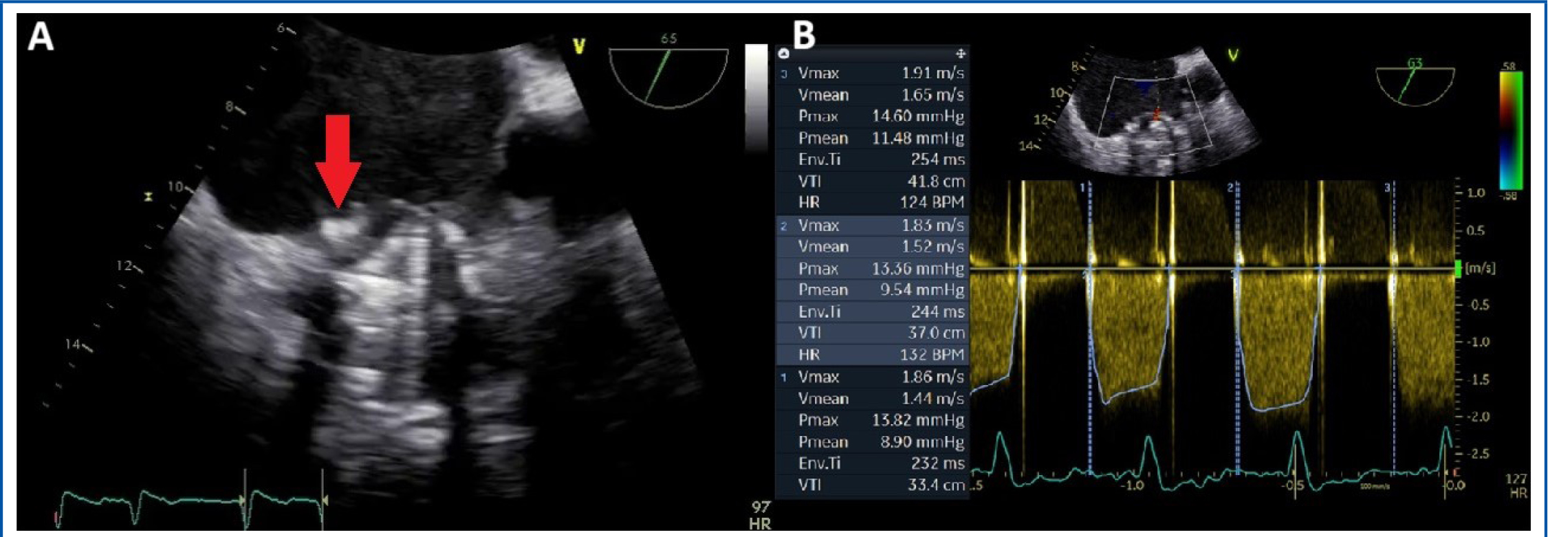
Figure 1
Transoesophageal echocardiography. A) 2D-image showing a stuck leaflet of the tricuspid prosthesis due to both pannus and thrombus (red arrow). B) CW Doppler flow interrogation of the tricuspid prosthesis revealing high mean transvalvular pressure gradient (examination in atrial fibrillation).
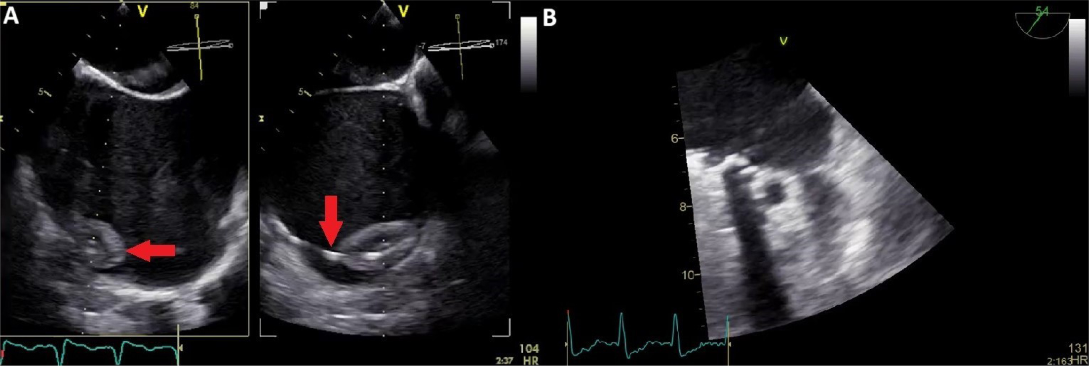
Figure 2
Transoesophageal echocardiography. A) Pacing lead thrombosis (red arrow). B) Left atrial appendage thrombosis.
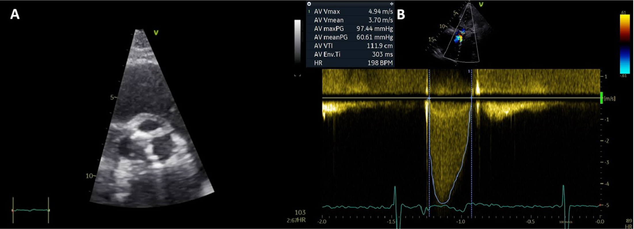
Figure 3
Transthoracic echocardiography. A) 2D parasternal short axis view of the aortic valve showing severe valvular calcifications. B) CW Doppler interrogation showing high velocity transvalvular aortic flow.
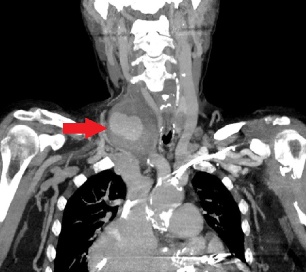
Figure 4
Computed tomography angiography shows a laterocervical pseudoaneurysm.
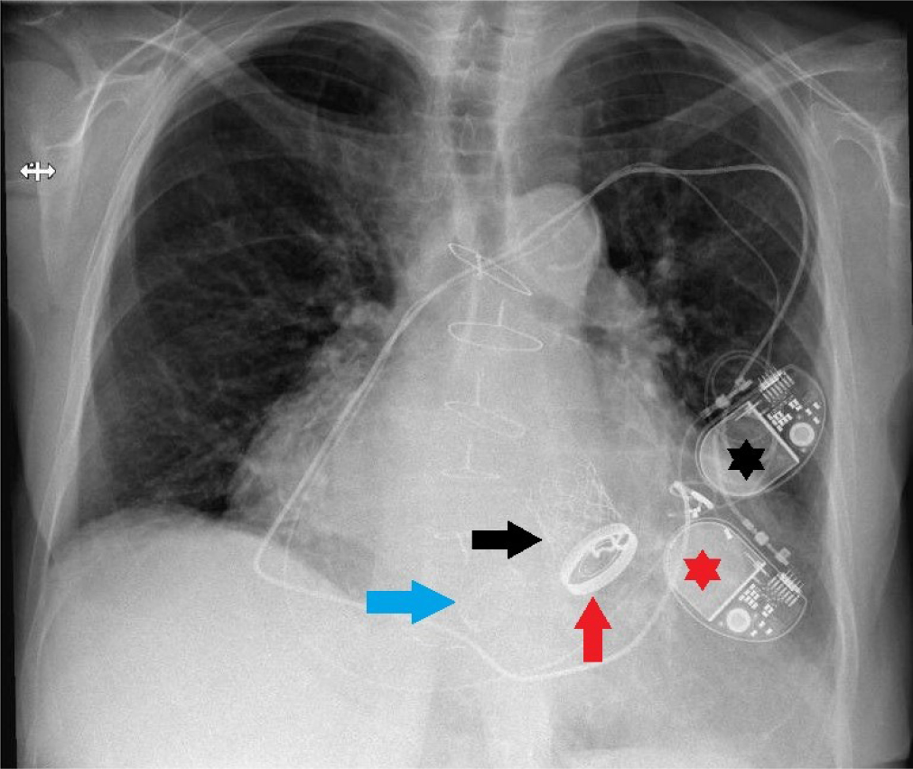
Figure 5
Chest X-Ray revealing the three different valve prosthese (blue arrow - tricuspid prosthesis, red arrow - mitral prosthesis, blac arrow - aortic prosthesis) and the two pacing systems (black asterisk first pacemaker, red asterisk - second pacemaker).