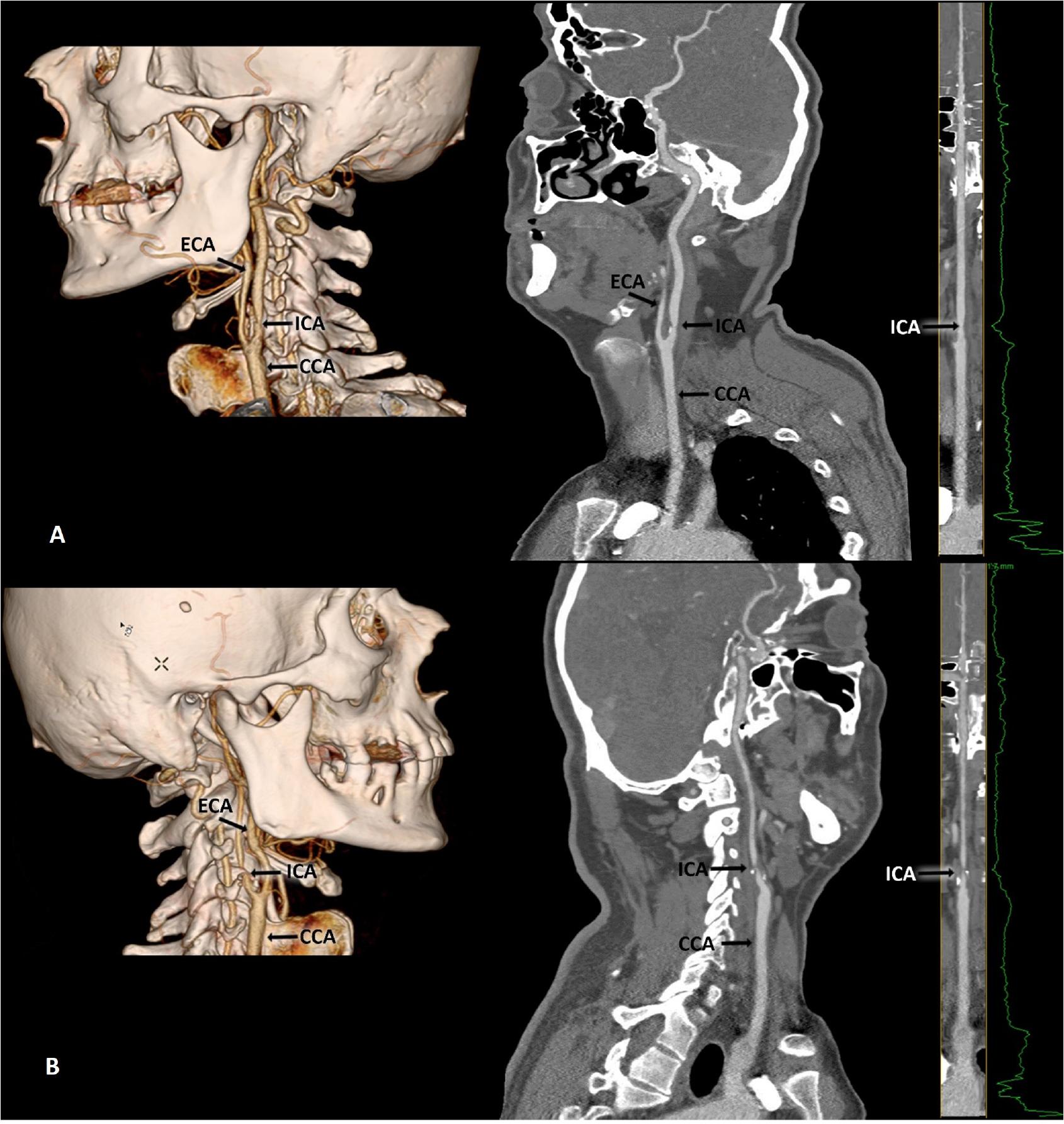
Figure 1
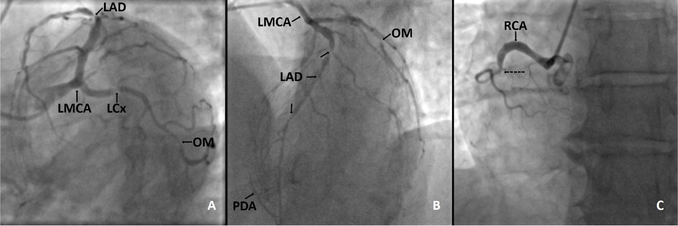
Figure 2
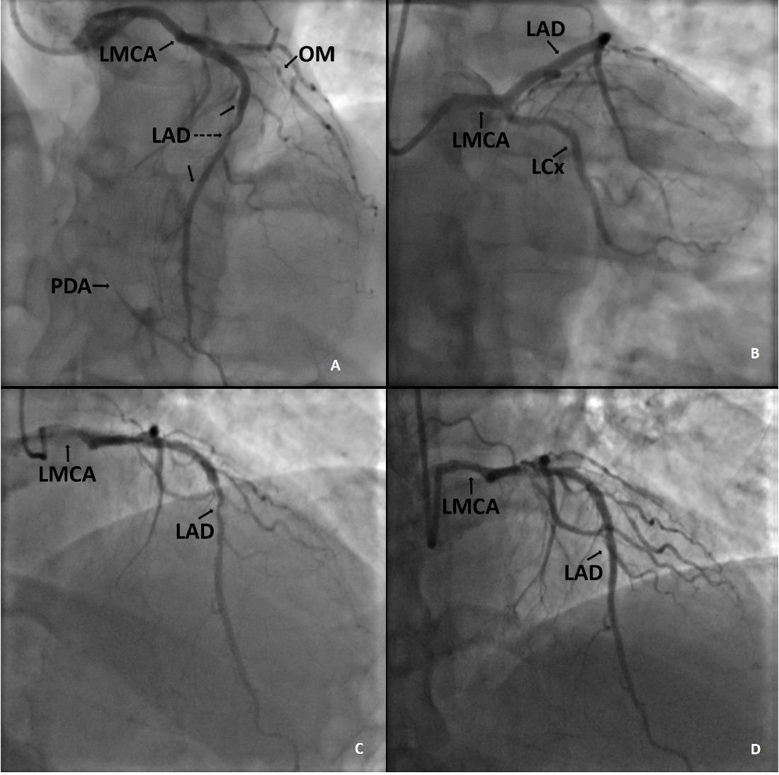
Figure 3
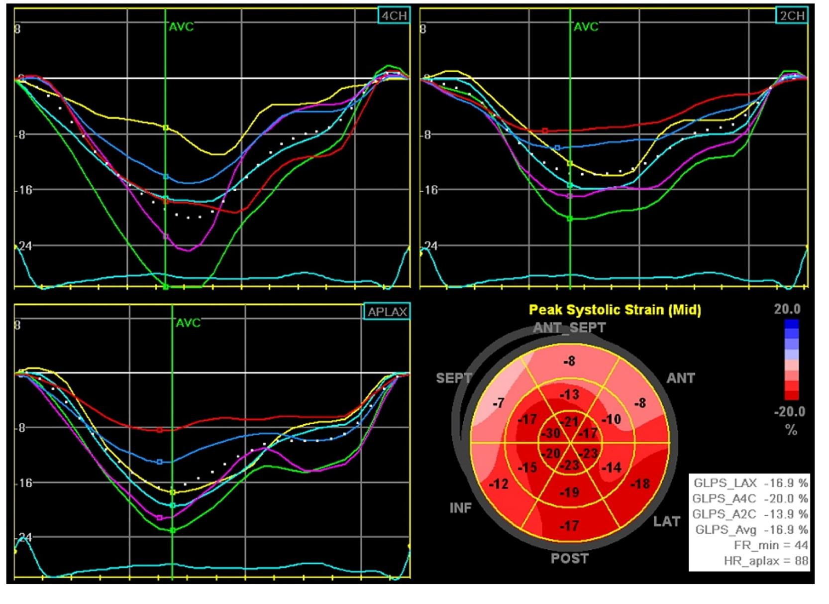
Figure 4
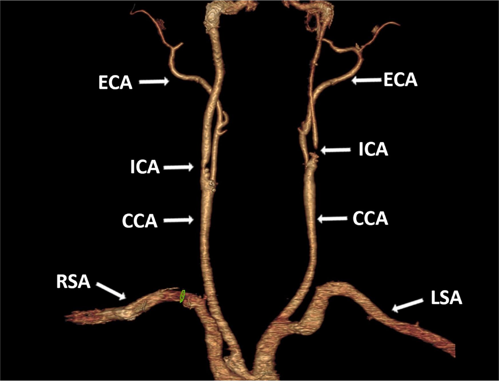
Figure 5
© 2025 Radu Adrian Suteu, Diana-Andreea Moldovan, Marian Pop, Ioan Tilea, Laurentiu Huma, Andreea Varga, published by Romanian Society of Cardiology
This work is licensed under the Creative Commons Attribution-NonCommercial-NoDerivatives 4.0 License.