
Table 1
The advantages and limitations of the current cardiac computed tomography methods for assessing coronary artery plaque burden.
| CT method | Description | Advantages | Limitations |
|---|---|---|---|
| SSS | Scores each segment based on stenosis severity | Quick estimation of plaque burden | Lacks detailed plaque composition |
| SIS | Scores the presence of plaque in each segment | Simple, rapid assessment | Does not quantify stenosis severity |
| Leaman Score | Considers plaque location, type, and stenosis degree | Detailed assessment | Time-consuming |
| Quantitative | Automated plaque volume and composition analysis | Accurate, detailed quantification | Requires advanced software |
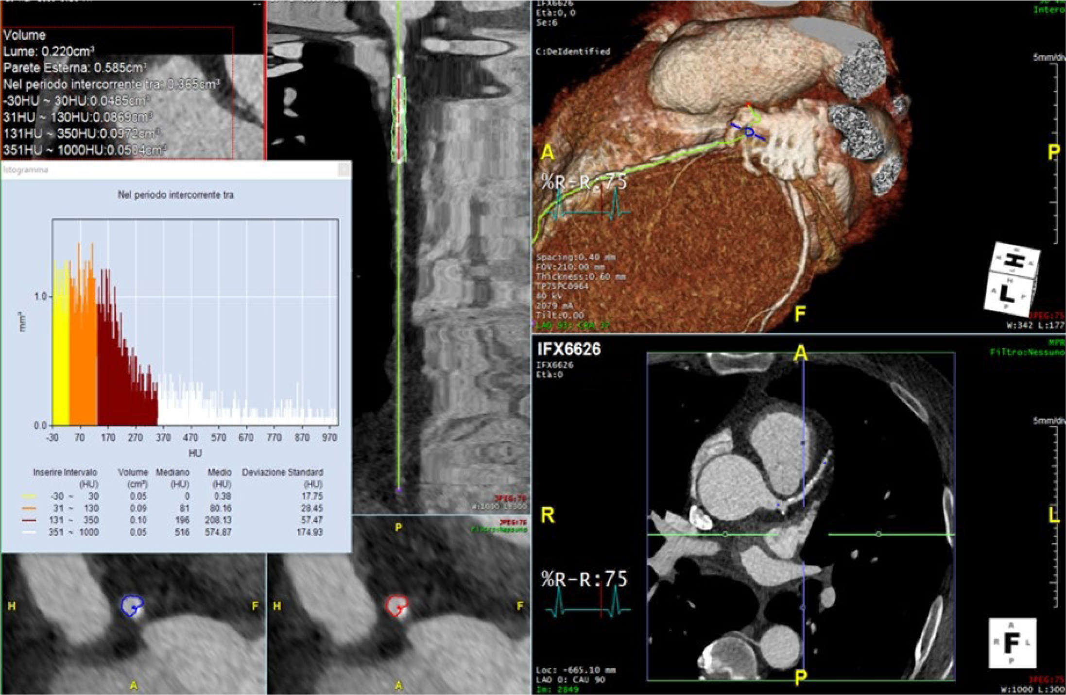
Figure 1
Methods of coronary plaque characterization in the left anterior descending artery using the software Aquarius TeraRecon (North Carolina, United States of America). The histogram of different plaque components is depicted on the left panel.
Table 2
Characteristics and clinical relevance of high-risk coronary plaque features detected by cardiac computed tomography angiography.[31]
| Plaque Feature | Description | Clinical Relevance |
|---|---|---|
| Positive Remodeling | Vessel enlargement at the plaque site | Indicator of plaque vulnerability |
| Spotty Calcifications | 1-3 mm calcifications in the context of a soft plaque | Indicator of plaque vulnerability |
| Low Attenuation Plaque | Low density (<30 HU) | Associated with increased risk of rupture |
| Napkin-Ring Sign | Hypoattenuating core with a hyperattenuating rim | A strong predictor of adverse events |
Table 3
Summary of the key conclusions of major trials evaluating the role of CCTA in cardiovascular risk assessment.
| Title | Journal, year | Key conclusions |
|---|---|---|
| Coronary CT Angiography versus Standard Evaluation in Acute Chest Pain (ROMICAT-II) [34] | N Engl J Med. 2012 |
|
| 1-Year Outcomes of FFRCT-Guided Care in Patients With Suspected Coronary Disease: The PLATFORM Study [46] | J Am Coll Cardiol. 2016 |
|
| Prognostic Value of Noninvasive Cardiovascular Testing in Patients With Stable Chest Pain: Insights From the PROMISE Trial (Prospective Multicenter Imaging Study for Evaluation of Chest Pain)[5] | Circulation. 2017 |
|
| Coronary CT Angiography and 5-Year Risk of Myocardial Infarction (SCOT-HEART trial)[4] | N Engl J Med. 2018 |
|
| Non-invasive detection of coronary inflammation using computed tomography and prediction of residual cardiovascular risk (the CRISP CT study): a post-hoc analysis of prospective outcome data[54] | Lancet. 2018 |
|
| CT or Invasive Coronary Angiography in Stable Chest Pain (DISCHARGE trial)[16] | N Engl J Med. 2022 |
|
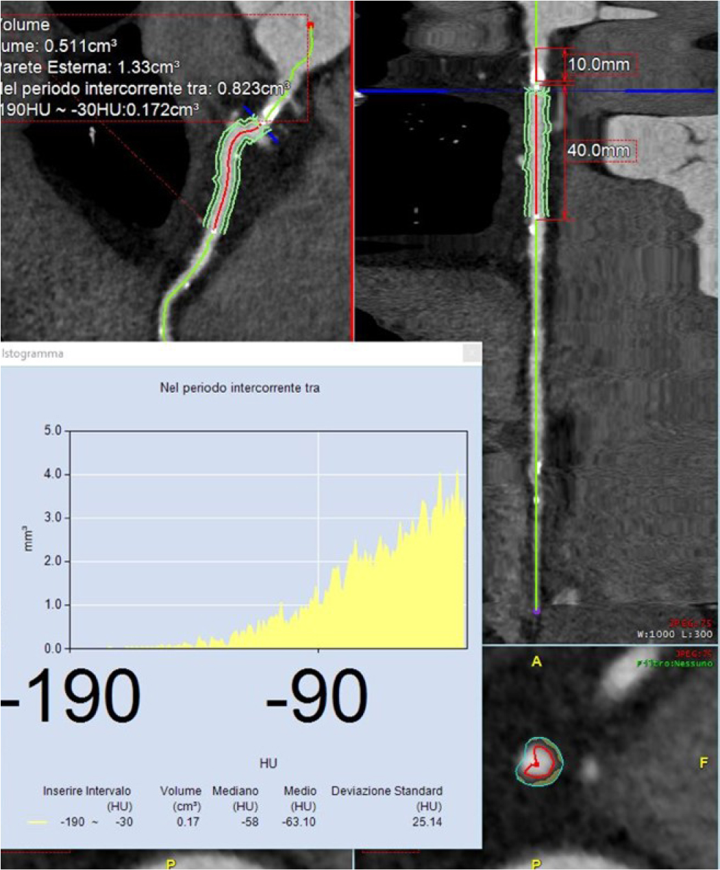
Figure 2
Pericoronary fat attenuation index (pFAI) analyses using the software Aquarius TeraRecon (North Carolina, United States of America) show gradients in the CT attenuation of peri coronary fat.
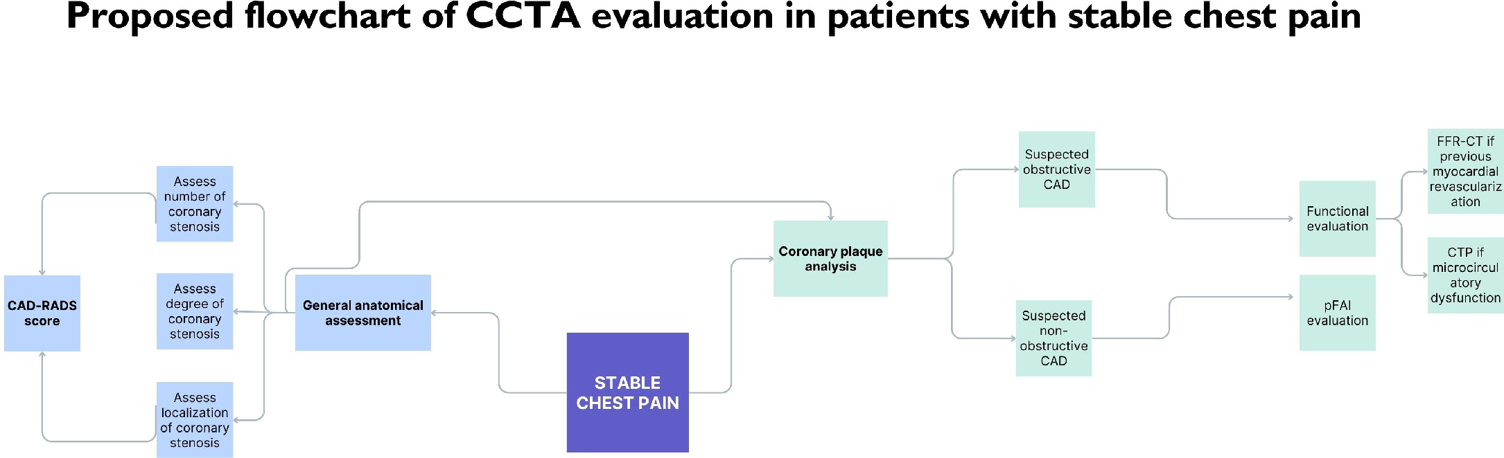
Figure 3
The proposed flowchart of the cardiac coronary computed tomography (CCTA) evaluation in patients with stable chest pain.