
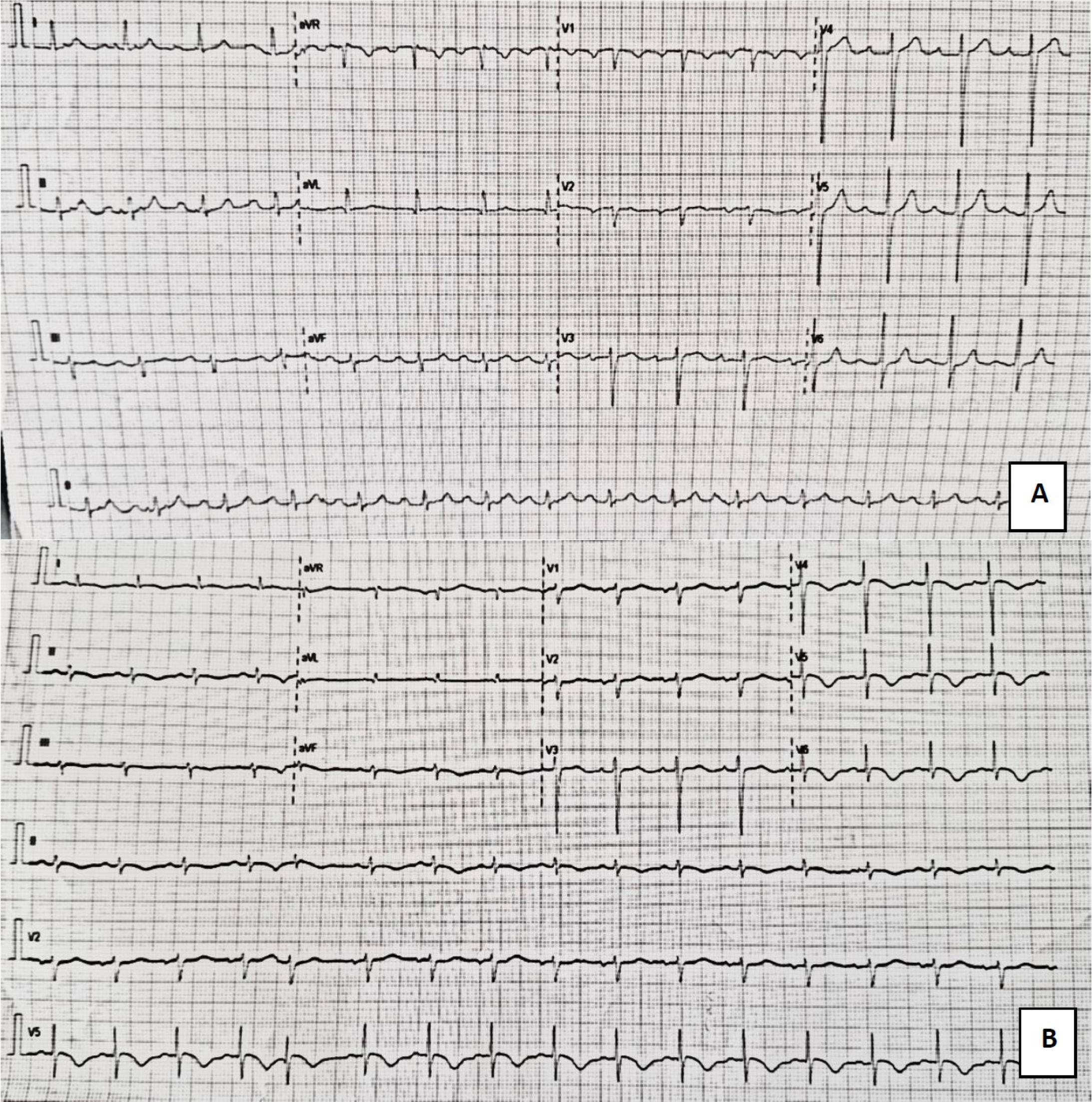
Figure 1 A.
Normal preoperative ECG; B. Day 2 postoperative ECG findings showing sinus tachycardia 105 bpm, new T wave inversion DI, aVL, V4-V6, QTc prolongation (QTc 516 ms)
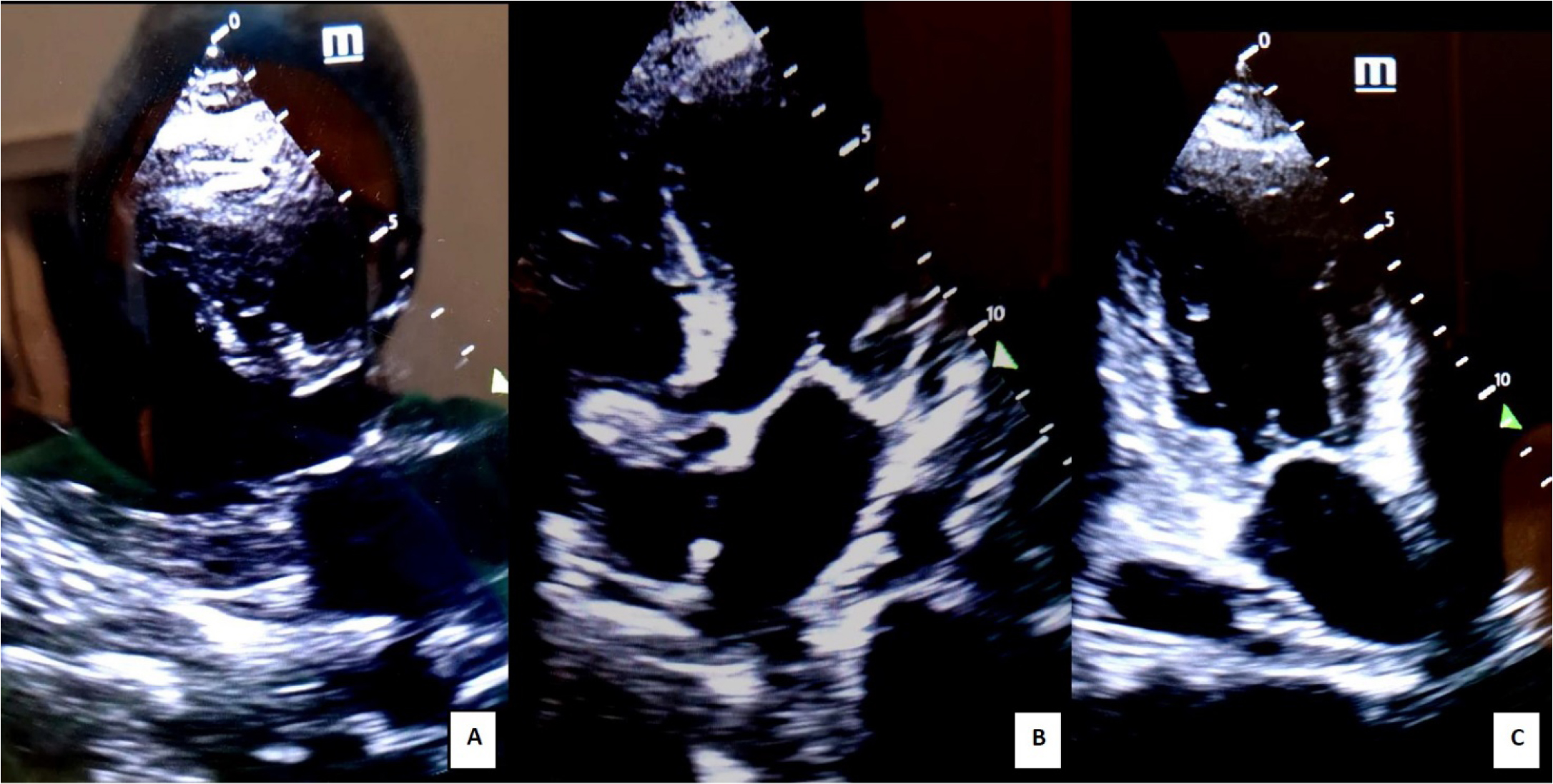
Figure 2
Transthoracic POCUS echocardiography 2nd day post-op: A. parasternal long axis view; B. apical four chamber view; C. apical 2 chamber view. Severe hypokinesia of 2/3 apical segments of anterior, inferior, and lateral walls (“apical ballooning” aspect of the left ventricle - dysfunctional myocardium extending beyond the territory of a single coronary artery); severe LV systolic dysfunction
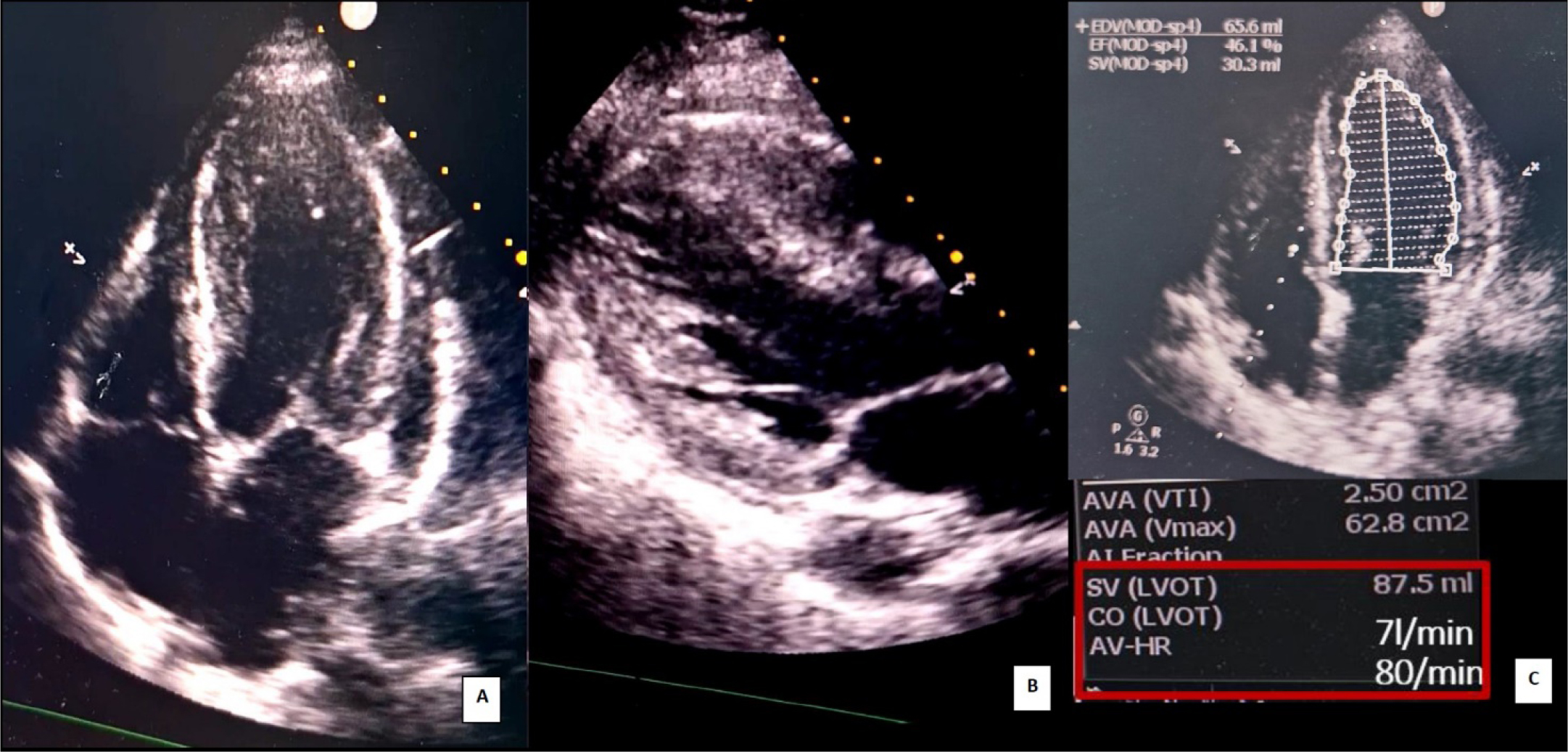
Figure 3
Transthoracic POCUS echocardiography 9th day post-op: A. apical four chamber view; B. parasternal long axis view; improved left ventricular systolic function, minimal hypokinesia of apical left ventricle walls; C. Left ventricular ejection fraction (LVEF) 46% (Simpson monoplane); Stroke volume (SV) and cardiac output (CO) determination using the continuity equation