
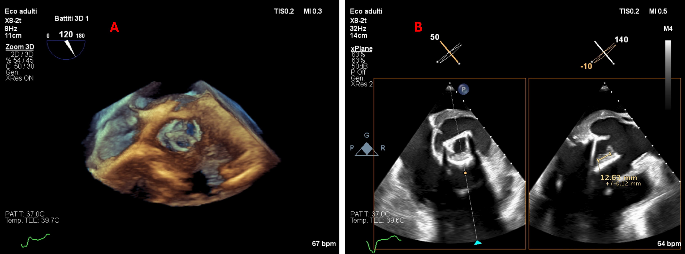
Figure 1
Transesophageal echocardiography using the three-dimensional (A), and the multi-plane method (B) to represent the aortic valve anatomy and identify the prosthetic valvular mass
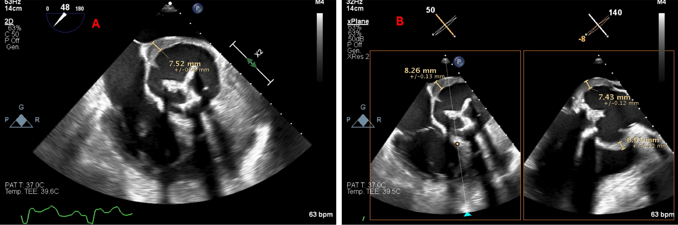
Figure 2
Transesophageal echocardiography in short-axis view identifying (A) the anterior thickening of the aortic wall, and (B) using the multi-plane modality, the inhomogeneous material both anteriorly and posteriorly (maximum 9 mm), extending up to the sino-tubular junction, highly suggestive of aortic root aneurysm and aortitis
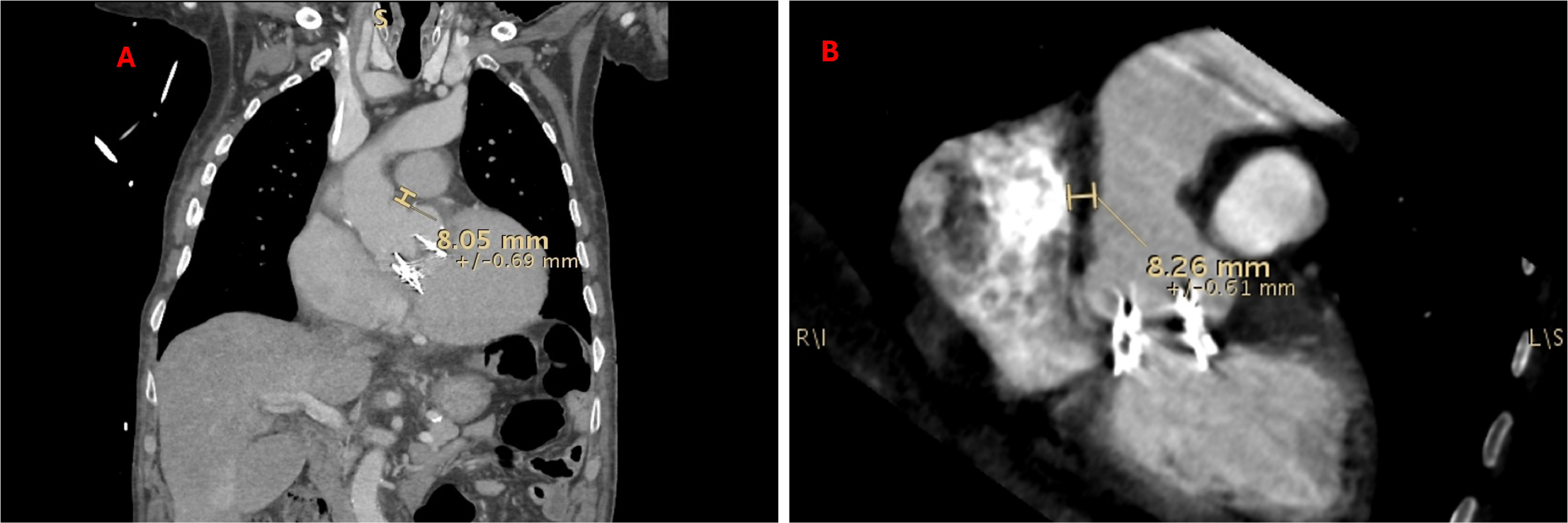
Figure 3
Computed tomography showing (A) in the frontal plane, the anterolateral thickening of the aortic wall; and (B) in the multiplanar reconstruction, depicting the inhomogeneous material with the irregular contour of the aortic root posteriorly